Sacrovertebral angle

Last updated
Sacrovertebral angle

Gray 111 - Vertebral column-coloured.png

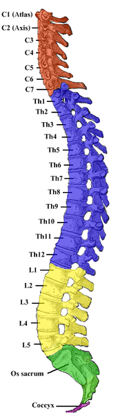
Lumbar vertebrae are yellow, and sacrum is green.
Identifiers

Anatomical terminology

The sacrum is curved upon itself and placed very obliquely, its base projecting forward and forming the prominent sacrovertebral angle when articulated with the last lumbar vertebra.

Sacrum bone of the pelvis

The sacrum, in human anatomy, is a large, triangular bone at the base of the spine that forms by the fusing of sacral vertebrae S1–S5 between 18 and 30 years of age.

It is also known as the "lumbosacral angle". [1]

See also

Related Research Articles

Scapula bone that connects the humerus (upper arm bone) with the clavicle (collar bone)

In anatomy, the scapula, also known as shoulder bone, shoulder blade, wing bone or blade bone, is the bone that connects the humerus with the clavicle. Like their connected bones the scapulae are paired, with the scapula on either side of the body being roughly a mirror image of the other. The name derives from early Roman times when it was thought that the bone resembled a trowel or small shovel.

Thoracic duct

In human anatomy, the thoracic duct is the larger of the two lymph ducts of the lymphatic system. It is also known as the left lymphatic duct, alimentary duct, chyliferous duct, and Van Hoorne's canal. The other duct is the right lymphatic duct. It carries chyle, a liquid containing both lymph and emulsified fats, rather than pure lymph. Thus when it ruptures, the resulting flood of liquid into the pleural cavity is known as chylothorax.

Mediastinum central compartment of the thoracic cavity that separates the lungs

The mediastinum is the central compartment of the thoracic cavity surrounded by loose connective tissue, as an undelineated region that contains a group of structures within the thorax. The mediastinum contains the heart and its vessels, the esophagus, the trachea, the phrenic and cardiac nerves, the thoracic duct, the thymus and the lymph nodes of the central chest.

Orbicularis oris muscle

In human anatomy, the orbicularis oris muscle is a complex of muscles in the lips that encircles the mouth. Until recently, it was misinterpreted as a sphincter, or circular muscle, but it is actually composed of four independent quadrants that interlace and give only an appearance of circularity.

External jugular vein

The external jugular vein receives the greater part of the blood from the exterior of the cranium and the deep parts of the face, being formed by the junction of the posterior division of the retromandibular vein with the posterior auricular vein.

Hip anatomical region

In vertebrate anatomy, hip refers to either an anatomical region or a joint.

Sternal angle

The sternal angle is the synarthrotic joint formed by the articulation of the manubrium and the body of the sternum.

Anatomical terms of motion Wikimedia list article

Motion, the process of movement, is described using specific anatomical terms. Motion includes movement of organs, joints, limbs, and specific sections of the body. The terminology used describes this motion according to its direction relative to the anatomical position of the joints. Anatomists use a unified set of terms to describe most of the movements, although other, more specialized terms are necessary for describing the uniqueness of the movements such as those of the hands, feet, and eyes.

Round ligament of uterus

The round ligament of the uterus originates at the uterine horns, in the parametrium. The round ligament exits the pelvis via the deep inguinal ring, passes through the inguinal canal and continues on to the labia majora where its fibers spread and mix with the tissue of the mons pubis.

Superior ophthalmic vein

The superior ophthalmic vein begins at the inner angle of the orbit in a vein named the nasofrontal which communicates anteriorly with the angular vein; it pursues the same course as the ophthalmic artery, and receives tributaries corresponding to the branches of that vessel.

Iliocostalis

The iliocostalis is the muscle immediately lateral to the longissimus that is the nearest to the furrow that separates the epaxial muscles from the hypaxial. It lies very deep to the fleshy portion of the serratus posterior muscle.

Transverse cervical artery Artery of the neck

The transverse cervical artery is an artery in the neck and a branch of the thyrocervical trunk, running at a higher level than the suprascapular artery.

Trochlea of humerus Trochlea of humerus

In the human arm, the humeral trochlea is the medial portion of the articular surface of the elbow joint which articulates with the trochlear notch on the ulna in the forearm.

Pubic arch

The pubic arch, also referred to as the ischiopubic arch, is part of the pelvis. It is formed by the convergence of the inferior rami of the ischium and pubis on either side, below the pubic symphysis. The angle at which they converge is known as the subpubic angle.

Lateral aperture

The lateral aperture is a paired structure in human anatomy. It is an opening in each lateral extremity of the lateral recess of the fourth ventricle of the human brain, which also has a single median aperture. The two lateral apertures provide a conduit for cerebrospinal fluid to flow from the brain's ventricular system into the subarachnoid space; specifically into the pontocerebellar cistern at the cerebellopontine angle. The structure is also called the lateral aperture of the fourth ventricle or the foramen of Luschka after anatomist Hubert von Luschka. Gross total resection of tumours that extend through foramen of Lushka is sometimes not possible due to bradycardia.

Zygomatic branches of the facial nerve

The zygomatic branches of the facial nerve run across the zygomatic bone to the lateral angle of the orbit, where they supply the orbicularis oculi, and join with filaments from the lacrimal nerve and the zygomaticofacial branch of the maxillary nerve.

Costodiaphragmatic recess

The costodiaphragmatic recess, also called the costophrenic recess or phrenicocostal sinus, is a potential space in the pleural cavity, at the posterior-most tips of the cavity, located at the junction of the costal pleura and diaphragmatic pleura. It measures approximately 5 cm vertically and extends from the eighth to the tenth rib along the mid-axillary line.

Angle of the mandible

The angle of the mandible is located at the posterior border at the junction of the lower border of the ramus of the mandible.

Infrasternal angle

The lower opening of the thorax is formed by the twelfth thoracic vertebra behind, by the eleventh and twelfth ribs at the sides, and in front by the cartilages of the tenth, ninth, eighth, and seventh ribs, which ascend on either side and form an angle, the infrasternal angle or subcostal angle, into the apex of which the xiphoid process projects.

Sternum flat bone in the middle front part of the rib cage

The sternum or breastbone is a long flat bone located in the central part of the chest. It connects to the ribs via cartilage and forms the front of the rib cage, thus helping to protect the heart, lungs, and major blood vessels from injury. Shaped roughly like a necktie, it is one of the largest and longest flat bones of the body. Its three regions are the manubrium, the body, and the xiphoid process. The word "sternum" originates from the Greek στέρνον, meaning "chest".

References

This article incorporates text in the public domain from page 106 of the 20th edition of Gray's Anatomy (1918)

The public domain consists of all the creative works to which no exclusive intellectual property rights apply. Those rights may have expired, been forfeited, expressly waived, or may be inapplicable.

<i>Grays Anatomy</i> English-language textbook of human anatomy

Gray's Anatomy is an English language textbook of human anatomy originally written by Henry Gray and illustrated by Henry Vandyke Carter. Earlier editions were called Anatomy: Descriptive and Surgical and Gray's Anatomy: Descriptive and Applied, but the book's name is commonly shortened to, and later editions are titled, Gray's Anatomy. The book is widely regarded as an extremely influential work on the subject, and has continued to be revised and republished from its initial publication in 1858 to the present day. The latest edition of the book, the 41st, was published in September 2015.

  1. A. Halim (2008). Human Anatomy:Volume Ii Abdomen And Lower Limb. I. K. International Pvt Ltd. pp. 4–. ISBN   978-81-906566-3-4 . Retrieved 19 May 2011.