Lateral pontine syndrome

Last updated
Lateral pontine syndrome
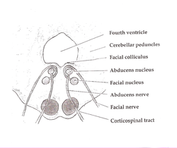
Pons section at facial colliculus.png
Pons
Specialty Neurology   OOjs UI icon edit-ltr-progressive.svg

Lateral pontine syndrome, also known as Marie-Foix syndrome or Marie-Foix-Alajouanine syndrome [1] is one of the brainstem stroke syndromes of the lateral aspect of the pons. A lateral pontine syndrome is a lesion which is similar to the lateral medullary syndrome, but because it occurs in the pons, it also involves the cranial nerve nuclei of the pons. [2]

Contents

History

Lateral pontine syndrome was first described in France by French neurologists Pierre Marie (1853-1940), Charles Foix (1882-1927), and Théophile Alajouanine (1890-1980) in 1922. They were the first to identify and describe the symptoms and causes of this syndrome.

In their original description, Pierre Marie, Charles Foix, and Théophile Alajouanine reported findings from autopsies that showed spinal cord necrosis and multiple tortuous and thickened blood vessels on the surface of the spinal cord. This condition was later called necrotizing myelopathy. [3]

They emphasized that in their two cases, no thrombosis was present. They considered the vascular component of the entity they reported to be a wall thickening, without luminal narrowing or obliteration of the cord vessels (arteries as well as veins). They addressed, and ruled out, the possibility of vascular malformations. [4]

These findings significantly contributed to the understanding of this syndrome

Symptoms

Damage to the following areas produces symptoms (from medial to lateral):

Structure affectedEffect
Lateral spinothalamic tract Contralateral loss of pain and temperature from the trunk and extremities.
Facial nucleus & facial Nerve (CN.VII)(1) Ipsilateral paralysis of the upper and lower face (lower motor neuron lesion). (2) Ipsilateral loss of lacrimation and reduced salivation. (3) Ipsilateral loss of taste from the anterior two-thirds of the tongue. (4) Loss of corneal reflex (efferent limb).
Principal sensory trigeminal nucleus and tractIpsilateral loss of all sensory modalities to the face (facial hemianesthesia)
Vestibular Nuclei and intraaxial nerve fibersNystagmus, nausea, vomiting, and vertigo
Cochlear nuclei and intraaxial nerve fibers Hearing loss - ipsilateral central deafness
Middle & inferior cerebellar peduncleIpsilateral limb and gait ataxia
Descending sympathetic tractIpsilateral Horner's syndrome (ptosis, miosis, & anhydrosis)

Causes

The syndrome occurs due to occlusion of perforating branches of the basilar and anterior inferior cerebellar (AICA) arteries. [5] [6] [7] It can also be caused by an interruption to the blood supply of the anterior inferior cerebellar artery or circumferential arteries. [8]

Treatment

The treatment for lateral pontine syndrome varies greatly, so there are different medications for different symptoms. [9] Sometimes blood thinning agents are prescribed to remove blood flow hindrance. [10] Other than these medications, physical therapy is also necessary [11]

References.

  1. Abdrabou, Ahmed. "Lateral pontine syndrome | Radiology Reference Article | Radiopaedia.org". Radiopaedia. Retrieved 2024-05-12.
  2. "Lateral pontine syndrome [Neurosurgerypaedia]". neurosurgerypaedia.org. Retrieved 2024-05-12.
  3. Pino-Camposeco, Jorge del; Villanueva-Castro, Eliezer; Ponce-Gómez, Juan Antonio; Ramírez-Aragón, Sergio; Hernández-Hernández, Alan; Arriada-Mendicoa, Juan Nicasio; Pino-Camposeco, Jorge Del; Villanueva-Castro, Eliezer; Ponce-Gómez, Juan Antonio; Ramírez-Aragón, Sergio; Hernández-Hernández, Alan; Arriada-Mendicoa, Juan Nicasio (2023-03-26). "Foix-Alajouanine Syndrome: A Case Report". Cureus. 15 (3): e36696. doi: 10.7759/cureus.36696 . ISSN   2168-8184. PMC   10128097 . PMID   37113340.
  4. https://www.ajnr.org/content/ajnr/11/6/1286.1.full.pdf [ bare URL PDF ]
  5. Abdrabou, Ahmed. "Lateral pontine syndrome | Radiology Reference Article | Radiopaedia.org". Radiopaedia. Retrieved 2024-05-12.
  6. "Lateral pontine syndrome [Neurosurgerypaedia]". neurosurgerypaedia.org. Retrieved 2024-05-12.
  7. "Lateral Pontine Syndrome Definition, Symptoms, Causes, Treatment". Health Care Tips and Natural Remedies. Retrieved 2024-05-12.
  8. Campbell, William W. (2012). DeJong's The Neurologic Examination. Lippincott, Williams & Wilkins. p. 338. ISBN   9781469817521.
  9. "Lateral Pontine Syndrome Definition, Symptoms, Causes, Treatment". Health Care Tips and Natural Remedies. Retrieved 2024-05-12.
  10. "Lateral Pontine Syndrome Definition, Symptoms, Causes, Treatment". Health Care Tips and Natural Remedies. Retrieved 2024-05-12.
  11. "Lateral Pontine Syndrome Definition, Symptoms, Causes, Treatment". Health Care Tips and Natural Remedies. Retrieved 2024-05-12.


Related Research Articles

<span class="mw-page-title-main">Arteriovenous malformation</span> Abnormal connection between arteries and veins, bypassing the capillaries

An arteriovenous malformation (AVM) is an abnormal connection between arteries and veins, bypassing the capillary system. Usually congenital, this vascular anomaly is widely known because of its occurrence in the central nervous system, but can appear anywhere in the body. The symptoms of AVMs can range from none at all to intense pain or bleeding, and they can lead to other serious medical problems.

<span class="mw-page-title-main">Foix–Alajouanine syndrome</span> Medical condition

Foix–Alajouanine syndrome, also called subacute ascending necrotizing myelitis, is a disease caused by an arteriovenous malformation of the spinal cord. In particular, most cases involve dural arteriovenous malformations that present in the lower thoracic or lumbar spinal cord. The condition is named after Charles Foix and Théophile Alajouanine who first described the condition in 1926.

<span class="mw-page-title-main">Cerebellum</span> Structure at the rear of the vertebrate brain, beneath the cerebrum

The cerebellum is a major feature of the hindbrain of all vertebrates. Although usually smaller than the cerebrum, in some animals such as the mormyrid fishes it may be as large as it or even larger. In humans, the cerebellum plays an important role in motor control and cognitive functions such as attention and language as well as emotional control such as regulating fear and pleasure responses, but its movement-related functions are the most solidly established. The human cerebellum does not initiate movement, but contributes to coordination, precision, and accurate timing: it receives input from sensory systems of the spinal cord and from other parts of the brain, and integrates these inputs to fine-tune motor activity. Cerebellar damage produces disorders in fine movement, equilibrium, posture, and motor learning in humans.

<span class="mw-page-title-main">Medulla oblongata</span> Structure of the brain stem

The medulla oblongata or simply medulla is a long stem-like structure which makes up the lower part of the brainstem. It is anterior and partially inferior to the cerebellum. It is a cone-shaped neuronal mass responsible for autonomic (involuntary) functions, ranging from vomiting to sneezing. The medulla contains the cardiovascular center, the respiratory center, vomiting and vasomotor centers, responsible for the autonomic functions of breathing, heart rate and blood pressure as well as the sleep–wake cycle. "Medulla" is from Latin, ‘pith or marrow’. And "oblongata" is from Latin, ‘lengthened or longish or elongated'.

<span class="mw-page-title-main">Brainstem</span> Posterior part of the brain, adjoining and structurally continuous

The brainstem is the posterior stalk-like part of the brain that connects the cerebrum with the spinal cord. In the human brain the brainstem is composed of the midbrain, the pons, and the medulla oblongata. The midbrain is continuous with the thalamus of the diencephalon through the tentorial notch, and sometimes the diencephalon is included in the brainstem.

<span class="mw-page-title-main">Dura mater</span> Outermost layer of the protective tissues around the central nervous system (meninges)

In neuroanatomy, dura mater is a thick membrane made of dense irregular connective tissue that surrounds the brain and spinal cord. It is the outermost of the three layers of membrane called the meninges that protect the central nervous system. The other two meningeal layers are the arachnoid mater and the pia mater. It envelops the arachnoid mater, which is responsible for keeping in the cerebrospinal fluid. It is derived primarily from the neural crest cell population, with postnatal contributions of the paraxial mesoderm.

<span class="mw-page-title-main">Lateral medullary syndrome</span> Medical condition

Lateral medullary syndrome is a neurological disorder causing a range of symptoms due to ischemia in the lateral part of the medulla oblongata in the brainstem. The ischemia is a result of a blockage most commonly in the vertebral artery or the posterior inferior cerebellar artery. Lateral medullary syndrome is also called Wallenberg's syndrome, posterior inferior cerebellar artery (PICA) syndrome and vertebral artery syndrome.

<span class="mw-page-title-main">Medial medullary syndrome</span> Medical condition

Medial medullary syndrome, also known as inferior alternating syndrome, hypoglossal alternating hemiplegia, lower alternating hemiplegia, or Dejerine syndrome, is a type of alternating hemiplegia characterized by a set of clinical features resulting from occlusion of the anterior spinal artery. This results in the infarction of medial part of the medulla oblongata.

<span class="mw-page-title-main">Charles Foix</span>

Charles Foix was a French internist and neurologist.

<span class="mw-page-title-main">Basilar artery</span> Artery that supplies the brain with blood

The basilar artery is one of the arteries that supplies the brain with oxygen-rich blood.

<span class="mw-page-title-main">Brain herniation</span> Potentially deadly side effect of very high pressure within the skull

Brain herniation is a potentially deadly side effect of very high pressure within the skull that occurs when a part of the brain is squeezed across structures within the skull. The brain can shift across such structures as the falx cerebri, the tentorium cerebelli, and even through the foramen magnum. Herniation can be caused by a number of factors that cause a mass effect and increase intracranial pressure (ICP): these include traumatic brain injury, intracranial hemorrhage, or brain tumor.

Vertebrobasilar insufficiency (VBI) describes a temporary set of symptoms due to decreased blood flow (ischemia) in the posterior circulation of the brain. The posterior circulation supplies the medulla, pons, midbrain, cerebellum and supplies the posterior cerebellar artery to the thalamus and occipital cortex. As a result, symptoms vary widely depending which brain region is predominantly affected.

<span class="mw-page-title-main">Anterior inferior cerebellar artery</span> Major blood supply to the cerebellum

The anterior inferior cerebellar artery (AICA) is one of three pairs of arteries that supplies blood to the cerebellum.

<span class="mw-page-title-main">Basilar part of pons</span>

The basilar part of pons, also known as basis pontis, or basilar pons, is the ventral part of the pons in the brainstem; the dorsal part is known as the pontine tegmentum.

Conjugate gaze palsies are neurological disorders affecting the ability to move both eyes in the same direction. These palsies can affect gaze in a horizontal, upward, or downward direction. These entities overlap with ophthalmoparesis and ophthalmoplegia.

<span class="mw-page-title-main">Posterior spinal artery syndrome</span> Human spinal cord disorder

Posterior spinal artery syndrome(PSAS), also known as posterior spinal cord syndrome, is a type of incomplete spinal cord injury. PSAS is the least commonly occurring of the six clinical spinal cord injury syndromes, with an incidence rate of less than 1%.

Vascular myelopathy refers to an abnormality of the spinal cord in regard to its blood supply. The blood supply is complicated and supplied by two major vessel groups: the posterior spinal arteries and the anterior spinal arteries—of which the Artery of Adamkiewicz is the largest. Both the posterior and anterior spinal arteries run the entire length of the spinal cord and receive anastomotic (conjoined) vessels in many places. The anterior spinal artery has a less efficient supply of blood and is therefore more susceptible to vascular disease. Whilst atherosclerosis of spinal arteries is rare, necrosis in the anterior artery can be caused by disease in vessels originating from the segmental arteries such as atheroma or aortic dissection.

<span class="mw-page-title-main">Spinal cord</span> Part of the vertebral column in animals

The spinal cord is a long, thin, tubular structure made up of nervous tissue that extends from the medulla oblongata in the lower brainstem to the lumbar region of the vertebral column (backbone) of vertebrate animals. The center of the spinal cord is hollow and contains a structure called the central canal, which contains cerebrospinal fluid. The spinal cord is also covered by meninges and enclosed by the neural arches. Together, the brain and spinal cord make up the central nervous system.

A brainstem stroke syndrome falls under the broader category of stroke syndromes, or specific symptoms caused by vascular injury to an area of brain. As the brainstem contains numerous cranial nuclei and white matter tracts, a stroke in this area can have a number of unique symptoms depending on the particular blood vessel that was injured and the group of cranial nerves and tracts that are no longer perfused. Symptoms of a brainstem stroke frequently include sudden vertigo and ataxia, with or without weakness. Brainstem stroke can also cause diplopia, slurred speech and decreased level of consciousness. A more serious outcome is locked-in syndrome.

<span class="mw-page-title-main">Mueller–Weiss syndrome</span> Osteonecrotic disease of the foot

Mueller–Weiss syndrome, also known as Mueller–Weiss disease, is a rare idiopathic degenerative disease of the adult navicular bone characterized by progressive collapse and fragmentation, leading to mid- and hindfoot pain and deformity. It is most commonly seen in females, ages 40–60. Characteristic imaging shows lateral navicular collapse. This disease had been historically considered to be a form of adult onset osteonecrosis, with blood flow cutoff to the navicular.