Cortical deafness

Last updated
Cortical deafness
Brodmann 41 42.png
Location of the primary auditory cortex in the brain
Specialty Neurology, otorhinolaryngology   OOjs UI icon edit-ltr-progressive.svg

Cortical deafness is a rare form of sensorineural hearing loss caused by damage to the primary auditory cortex. Cortical deafness is an auditory disorder where the patient is unable to hear sounds but has no apparent damage to the structures of the ear (see auditory system). It has been argued to be as the combination of auditory verbal agnosia and auditory agnosia. Patients with cortical deafness cannot hear any sounds, that is, they are not aware of sounds including non-speech, voices, and speech sounds. [1] Although patients appear and feel completely deaf, they can still exhibit some reflex responses such as turning their head towards a loud sound. [2]

Contents

Cause

Cortical deafness is caused by bilateral cortical lesions in the primary auditory cortex located in the temporal lobes of the brain. [3] The ascending auditory pathways are damaged, causing a loss of perception of sound. Inner ear functions, however, remains intact. Cortical deafness is most often caused by stroke, but can also result from brain injury or birth defects. [4] [5] More specifically, a common cause is bilateral embolic stroke to the area of Heschl's gyri.[ citation needed ] Cortical deafness is extremely rare, with only twelve reported cases. Each case has a distinct context and different rates of recovery.

It is thought that cortical deafness could be a part of a spectrum of an overall cortical hearing disorder. [3] In some cases, patients with cortical deafness have had recovery of some hearing function, resulting in partial auditory deficits such as auditory verbal agnosia. [3] [6] This syndrome might be difficult to distinguish from a bilateral temporal lesion such as described above.

Diagnosis

Since cortical deafness and auditory agnosia have many similarities, diagnosing the disorder proves to be difficult. Bilateral lesions near the primary auditory cortex in the temporal lobe are important criteria. Cortical deafness requires demonstration that brainstem auditory responses are normal, but cortical evoked potentials are impaired. Brainstem auditory evoked potentials, also called brainstem auditory evoked responses, show the neuronal activity in the auditory nerve, cochlear nucleus, superior olive, and inferior colliculus of the brainstem. They typically have a response latency of no more than six milliseconds with an amplitude of approximately one microvolt. The latency of the responses gives critical information: if cortical deafness is applicable, long latency responses are completely abolished and middle latency responses are either abolished or significantly impaired. [2] In auditory agnosia, long and middle latency responses are preserved.

Another important aspect of cortical deafness that is often overlooked is that patients feel deaf. They are aware of their inability to hear environmental sounds, non-speech and speech sounds. Patients with auditory agnosia can be unaware of their deficit, and insist that they are not deaf. [7] Verbal deafness and auditory agnosia are disorders of a selective, perceptive and associative nature whereas cortical deafness relies on the anatomic and functional disconnection of the auditory cortex from acoustic impulses.

Case examples

Although cortical deafness has very specific parameters of diagnosis, its causes on the other hand can vary tremendously. The following are three case studies with different reasons for cortical deafness.

  1. A case published in 2001 describes the patient as 20-year-old man referred for cochlear implants because of bilateral deafness following a motorcycle accident two years earlier. [2] His CT shows hemorrhagic lesions involving both internal capsules. He was comatose for several weeks and awoke quadriparetic, cognitively impaired and completely deaf. He exhibited a response towards the occasional sudden, loud sound, however, by turning his head. Reading and writing capabilities were maintained, and he was able to communicate by lip-reading. His own speech was dysarthric, but comprehensible. Normal tympanograms and stapedial reflexes imply that the middle and inner ear remained functioning and the auditory nerve was intact. His auditory nerve was tested by evoking responses with normal auditory nerve potentials at 10 dB bilaterally. The results of the brainstem auditory evoked responses waves were normal, but an abnormal complex IV-V suggested that the pathways were functioning through the brainstem, but there was a lesion present in the mid-brain. With these findings, it was determined the patient had cortical deafness due to bilateral interruption of the ascending auditory pathway associated with hemorrhagic lesions of both internal capsules. Therefore, cochlear implantation was not performed. [2]
  2. Published in 1994, this patient was monitored over the course of almost 20 years after exhibiting signs of hearing impairment as an infant. [4] Audiologic and related test results in concurrence with MRI confirmed bilateral absence of considerable portions of her temporal lobes resulting in cortical deafness. Although physiologic measures demonstrate normal peripheral hearing sensitivity, this patient's speech has the inflection and prosodic characteristics associated with profound peripheral hearing loss, and she is unable to understand spoken communication. Behaviorally obtained pure-tone thresholds were variable, ranging from normal to moderate hearing loss with normal middle ear muscle reflexes and normal ABRs to high- and low-intensity stimuli. Auditory middle latency and cortical evoked potentials were grossly abnormal, consistent with the central nature of cortical deafness. Because of her inability to communicate auditorily, this patient was ultimately taught American Sign Language and educated at the Louisiana School for the Deaf. At the completion of the case study, the patient was married and expecting a child. [4]
  3. A more recent study, published in 2013 the patient described is a 56-year-old woman a history of hypertension, hypercholesterolemia, and multiple strokes who presented with a complaint of complete bilateral hearing loss. In March 2009, she experienced an acute right-sided insulotemporal intracerebral hemorrhage. Immediately after this event, the patient complained of hearing loss with the inability to hear all sounds except for severe bilateral tinnitus. Imaging revealed sequelae in the left cerebral cortex from her previous strokes. The new right-sided hemorrhage was centered on the posterior putamen with surrounding edema involving the posterior portion of the posterior limbs of the internal, external, and extreme capsules. Signal abnormalities extended into the right temporal lobe. The patient had no other neurologic deficits and spoke fluently, although with poor internal volume control of her voice. Otoscopic examination revealed normal-appearing external auditory canals, intact tympanic membranes bilaterally, and normal middle ear anatomy. Audiogram at that time showed bilateral profound hearing loss with no responses to pure-tone or speech testing.
  4. In a case study by Sasidharan et al. (2020), a patient developed cortical deafness following bacterial meningitis at 5 months old. The case was evaluated when the patient was 7 years old. Objective tests showed normal peripheral hearing, but the patient did not respond to sounds during pure-tone audiometry. Late latency response tests showed absent bilateral responses, confirming cortical deafness. This case highlights that meningitis can lead to cortical deafness in addition to peripheral hearing loss. [8]

Treatment

Auditory perception can improve with time. There seems to be a level of neuroplasticity that allows patients to recover the ability to perceive environmental and certain musical sounds. [9] Patients presenting with cortical hearing loss and no other associated symptoms recover to a variable degree, depending on the size and type of the cerebral lesion. Patients whose symptoms include both motor deficits and aphasias often have larger lesions with an associated poorer prognosis in regard to functional status and recovery. [9]

Cochlear or auditory brainstem implantation could also be treatment options. Electrical stimulation of the peripheral auditory system may result in improved sound perception or cortical remapping in patients with cortical deafness. [3] However, hearing aids are an inappropriate answer for cases like these. Any auditory signal, regardless if has been amplified to normal or high intensities, is useless to a system unable to complete its processing. [4] Ideally, patients should be directed toward resources to aid them in lip-reading, learning American Sign Language, as well as speech and occupational therapy. Patients should follow-up regularly to evaluate for any long-term recovery. [9]

History

Early reports, published in the late 19th century, describe patients with acute onset of deafness after experiencing symptoms described as apoplexy. The only means of definitive diagnosis in these reports were postmortem dissections. [9] Subsequent cases throughout the 20th century reflect advancements in diagnoses of both hearing loss and stroke. With the advent of audiometric and electrophysiologic studies, investigators could diagnose cortical deafness with increasing precision. Advances in imaging techniques, such as MRI, greatly improved the diagnosis and localization of cerebral infarcts that coincide with primary or secondary auditory centers. [9] Neurological and cognitive testing help to distinguish between total cortical deafness and auditory agnosia, resulting in the inability to perceive words, music, or specific environmental sounds.

Related Research Articles

<span class="mw-page-title-main">Agnosia</span> Medical condition

Agnosia is the inability to process sensory information. Often there is a loss of ability to recognize objects, persons, sounds, shapes, or smells while the specific sense is not defective nor is there any significant memory loss. It is usually associated with brain injury or neurological illness, particularly after damage to the occipitotemporal border, which is part of the ventral stream. Agnosia only affects a single modality, such as vision or hearing. More recently, a top-down interruption is considered to cause the disturbance of handling perceptual information.

<span class="mw-page-title-main">Temporal lobe</span> One of the four lobes of the mammalian brain

The temporal lobe is one of the four major lobes of the cerebral cortex in the brain of mammals. The temporal lobe is located beneath the lateral fissure on both cerebral hemispheres of the mammalian brain.

<span class="mw-page-title-main">Auditory cortex</span> Part of the temporal lobe of the brain

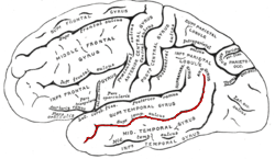
The auditory cortex is the part of the temporal lobe that processes auditory information in humans and many other vertebrates. It is a part of the auditory system, performing basic and higher functions in hearing, such as possible relations to language switching. It is located bilaterally, roughly at the upper sides of the temporal lobes – in humans, curving down and onto the medial surface, on the superior temporal plane, within the lateral sulcus and comprising parts of the transverse temporal gyri, and the superior temporal gyrus, including the planum polare and planum temporale.

Auditory neuropathy (AN) is a hearing disorder in which the outer hair cells of the cochlea are present and functional, but sound information is not transmitted sufficiently by the auditory nerve to the brain. Hearing loss with AN can range from normal hearing sensitivity to profound hearing loss.

Amusia is a musical disorder that appears mainly as a defect in processing pitch but also encompasses musical memory and recognition. Two main classifications of amusia exist: acquired amusia, which occurs as a result of brain damage, and congenital amusia, which results from a music-processing anomaly present since birth.

<span class="mw-page-title-main">Language processing in the brain</span> How humans use words to communicate

In psycholinguistics, language processing refers to the way humans use words to communicate ideas and feelings, and how such communications are processed and understood. Language processing is considered to be a uniquely human ability that is not produced with the same grammatical understanding or systematicity in even human's closest primate relatives.

Auditory verbal agnosia (AVA), also known as pure word deafness, is the inability to comprehend speech. Individuals with this disorder lose the ability to understand language, repeat words, and write from dictation. Some patients with AVA describe hearing spoken language as meaningless noise, often as though the person speaking was doing so in a foreign language. However, spontaneous speaking, reading, and writing are preserved. The maintenance of the ability to process non-speech auditory information, including music, also remains relatively more intact than spoken language comprehension. Individuals who exhibit pure word deafness are also still able to recognize non-verbal sounds. The ability to interpret language via lip reading, hand gestures, and context clues is preserved as well. Sometimes, this agnosia is preceded by cortical deafness; however, this is not always the case. Researchers have documented that in most patients exhibiting auditory verbal agnosia, the discrimination of consonants is more difficult than that of vowels, but as with most neurological disorders, there is variation among patients.

<span class="mw-page-title-main">Foix–Chavany–Marie syndrome</span> Medical condition

Foix–Chavany–Marie Syndrome (FCMS), also known as bilateral opercular syndrome, is a neuropathological disorder characterized by paralysis of the facial, tongue, pharynx, and masticatory muscles of the mouth that aid in chewing. The disorder is primarily caused by thrombotic and embolic strokes, which cause a deficiency of oxygen in the brain. As a result, bilateral lesions may form in the junctions between the frontal lobe and temporal lobe, the parietal lobe and cortical lobe, or the subcortical region of the brain. FCMS may also arise from defects existing at birth that may be inherited or nonhereditary. Symptoms of FCMS can be present in a person of any age and it is diagnosed using automatic-voluntary dissociation assessment, psycholinguistic testing, neuropsychological testing, and brain scanning. Treatment for FCMS depends on the onset, as well as on the severity of symptoms, and it involves a multidisciplinary approach.

The auditory brainstem response (ABR), also called brainstem evoked response audiometry (BERA), is an auditory evoked potential extracted from ongoing electrical activity in the brain and recorded via electrodes placed on the scalp. The measured recording is a series of six to seven vertex positive waves of which I through V are evaluated. These waves, labeled with Roman numerals in Jewett and Williston convention, occur in the first 10 milliseconds after onset of an auditory stimulus. The ABR is considered an exogenous response because it is dependent upon external factors.

Auditory agnosia is a form of agnosia that manifests itself primarily in the inability to recognize or differentiate between sounds. It is not a defect of the ear or "hearing", but rather a neurological inability of the brain to process sound meaning. While auditory agnosia impairs the understanding of sounds, other abilities such as reading, writing, and speaking are not hindered. It is caused by bilateral damage to the anterior superior temporal gyrus, which is part of the auditory pathway responsible for sound recognition, the auditory "what" pathway.

<span class="mw-page-title-main">Hearing</span> Sensory perception of sound by living organisms

Hearing, or auditory perception, is the ability to perceive sounds through an organ, such as an ear, by detecting vibrations as periodic changes in the pressure of a surrounding medium. The academic field concerned with hearing is auditory science.

<span class="mw-page-title-main">Superior temporal sulcus</span> Part of the brains temporal lobe

In the human brain, the superior temporal sulcus (STS) is the sulcus separating the superior temporal gyrus from the middle temporal gyrus in the temporal lobe of the brain. A sulcus is a deep groove that curves into the largest part of the brain, the cerebrum, and a gyrus is a ridge that curves outward of the cerebrum.

The neuroscience of music is the scientific study of brain-based mechanisms involved in the cognitive processes underlying music. These behaviours include music listening, performing, composing, reading, writing, and ancillary activities. It also is increasingly concerned with the brain basis for musical aesthetics and musical emotion. Scientists working in this field may have training in cognitive neuroscience, neurology, neuroanatomy, psychology, music theory, computer science, and other relevant fields.

Neuroscientists have learned much about the role of the brain in numerous cognitive mechanisms by understanding corresponding disorders. Similarly, neuroscientists have come to learn much about music cognition by studying music-specific disorders. Even though music is most often viewed from a "historical perspective rather than a biological one" music has significantly gained the attention of neuroscientists all around the world. For many centuries music has been strongly associated with art and culture. The reason for this increased interest in music is because it "provides a tool to study numerous aspects of neuroscience, from motor skill learning to emotion".

Phonagnosia is a type of agnosia, or loss of knowledge, that involves a disturbance in the recognition of familiar voices and the impairment of voice discrimination abilities in which the affected individual does not suffer from comprehension deficits. Phonagnosia is an auditory agnosia, an acquired auditory processing disorder resulting from brain damage, other auditory agnosias include cortical deafness and auditory verbal agnosia also known as pure word deafness.

Spatial hearing loss refers to a form of deafness that is an inability to use spatial cues about where a sound originates from in space. Poor sound localization in turn affects the ability to understand speech in the presence of background noise.

Musical hallucinations describes a neurological disorder in which the patient will hallucinate songs, tunes, instruments and melodies. The source of these hallucinations are derived from underlying psychotic illness or hearing impairment. These hallucinations are often rare and are followed by mental decline. A majority of patients who have symptoms of musical hallucinations are older and have onset conditions predisposing them to the disease. While there is no set form of treatment, research has discovered medications and alternative therapies to be successful in alleviating the hallucinations.

<span class="mw-page-title-main">Sign language in the brain</span>

Sign language refers to any natural language which uses visual gestures produced by the hands and body language to express meaning. The brain's left side is the dominant side utilized for producing and understanding sign language, just as it is for speech. In 1861, Paul Broca studied patients with the ability to understand spoken languages but the inability to produce them. The damaged area was named Broca's area, and located in the left hemisphere’s inferior frontal gyrus. Soon after, in 1874, Carl Wernicke studied patients with the reverse deficits: patients could produce spoken language, but could not comprehend it. The damaged area was named Wernicke's area, and is located in the left hemisphere’s posterior superior temporal gyrus.

<span class="mw-page-title-main">Diagnosis of hearing loss</span>

Identification of a hearing loss is usually conducted by a general practitioner medical doctor, otolaryngologist, certified and licensed audiologist, school or industrial audiometrist, or other audiometric technician. Diagnosis of the cause of a hearing loss is carried out by a specialist physician or otorhinolaryngologist.

<span class="mw-page-title-main">Auditosensory cortex</span>

Auditosensory cortex is the part of the auditory system that is associated with the sense of hearing in humans. It occupies the bilateral primary auditory cortex in the temporal lobe of the mammalian brain. The term is used to describe Brodmann area 42 together with the transverse temporal gyri of Heschl. The auditosensory cortex takes part in the reception and processing of auditory nerve impulses, which passes sound information from the thalamus to the brain. Abnormalities in this region are responsible for many disorders in auditory abilities, such as congenital deafness, true cortical deafness, primary progressive aphasia and auditory hallucination.

References

  1. Ingram, John Henry (2007). Neurolinguistics: an introduction to spoken language processing and its disorders . Cambridge, UK: Cambridge University Press. pp.  160–171. ISBN   978-0-521-79190-8. OCLC   297335127.
  2. 1 2 3 4 Bogousslavsky, Julien (2001). Stroke Syndromes . Cambridge: Cambridge UP. pp.  153.
  3. 1 2 3 4 Graham J, Greenwood R, Lecky B (October 1980). "Cortical deafness—a case report and review of the literature". J. Neurol. Sci. 48 (1): 35–49. doi:10.1016/0022-510X(80)90148-3. PMID   7420124. S2CID   36026035.
  4. 1 2 3 4 Hood, Linda (1999). "Cortical Deafness: A longitudinal study" (PDF). The American Journal of Nursing.
  5. Silva, Júlia; Sousa, Marisa; Mestre, Susana; Nzwalo, Isa; Nzwalo, Hipólito (July 2020). "Cortical deafness of following bilateral temporal lobe stroke". Journal of Stroke and Cerebrovascular Diseases. 29 (7): 104827. doi:10.1016/j.jstrokecerebrovasdis.2020.104827. PMID   32386850. S2CID   218585923.
  6. Cavinato M, Rigon J, Volpato C, Semenza C, Piccione F (2012). "Preservation of auditory P300-like potentials in cortical deafness". PLOS One. 7 (1): e29909. Bibcode:2012PLoSO...729909C. doi: 10.1371/journal.pone.0029909 . PMC   3260175 . PMID   22272260.
  7. Denes, G.; Pizzamiglio, P. "Comprehension Disorders." Handbook of Clinical and Experimental Neuropsychology. Hove, East Sussex, UK: Psychology, 1999. 210. Print.
  8. Sasidharan, Megha; Elsa Itty, Jinsi; Hinduja, Ghanta; Hasna, Shabnam; Priya, Deena (31 October 2020). "Cortical deafness as a sequela to meningitis: a single case study". Intractable & Rare Diseases Research. 9 (4): 247–250. doi:10.5582/irdr.2020.03072. PMC   7586874 . PMID   33139984.
  9. 1 2 3 4 5 Brody, Robert M., Brian D. Nicholas, Michael J. Wolf, Paula B. Marcinkevich, and Gregory J. Artz. "Cortical Deafness: A Case Report and Review of the Literature." Otology and Neurology 34.7 (2013): 1226–229. Ovid. Web.

Further reading