Constructional apraxia

Last updated

Constructional apraxia is a neurological disorder in which people are unable to perform tasks or movements even though they understand the task, are willing to complete it, and have the physical ability to perform the movements. [1] It is characterized by an inability or difficulty to build, assemble, or draw objects. [2] [3] Constructional apraxia may be caused by lesions in the parietal lobe following stroke or it may serve as an indicator for Alzheimer's disease.

Contents

Signs and symptoms

A key deficit in constructional apraxia patients is the inability to correctly copy or draw an image. There are qualitative differences between patients with left hemisphere damage, right hemisphere damage, and Alzheimer's disease. [2] [4]

Left hemisphere damage

Patients with damage to their left hemisphere tend to preserve items, oversimplify drawing features [5] and omit details when drawing from memory. In addition, left hemisphere patients are less likely to systematically arrange the parts of their drawing. [6]

Right hemisphere damage

Patients with damage to their right hemisphere have trouble correctly replicating spatial relationships of complex figures. Drawing elements are often piecemeal, transposed to different positions or orientations, or shown diagonally on the page. [5] As a result, right hemisphere patients tend to produce asymmetric or distorted drawings [6] characterized by hemispatial neglect, the omission of elements from one side of the model. [7] It was once thought that right hemisphere patients were twice as likely to make mistakes in 3D construction tasks as left hemisphere patients but this inaccurate conclusion was attributable to participant selection bias in that researchers excluded from studies individuals with severe left hemisphere lesions due to the debilitating language impairments of those individuals. However, included in studies were individuals with severe right hemisphere lesions. [8] Subsequent research has substantiated the absence of a marked difference in performance between left and right hemisphere patients on 3D construction tasks. [9]

Alzheimer's disease

Alzheimer's disease patients with constructional apraxia have unique symptoms. Their drawings contain fewer angles, spatial alterations, a lack of perspective and simplifications, which are uncharacteristic of left hemisphere or right hemisphere patients. Constructional disabilities are present early on in the disease and get progressively worse over time; [4] however even patients with advanced Alzheimer's disease may be able to do some constructional tasks. [10] Spontaneous drawing is affected early and is heavily dependent upon semantic memory; therefore simplifications in the drawing may be due to impaired access to semantic knowledge. As Alzheimer's disease progresses, the patient's ability to copy objects becomes increasingly impaired and they may lose the ability to draw correctly a simple figures due to a motor loss in routine memories. [4]

Causes

Constructional apraxia cannot be localized to a specific hemisphere or cerebral area because drawing and constructional tasks require both perceptual and motor functioning. [7] [9] It has been linked to parietal lesions in the left and right hemisphere, stroke and Alzheimer's disease.

Initially, researchers tried to isolate the cause to left hemisphere lesions in the parietal lobe because of its similarities to Gerstmann syndrome; however, lesions in the dorsal stream also result in visual agnosia and a piecemeal drawing. [2] Although constructional apraxia can result from lesions in any part of the brain, it is most commonly associated with lesions in the parietal-occipital lobes. Constructional apraxia is common after right parietal stroke and it continues after visuospatial symptoms have subsided. [5] Patients with posterior and parietal lobe lesions tend to have the most severe symptoms. [9]

In Alzheimer's disease research, the AT8 antibody has proven to be an early indicator of tau protein pathology. Constructional apraxia patients with the most AT8 pathology were least able to copy an image, while those best able to had the least neuritic pathology. Therefore, figure copying ability is highly correlated with Alzheimer's disease pathology. [10]

Mechanisms

Drawing

As the study of constructional apraxia impairments narrows, research is concentrating on analyzing drawing abilities. Drawing abilities may be decomposed into three steps: visual perception, visual imagery, and graphic production.

According to the two-streams hypothesis, as information exits the occipital lobe it follows two pathways. The dorsal stream ("where pathway") ends in the parietal lobe while the ventral stream ("what pathway") terminates in the temporal lobe. [7] [11] Damage to the parietal lobe is highly correlated with constructional apraxia since it is involved in drawing and copying. The parietal lobe is also critical for remapping spatial position across saccades. [5] There is an attentional subsystem responsible for moving the eyes, head, and body to focus on different images. Damage at various levels of this system could lead to trouble localizing a stimulus or hemispatial neglect which manifests as perseverative errors on the drawing. [12] There are a couple theories used to describe the neurological mechanisms behind drawing.

Kosslyn and Koeing model

Kosslyn proposes that there is an early split of information in the dorsal stream. The first pathway captures coordinate relations by defining distances between points in space. These points become a continuum which can transform into other points through intermediate points. This coding of points would allow space to be perceived qualitatively, which would also help with movement. [7]

The other pathway encodes "categorical" information, which synthesizes information about the shape and spatial arrangement of an objects parts. It decomposes objects into their most basic form, by looking for boundaries, lines, or patches. These categorical relations in turn lead to abstract spatial relations that allow one to perceive objects as being "on top," "inside," "between," "beside," etc. [7]

Van Sommers model

The Van Sommers model describes two hierarchical systems for drawing: one for visual perception, another for graphic production. The visual perception model utilizes David Marr's three stage system to describe visual perception in copying. In the first stage, an image us represented in 2D based on changes in intensity. Foreground and background are not distinguished. In the second stage, a 2.5D representation is formed which encodes the object in a viewer-centered coordinate system. Finally a 3D object-centered representation is established making it possible to appreciate volume. Visual representations of familiar drawings are stored in memory. This representation sends feedback to the other areas of the brain which encoded the spatial and physical properties of the object. Feedback from these areas allows the drawer to successfully encode the coordinate and categorical relations. [2] [13]

In the graphic production model, the viewer begins by making a series of depiction decisions about the dimensions, amount of detail to include, etc. Depiction decisions are not used when copying a drawing because they're dictated by the situation. Next, the production strategy is formed. If the drawing is unfamiliar, then the drawer will divide and rank the different portions of the drawings. If the drawing is familiar (e.g. a sun), then the drawer will reproduce the item line by lie regardless of the pictures organization due to automatic execution. The third component, contingent planning, reflects the importance of planning in drawing. Contingent planning is a result of production strategy. If the drawing is unfamiliar and requires a segmented approach, then the most appropriate sequence is determined before the drawing. At this point, the drawing task becomes a problem solving task. The fourth and final component of the model refers to the articulatory and economic constraints placed on the drawer by using a pencil. Certain directions are favored due to the orientation of the hand and fingers, etc. However, some feel the Van Sommers model does not adequately account for all aspects of drawing. [2] [13]

Other theories

Drawing from memory in response to a verbal order requires the image to be recalled from associative memory and brought into the visual buffer. Once there, it can be successfully drawn and copied from memory. Familiar images (like the sun) may not require visual imagery to draw, as the production schemes and action programming stored in the associative memory and procedural memory may be sufficient to reproduce the drawing.

Construction

Construction problems are usually caused by visual perception deficits. They require normal vision and the ability to execute a series of motor activities. When looking at performance, it is important to consider perceptual and executive functioning. A patient with trouble visually recognizing patterns or spatial relations may have difficulty correctly building a model. In addition, problems planning, organizing, or carrying out action may impede the ability to solve a construction problem. [9]

Neuropsychological mechanisms

Re-created geometric drawings from patients with left and right hemisphere damage, hemineglect, and dementia.png

Modern attempts to understand constructional apraxia have moved away from anatomical functions towards a cognitive neuropsychological approach. Both adults and children alike experience difficulty reproducing oblique lines. Some feel that these deficiencies may be attributed to planning since it is easier to plan horizontal and vertical lines than oblique lines. Research indicates that both adults and children are more able to draw squares than diamonds, although as children grow into adults they are more accurately able to depict diamonds. [6]

One study showed that constructional apraxia patients were significantly less accurate than the control patients in producing angles with vertical and horizontal orientations. In this study, constructional apraxia patients drew patterns usually found in children 8 and younger. Gregory argues that ontogenetically and phylogenetically earlier behavioral traits are present in the brain, but inhibited. When these inhibitory mechanisms become compromised, then the childlike behavior patterns re-emerge. Therefore, according to this theory, the inhibitory mechanisms in patients with constructional apraxia have failed, causing them to draw like young children who have difficulty drawing oblique lines. [6]

Diagnosis

Constructional disabilities are often tested by asking the patient to draw a 2D model or assemble an object. Some researchers feel that neuronal mechanisms involved in drawing and copying differ, thus they should be tested individually. Free drawing is a commonly used test in which the patient is asked to draw a named object. It can be an effective tool in measuring the patient's ability to maintain spatial relations, organize the drawing, and draw complete shapes. The complexity of the task should be noted as such tasks often require lexical-semantic abilities as well as imagery abilities. [4]

Treatment

Motor imagery has been explored as a potential therapy for constructional apraxia patients. Motor imagery is a process by which a specific action is mimicked in the working memory without a corresponding motor output. Since constructional apraxia is a visuospatial problem not a motor problem, rehabilitation-treatment based on motor imagery has not proven to be an effective in patients with right hemisphere stroke or hemispatial neglect. [14]

History

In 1934, Karl Kleist characterized constructional apraxia as a disturbance "in formative activities such as assembling, building and drawing, in which the spatial form of the product proves to be unsuccessful, without there being an apraxia for single movements." [15] [16] In the years following, the definition of constructional apraxia diverged. There were those who felt it was an executive processing order and those who felt it was a visuospatial disorder. Due to discrepancies in definitions, constructional apraxia became a blanket term to describe any kind of constructional impairment. Modern researchers question whether the term "apraxia" is appropriate to describe this condition. [4]

Related Research Articles

<span class="mw-page-title-main">Apraxia</span> Medical condition

Apraxia is a motor disorder caused by damage to the brain, which causes difficulty with motor planning to perform tasks or movements. The nature of the damage determines the disorder's severity, and the absence of sensory loss or paralysis helps to explain the level of difficulty. Children may be born with apraxia; its cause is unknown, and symptoms are usually noticed in the early stages of development. Apraxia occurring later in life, known as acquired apraxia, is typically caused by traumatic brain injury, stroke, dementia, Alzheimer's disease, brain tumor, or other neurodegenerative disorders. The multiple types of apraxia are categorized by the specific ability and/or body part affected.

Agraphia is an acquired neurological disorder causing a loss in the ability to communicate through writing, either due to some form of motor dysfunction or an inability to spell. The loss of writing ability may present with other language or neurological disorders; disorders appearing commonly with agraphia are alexia, aphasia, dysarthria, agnosia, acalculia and apraxia. The study of individuals with agraphia may provide more information about the pathways involved in writing, both language related and motoric. Agraphia cannot be directly treated, but individuals can learn techniques to help regain and rehabilitate some of their previous writing abilities. These techniques differ depending on the type of agraphia.

Alien hand syndrome (AHS) or Dr. Strangelove syndrome is a category of conditions in which a person experiences their limbs acting seemingly on their own, without conscious control over the actions. There are a variety of clinical conditions that fall under this category, which most commonly affects the left hand. There are many similar terms for the various forms of the condition, but they are often used inappropriately. The affected person may sometimes reach for objects and manipulate them without wanting to do so, even to the point of having to use the controllable hand to restrain the alien hand. Under normal circumstances however, given that intent and action can be assumed to be deeply mutually entangled, the occurrence of alien hand syndrome can be usefully conceptualized as a phenomenon reflecting a functional "disentanglement" between thought and action.

<span class="mw-page-title-main">Parietal lobe</span> Part of the brain responsible for sensory input and some language processing

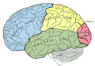
The parietal lobe is one of the four major lobes of the cerebral cortex in the brain of mammals. The parietal lobe is positioned above the temporal lobe and behind the frontal lobe and central sulcus.

Anosognosia is a condition in which a person with a disability is cognitively unaware of having it due to an underlying physical condition. Anosognosia results from physiological damage to brain structures, typically to the parietal lobe or a diffuse lesion on the fronto-temporal-parietal area in the right hemisphere, and is thus a neuropsychiatric disorder. A deficit of self-awareness, it was first named by the neurologist Joseph Babinski in 1914. Phenomenologically, anosognosia has similarities to denial, which is a psychological defense mechanism; attempts have been made at a unified explanation. Anosognosia is sometimes accompanied by asomatognosia, a form of neglect in which patients deny ownership of body parts such as their limbs. The term is from Ancient Greek ἀ- a-, 'without', νόσος nosos, 'disease' and γνῶσις gnōsis, 'knowledge'. It is also considered a disorder that makes the treatment of the patient more difficult, since it may affect negatively the therapeutic relationship.

Astereognosis is the inability to identify an object by active touch of the hands without other sensory input, such as visual or sensory information. An individual with astereognosis is unable to identify objects by handling them, despite intact elementary tactile, proprioceptive, and thermal sensation. With the absence of vision, an individual with astereognosis is unable to identify what is placed in their hand based on cues such as texture, size, spatial properties, and temperature. As opposed to agnosia, when the object is observed visually, one should be able to successfully identify the object.

<span class="mw-page-title-main">Hemispatial neglect</span> Medical condition

Hemispatial neglect is a neuropsychological condition in which, after damage to one hemisphere of the brain, a deficit in attention and awareness towards the side of space opposite brain damage is observed. It is defined by the inability of a person to process and perceive stimuli towards the contralesional side of the body or environment. Hemispatial neglect is very commonly contralateral to the damaged hemisphere, but instances of ipsilesional neglect have been reported.

<span class="mw-page-title-main">Bálint's syndrome</span> Medical condition

Bálint's syndrome is an uncommon and incompletely understood triad of severe neuropsychological impairments: inability to perceive the visual field as a whole (simultanagnosia), difficulty in fixating the eyes, and inability to move the hand to a specific object by using vision. It was named in 1909 for the Austro-Hungarian neurologist and psychiatrist Rezső Bálint who first identified it.

<span class="mw-page-title-main">Visual memory</span> Ability to process visual and spatial information

Visual memory describes the relationship between perceptual processing and the encoding, storage and retrieval of the resulting neural representations. Visual memory occurs over a broad time range spanning from eye movements to years in order to visually navigate to a previously visited location. Visual memory is a form of memory which preserves some characteristics of our senses pertaining to visual experience. We are able to place in memory visual information which resembles objects, places, animals or people in a mental image. The experience of visual memory is also referred to as the mind's eye through which we can retrieve from our memory a mental image of original objects, places, animals or people. Visual memory is one of several cognitive systems, which are all interconnected parts that combine to form the human memory. Types of palinopsia, the persistence or recurrence of a visual image after the stimulus has been removed, is a dysfunction of visual memory.

<span class="mw-page-title-main">Posterior parietal cortex</span>

The posterior parietal cortex plays an important role in planned movements, spatial reasoning, and attention.

Ideomotor Apraxia, often IMA, is a neurological disorder characterized by the inability to correctly imitate hand gestures and voluntarily mime tool use, e.g. pretend to brush one's hair. The ability to spontaneously use tools, such as brushing one's hair in the morning without being instructed to do so, may remain intact, but is often lost. The general concept of apraxia and the classification of ideomotor apraxia were developed in Germany in the late 19th and early 20th centuries by the work of Hugo Liepmann, Adolph Kussmaul, Arnold Pick, Paul Flechsig, Hermann Munk, Carl Nothnagel, Theodor Meynert, and linguist Heymann Steinthal, among others. Ideomotor apraxia was classified as "ideo-kinetic apraxia" by Liepmann due to the apparent dissociation of the idea of the action with its execution. The classifications of the various subtypes are not well defined at present, however, owing to issues of diagnosis and pathophysiology. Ideomotor apraxia is hypothesized to result from a disruption of the system that relates stored tool use and gesture information with the state of the body to produce the proper motor output. This system is thought to be related to the areas of the brain most often seen to be damaged when ideomotor apraxia is present: the left parietal lobe and the premotor cortex. Little can be done at present to reverse the motor deficit seen in ideomotor apraxia, although the extent of dysfunction it induces is not entirely clear.

<span class="mw-page-title-main">Integrative agnosia</span> Medical condition

Integrative agnosia is a sub-disease of agnosia, meaning the lack of integrating perceptual wholes within knowledge. Integrative agnosia can be assessed by several experimental tests such as the Efron shape test, which determines the specificity of the disease being Integrative. This disease is often caused by brain trauma, producing medial ventral lesions to the extrastriate cortex. Affecting this region of the brain produces learning impairments: the inability to integrate parts such as spatial distances or producing visual images from short or long-term memory.

The neuroanatomy of memory encompasses a wide variety of anatomical structures in the brain.

<span class="mw-page-title-main">Gerstmann syndrome</span> Neuropsychological disorder caused by damage to the inferior parietal lobule

Gerstmann syndrome is a neuropsychological disorder that is characterized by a constellation of symptoms that suggests the presence of a lesion usually near the junction of the temporal and parietal lobes at or near the angular gyrus. Gerstmann syndrome is typically associated with damage to the inferior parietal lobule of the dominant hemisphere. It is classically considered a left-hemisphere disorder, although right-hemisphere damage has also been associated with components of the syndrome.

<span class="mw-page-title-main">Allochiria</span> Medical condition

Allochiria is a neurological disorder in which the patient responds to stimuli presented to one side of their body as if the stimuli had been presented at the opposite side. It is associated with spatial transpositions, usually symmetrical, of stimuli from one side of the body to the opposite one. Thus a touch to the left side of the body will be reported as a touch to the right side, which is also known as somatosensory allochiria. If the auditory or visual senses are affected, sounds will be reported as being heard on the opposite side to that on which they occur and objects presented visually will be reported as having been presented on the opposite side. Often patients may express allochiria in their drawing while copying an image. Allochiria often co-occurs with unilateral neglect and, like hemispatial neglect, the disorder arises commonly from damage to the right parietal lobe.

Amorphosynthesis, also called a hemi-sensory deficit, is a neuropsychological condition in which a patient experiences unilateral inattention to sensory input. This phenomenon is frequently associated with damage to the right cerebral hemisphere resulting in severe sensory deficits that are observed on the contralesional (left) side of the body. A right-sided deficit is less commonly observed and the effects are reported to be temporary and minor. Evidence suggests that the right cerebral hemisphere has a dominant role in attention and awareness to somatic sensations through ipsilateral and contralateral stimulation. In contrast, the left cerebral hemisphere is activated only by contralateral stimuli. Thus, the left and right cerebral hemispheres exhibit redundant processing to the right-side of the body and a lesion to the left cerebral hemisphere can be compensated by the ipsiversive processes of the right cerebral hemisphere. For this reason, right-sided amorphosynthesis is less often observed and is generally associated with bilateral lesions.

Visuospatial dysgnosia is a loss of the sense of "whereness" in the relation of oneself to one's environment and in the relation of objects to each other. Visuospatial dysgnosia is often linked with topographical disorientation.

<span class="mw-page-title-main">Disconnection syndrome</span> Collection of neurological symptoms

Disconnection syndrome is a general term for a collection of neurological symptoms caused – via lesions to associational or commissural nerve fibres – by damage to the white matter axons of communication pathways in the cerebrum, independent of any lesions to the cortex. The behavioral effects of such disconnections are relatively predictable in adults. Disconnection syndromes usually reflect circumstances where regions A and B still have their functional specializations except in domains that depend on the interconnections between the two regions.

Visual spatial attention is a form of visual attention that involves directing attention to a location in space. Similar to its temporal counterpart visual temporal attention, these attention modules have been widely implemented in video analytics in computer vision to provide enhanced performance and human interpretable explanation of deep learning models.

<span class="mw-page-title-main">Masud Husain</span> British professor of neuroscience, editor of Brain

Masud Husain FMedSci is a clinical neurologist and neuroscientist working in the UK. He is Professor of Neurology & Cognitive Neuroscience at the Nuffield Department of Clinical Neurosciences and Departmental of Experimental Psychology, University of Oxford, a Professorial Fellow at New College, Oxford, and Editor-in-Chief of the journal Brain. He was born in East Pakistan.

References

  1. " apraxia " at Dorland's Medical Dictionary
  2. 1 2 3 4 5 Guérin F, Ska B, Belleville S (August 1999). "Cognitive processing of drawing abilities". Brain Cogn. 40 (3): 464–78. doi:10.1006/brcg.1999.1079. PMID   10415132. S2CID   41074859.
  3. Caminiti R, Chafee MV, Battaglia-Mayer A, Averbeck BB, Crowe DA, Georgopoulos AP (June 2010). "Understanding the parietal lobe syndrome from a neurophysiological and evolutionary perspective". Eur. J. Neurosci. 31 (12): 2320–40. doi:10.1111/j.1460-9568.2010.07291.x. PMC   2900452 . PMID   20550568.
  4. 1 2 3 4 5 Behrmann, Marlene; Boller, François; Grafman, Jordan (2001). Disorders of visual behavior. Amsterdam: Elsevier. pp. 99–118. ISBN   0-444-50360-9. OCLC   47703916.
  5. 1 2 3 4 Russell C, Deidda C, Malhotra P, Crinion JT, Merola S, Husain M (April 2010). "A deficit of spatial remapping in constructional apraxia after right-hemisphere stroke". Brain. 133 (Pt 4): 1239–51. doi: 10.1093/brain/awq052 . PMID   20375139.
  6. 1 2 3 4 Smith AD, Gilchrist ID (April 2005). "Drawing from childhood experience: constructional apraxia and the production of oblique lines" (PDF). Cortex. 41 (2): 195–204. doi:10.1016/S0010-9452(08)70894-3. PMID   15714902. S2CID   4480723. Archived from the original (PDF) on 2016-03-04. Retrieved 2013-08-14.
  7. 1 2 3 4 5 Laeng B (2006). "Constructional apraxia after left or right unilateral stroke". Neuropsychologia. 44 (9): 1595–606. doi:10.1016/j.neuropsychologia.2006.01.023. PMID   16516249. S2CID   25691047.
  8. Bonato M, Sella F, Verteltti I, Umilta C (June 2011). "Neuropsychology is nothing with out control: A potential fallacy hidden in clinical studies". Cortex. 48 (6): 353–355. doi:10.1016/j.cortex.2011.06.017. PMID   21783188. S2CID   206984237.
  9. 1 2 3 4 Capruso DX; Hamsher Kd (June 2011). "Constructional ability in two- versus three-dimensions: relationship to spatial vision and locus of cerebrovascular lesion". Cortex. 47 (6): 696–705. doi:10.1016/j.cortex.2010.05.001. PMID   20547388. S2CID   206983724.
  10. 1 2 Nielson KA, Cummings BJ, Cotman CW (November 1996). "Constructional apraxia in Alzheimer's disease correlates with neuritic neuropathology in occipital cortex". Brain Res. 741 (1–2): 284–93. doi:10.1016/S0006-8993(96)00983-3. PMID   9001734. S2CID   31103796.
  11. Milner, A. David (1995). The Visual Brain in Action . p.  128. ISBN   0198521367.
  12. Makuuchi M, Kaminaga T, Sugishita M (May 2003). "Both parietal lobes are involved in drawing: a functional MRI study and implications for constructional apraxia". Brain Res Cogn Brain Res. 16 (3): 338–47. doi:10.1016/S0926-6410(02)00302-6. PMID   12706214.
  13. 1 2 Bouaziz, Serge; Magnan, Annie (2007). "Contribution of the visual perception and graphic production systems to the copying of complex geometrical drawings: A developmental study". Cognitive Development. 22 (1): 5–15. doi:10.1016/j.cogdev.2006.10.002. ISSN   0885-2014.
  14. Vromen A, Verbunt JA, Rasquin S, Wade DT (2011). "Motor imagery in patients with a right hemisphere stroke and unilateral neglect". Brain Inj. 25 (4): 387–93. doi:10.3109/02699052.2011.558041. PMID   21355672. S2CID   21780736.
  15. Kleist K (1934). "Gehirnpathologie" (in German). Leipzig: Barth. Konstruktive (optische) Apraxie.{{cite journal}}: Cite journal requires |journal= (help)
  16. Benton, AL (2000). "6, Spatial thinking in neurological patients: Historical aspects". Exploring the history of neuropsychology: selected papers. Oxford [Oxfordshire]: Oxford University Press. ISBN   0-19-513808-2. OCLC   42935803.

Further reading