Related Research Articles

A saccade is a quick, simultaneous movement of both eyes between two or more phases of focal points in the same direction. In contrast, in smooth-pursuit movements, the eyes move smoothly instead of in jumps; it could be associated with a shift in frequency of an emitted signal or a movement of a body part or device. Controlled cortically by the frontal eye fields (FEF), or subcortically by the superior colliculus, saccades serve as a mechanism for focal points, rapid eye movement, and the fast phase of optokinetic nystagmus. The word appears to have been coined in the 1880s by French ophthalmologist Émile Javal, who used a mirror on one side of a page to observe eye movement in silent reading, and found that it involves a succession of discontinuous individual movements.
Saccadic masking, also known as (visual) saccadic suppression, is the phenomenon in visual perception where the brain selectively blocks visual processing during eye movements in such a way that neither the motion of the eye nor the gap in visual perception is noticeable to the viewer.

The vestibulo-ocular reflex (VOR) is a reflex that acts to stabilize gaze during head movement, with eye movement due to activation of the vestibular system, it is also known as the Cervico-ocular reflex. The reflex acts to stabilize images on the retinas of the eye during head movement. Gaze is held steadily on a location by producing eye movements in the direction opposite that of head movement. For example, when the head moves to the right, the eyes move to the left, meaning the image a person sees stays the same even though the head has turned. Since slight head movement is present all the time, VOR is necessary for stabilizing vision: people with an impaired reflex find it difficult to read using print, because the eyes do not stabilise during small head tremors, and also because damage to reflex can cause nystagmus.
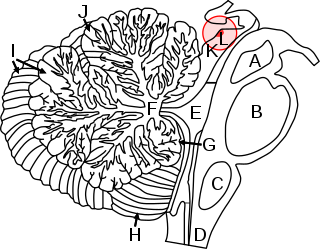
In neuroanatomy, the superior colliculus is a structure lying on the roof of the mammalian midbrain. In non-mammalian vertebrates, the homologous structure is known as the optic tectum or optic lobe. The adjective form tectal is commonly used for both structures.

Eye movement includes the voluntary or involuntary movement of the eyes. Eye movements are used by a number of organisms to fixate, inspect and track visual objects of interests. A special type of eye movement, rapid eye movement, occurs during REM sleep.
Oscillopsia is a visual disturbance in which objects in the visual field appear to oscillate. The severity of the effect may range from a mild blurring to rapid and periodic jumping. Oscillopsia is an incapacitating condition experienced by many patients with neurological disorders. It may be the result of ocular instability occurring after the oculomotor system is affected, no longer holding images steady on the retina. A change in the magnitude of the vestibulo-ocular reflex due to vestibular disease can also lead to oscillopsia during rapid head movements. Oscillopsia may also be caused by involuntary eye movements such as nystagmus, or impaired coordination in the visual cortex and is one of the symptoms of superior canal dehiscence syndrome. Those affected may experience dizziness and nausea. Oscillopsia can also be used as a quantitative test to document aminoglycoside toxicity. Permanent oscillopsia can arise from an impairment of the ocular system that serves to maintain ocular stability. Paroxysmal oscillopsia can be due to an abnormal hyperactivity in the peripheral ocular or vestibular system.
The pars reticulata (SNpr) is a portion of the substantia nigra and is located lateral to the pars compacta. Most of the neurons that project out of the pars reticulata are inhibitory GABAergic neurons.
Dysmetria is a lack of coordination of movement typified by the undershoot or overshoot of intended position with the hand, arm, leg, or eye. It is a type of ataxia. It can also include an inability to judge distance or scale.
Microsaccades are a kind of fixational eye movement. They are small, jerk-like, involuntary eye movements, similar to miniature versions of voluntary saccades. They typically occur during prolonged visual fixation, not only in humans, but also in animals with foveal vision. Microsaccade amplitudes vary from 2 to 120 arcminutes. The first empirical evidence for their existence was provided by Robert Darwin, the father of Charles Darwin.

The frontal eye fields (FEF) are a region located in the frontal cortex, more specifically in Brodmann area 8 or BA8, of the primate brain. In humans, it can be more accurately said to lie in a region around the intersection of the middle frontal gyrus with the precentral gyrus, consisting of a frontal and parietal portion. The FEF is responsible for saccadic eye movements for the purpose of visual field perception and awareness, as well as for voluntary eye movement. The FEF communicates with extraocular muscles indirectly via the paramedian pontine reticular formation. Destruction of the FEF causes deviation of the eyes to the ipsilateral side.

Supplementary eye field (SEF) is the name for the anatomical area of the dorsal medial frontal lobe of the primate cerebral cortex that is indirectly involved in the control of saccadic eye movements. Evidence for a supplementary eye field was first shown by Schlag, and Schlag-Rey. Current research strives to explore the SEF's contribution to visual search and its role in visual salience. The SEF constitutes together with the frontal eye fields (FEF), the intraparietal sulcus (IPS), and the superior colliculus (SC) one of the most important brain areas involved in the generation and control of eye movements, particularly in the direction contralateral to their location. Its precise function is not yet fully known. Neural recordings in the SEF show signals related to both vision and saccades somewhat like the frontal eye fields and superior colliculus, but currently most investigators think that the SEF has a special role in high level aspects of saccade control, like complex spatial transformations, learned transformations, and executive cognitive functions.

Fixation or visual fixation is the maintaining of the gaze on a single location. An animal can exhibit visual fixation if it possess a fovea in the anatomy of their eye. The fovea is typically located at the center of the retina and is the point of clearest vision. The species in which fixational eye movement has been verified thus far include humans, primates, cats, rabbits, turtles, salamanders, and owls. Regular eye movement alternates between saccades and visual fixations, the notable exception being in smooth pursuit, controlled by a different neural substrate that appears to have developed for hunting prey. The term "fixation" can either be used to refer to the point in time and space of focus or the act of fixating. Fixation, in the act of fixating, is the point between any two saccades, during which the eyes are relatively stationary and virtually all visual input occurs. In the absence of retinal jitter, a laboratory condition known as retinal stabilization, perceptions tend to rapidly pass away. To maintain visibility, the nervous system carries out a procedure called fixational eye movement, which continuously stimulates neurons in the early visual areas of the brain responding to transient stimuli. There are three categories of fixational eye movement: microsaccades, ocular drifts, and ocular microtremor. At small amplitudes the boundaries between categories become unclear, particularly between drift and tremor.
Within computer technology, the gaze-contingency paradigm is a general term for techniques allowing a computer screen display to change in function depending on where the viewer is looking. Gaze-contingent techniques are part of the eye movement field of study in psychology.
Eye–hand coordination is the coordinated motor control of eye movement with hand movement and the processing of visual input to guide reaching and grasping along with the use of proprioception of the hands to guide the eyes, a modality of multisensory integration. Eye–hand coordination has been studied in activities as diverse as the movement of solid objects such as wooden blocks, archery, sporting performance, music reading, computer gaming, copy-typing, and even tea-making. It is part of the mechanisms of performing everyday tasks; in its absence, most people would not be able to carry out even the simplest of actions such as picking up a book from a table.
Listing's law, named after German mathematician Johann Benedict Listing (1808–1882), describes the three-dimensional orientation of the eye and its axes of rotation. Listing's law has been shown to hold when the head is stationary and upright and gaze is directed toward far targets, i.e., when the eyes are either fixating, making saccades, or pursuing moving visual targets.

Visual processing abnormalities in schizophrenia are commonly found, and contribute to poor social function.
The anti-saccade (AS) task is a way of measuring how well the frontal lobe of the brain can control the reflexive saccade, or eye movement. Saccadic eye movement is primarily controlled by the frontal cortex.

John Douglas (Doug) Crawford is a Canadian neuroscientist and the Scientific Director of the Connected Minds program. He is a professor at York University where he holds the York Research Chair in Visuomotor Neuroscience and the title of Distinguished Research Professor in Neuroscience.

Peter H. Schiller was a German-born neuroscientist. At the time of his death, he was a professor emeritus of Neuroscience in the Department of Brain and Cognitive Sciences at the Massachusetts Institute of Technology (MIT). Schiller is well known for his work on the behavioral, neurophysiological and pharmacological studies of the primate visual and oculomotor systems.
The tectopulvinar pathway and the geniculostriate pathway are the two visual pathways that travel from the retina to the early visual cortical areas. From the optic tract, the tectopulvinar pathway sends neuronal radiations to the superior colliculus in the tectum, then to the lateral posterior-pulvinar thalamic complex. Approximately 10% of retinal ganglion cells project onto the tectopulvinar pathway.
References
- ↑ Grasse, K. L; Lisberger, S. G (1992). "Analysis of a naturally occurring asymmetry in vertical smooth pursuit eye movements in a monkey". Journal of Neurophysiology. 67 (1): 164–79. doi:10.1152/jn.1992.67.1.164. PMID 1552317.
- ↑ "What is Eye Tracking and How Does it Work? - iMotions". Imotions Publish. 2019-04-01. Retrieved 30 June 2021.
- ↑ Orban de Xivry, Jean-Jacques; Lefèvre, Philippe (2007-10-01). "Saccades and pursuit: two outcomes of a single sensorimotor process: Saccades and smooth pursuit eye movements". The Journal of Physiology. 584 (1): 11–23. doi:10.1113/jphysiol.2007.139881. PMC 2277072 . PMID 17690138.
- ↑ Newsome, W. T; Wurtz, R. H; Dürsteler, M. R; Mikami, A (1985). "Deficits in visual motion processing following ibotenic acid lesions of the middle temporal visual area of the macaque monkey". The Journal of Neuroscience. 5 (3): 825–40. doi: 10.1523/JNEUROSCI.05-03-00825.1985 . PMC 6565029 . PMID 3973698.
- ↑ Tian, J. R; Lynch, J. C (1996). "Corticocortical input to the smooth and saccadic eye movement subregions of the frontal eye field in Cebus monkeys". Journal of Neurophysiology. 76 (4): 2754–71. doi:10.1152/jn.1996.76.4.2754. PMID 8899643.
- ↑ Krauzlis, R. J (2003). "Neuronal activity in the rostral superior colliculus related to the initiation of pursuit and saccadic eye movements". The Journal of Neuroscience. 23 (10): 4333–44. doi: 10.1523/JNEUROSCI.23-10-04333.2003 . PMC 6741111 . PMID 12764122.
- ↑ Leigh, RJ; Zee, DS (2006). The Neurology of Eye Movements (4th ed.). Oxford University Press. pp. 209–11.
- ↑ Coltz, J. D; Johnson, M. T; Ebner, T. J (2000). "Population code for tracking velocity based on cerebellar Purkinje cell simple spike firing in monkeys". Neuroscience Letters. 296 (1): 1–4. doi:10.1016/S0304-3940(00)01571-8. PMID 11099819. S2CID 40363291.
- ↑ Krauzlis, R. J; Lisberger, S. G (1994). "Temporal properties of visual motion signals for the initiation of smooth pursuit eye movements in monkeys". Journal of Neurophysiology. 72 (1): 150–62. doi:10.1152/jn.1994.72.1.150. PMID 7965001.
- ↑ Khurana, Beena; Kowler, Eileen (1987). "Shared attentional control of smooth eye movement and perception". Vision Research. 27 (9): 1603–18. doi:10.1016/0042-6989(87)90168-4. PMID 3445492. S2CID 32373643.
- ↑ Souto, D; Kerzel, D (2008). "Dynamics of attention during the initiation of smooth pursuit eye movements". Journal of Vision. 8 (14): 3.1–16. doi: 10.1167/8.14.3 . PMID 19146304.
- 1 2 Joiner, Wilsaan M; Shelhamer, Mark (2006). "Pursuit and saccadic tracking exhibit a similar dependence on movement preparation time". Experimental Brain Research. 173 (4): 572–86. doi:10.1007/s00221-006-0400-3. PMID 16550393. S2CID 19126627.
- ↑ Krauzlis, Richard J (2016). "The Control of Voluntary Eye Movements: New Perspectives". The Neuroscientist. 11 (2): 124–37. CiteSeerX 10.1.1.135.8577 . doi:10.1177/1073858404271196. PMID 15746381. S2CID 1439113.
- 1 2 Barnes, G.R (2008). "Cognitive processes involved in smooth pursuit eye movements". Brain and Cognition. 68 (3): 309–26. doi:10.1016/j.bandc.2008.08.020. PMID 18848744. S2CID 34777376.
- ↑ Berryhill, Marian E; Chiu, Tanya; Hughes, Howard C (2006). "Smooth Pursuit of Nonvisual Motion". Journal of Neurophysiology. 96 (1): 461–5. doi:10.1152/jn.00152.2006. PMID 16672304.
- ↑ "Sensory Reception: Human Vision: Structure and function of the Human Eye" vol. 27, p. 179 Encyclopædia Britannica, 1987
- ↑ Miles, F. A; Kawano, K; Optican, L. M (1986). "Short-latency ocular following responses of monkey. I. Dependence on temporospatial properties of visual input". Journal of Neurophysiology. 56 (5): 1321–54. doi:10.1152/jn.1986.56.5.1321. PMID 3794772.
- ↑ Ilg, Uwe J (1997). "Slow eye movements". Progress in Neurobiology. 53 (3): 293–329. doi:10.1016/S0301-0082(97)00039-7. PMID 9364615. S2CID 39852528.
- ↑ Krauzlis, Richard J (2004). "Recasting the Smooth Pursuit Eye Movement System". Journal of Neurophysiology. 91 (2): 591–603. doi:10.1152/jn.00801.2003. PMID 14762145.
- ↑ Hong, L. Elliot; Tagamets, Malle; Avila, Matthew; Wonodi, Ikwunga; Holcomb, Henry; Thaker, Gunvant K (2005). "Specific motion processing pathway deficit during eye tracking in schizophrenia: A performance-matched functional magnetic resonance imaging study". Biological Psychiatry. 57 (7): 726–32. doi:10.1016/j.biopsych.2004.12.015. PMID 15820229. S2CID 20560856.
- ↑ Avila, Matthew T; Hong, L. Elliot; Moates, Amanda; Turano, Kathleen A; Thaker, Gunvant K (2006). "Role of Anticipation in Schizophrenia-Related Pursuit Initiation Deficits". Journal of Neurophysiology. 95 (2): 593–601. doi:10.1152/jn.00369.2005. PMID 16267121.
- ↑ Takarae, Y (2004). "Pursuit eye movement deficits in autism". Brain. 127 (12): 2584–94. CiteSeerX 10.1.1.580.5909 . doi: 10.1093/brain/awh307 . PMID 15509622.
- ↑ Cerbone, A; Sautter, F. J; Manguno-Mire, G; Evans, W. E; Tomlin, H; Schwartz, B; Myers, L (2003). "Differences in smooth pursuit eye movement between posttraumatic stress disorder with secondary psychotic symptoms and schizophrenia". Schizophrenia Research. 63 (1–2): 59–62. doi:10.1016/S0920-9964(02)00341-9. PMID 12892858. S2CID 34234246.
- ↑ Irwin, Harvey J; Green, Melissa J; Marsh, Pamela J (2016). "Dysfunction in Smooth Pursuit Eye Movements and History of Childhood Trauma". Perceptual and Motor Skills. 89 (3 Pt 2): 1230–6. doi:10.2466/pms.1999.89.3f.1230. PMID 10710773. S2CID 24180179.
- ↑ Strand-Brodd, Katarina; Ewald, Uwe; Grönqvist, Helena; Holmström, Gerd; Strömberg, Bo; Grönqvist, Erik; von Hofsten, Claes; Rosander, Kerstin (2011). "Development of smooth pursuit eye movements in very preterm infants: 1. General aspects". Acta Paediatrica. 100 (7): 983–91. doi:10.1111/j.1651-2227.2011.02218.x. PMC 3123744 . PMID 21332783.
- ↑ Kaul, Ylva Fredriksson; Rosander, Kerstin; von Hofsten, Claes; Brodd, Katarina Strand; Holmström, Gerd; Kaul, Alexander; Böhm, Birgitta; Hellström-Westas, Lena (2016). "Visual tracking in very preterm infants at 4 mo predicts neurodevelopment at 3 y of age". Pediatric Research. 80 (1): 35–42. doi: 10.1038/pr.2016.37 . PMID 27027722.