Corpora arenacea

Last updated
Brain sand
Histopathology of brain sand (corpus arenaceum) in cerebral white matter.jpg
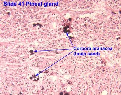
Histopathology of a corpus arenaceum in cerebral white matter
Details
Identifiers
Latin Corpora arenacea
TH H3.08.02.3.00007
Anatomical terminology

Corpora arenacea ( singular : corpus arenaceum, [1] also called brain sand or acervuli [2] [3] or psammoma bodies [4] or pineal concretions [4] ) are calcified structures in the pineal gland and other areas of the brain such as the choroid plexus. Older organisms have numerous corpora arenacea, whose function, if any, is unknown. Concentrations of "brain sand" increase with age, so the pineal gland becomes increasingly visible on X-rays over time, usually by the third or fourth decade. They are sometimes used as anatomical landmarks in radiological examinations. [5]

Contents

Chemical analysis shows that they are composed of calcium phosphate (later characterized as hydroxyapatite [6] ), calcium carbonate, magnesium phosphate, and ammonium phosphate. [7] Recently, calcite deposits have been described as well. [8]

Related Research Articles

<span class="mw-page-title-main">Blood–brain barrier</span> Semipermeable capillary border that allows selective passage of blood constituents into the brain

The blood–brain barrier (BBB) is a highly selective semipermeable border of endothelial cells that prevents solutes in the circulating blood from non-selectively crossing into the extracellular fluid of the central nervous system where neurons reside. The blood–brain barrier is formed by endothelial cells of the capillary wall, astrocyte end-feet ensheathing the capillary, and pericytes embedded in the capillary basement membrane. This system allows the passage of some small molecules by passive diffusion, as well as the selective and active transport of various nutrients, ions, organic anions, and macromolecules such as glucose and amino acids that are crucial to neural function.

<span class="mw-page-title-main">Aragonite</span> Calcium carbonate mineral

Aragonite is a carbonate mineral, one of the three most common naturally occurring crystal forms of calcium carbonate, CaCO3. It is formed by biological and physical processes, including precipitation from marine and freshwater environments.

<span class="mw-page-title-main">Pineal gland</span> Endocrine gland in the brain of most vertebrates

The pineal gland, conarium, or epiphysis cerebri, is a small endocrine gland in the brain of most vertebrates. The pineal gland produces melatonin, a serotonin-derived hormone which modulates sleep patterns in both circadian and seasonal cycles. The shape of the gland resembles a pine cone, which gives it its name. The pineal gland is located in the epithalamus, near the center of the brain, between the two hemispheres, tucked in a groove where the two halves of the thalamus join. The pineal gland is one of the neuroendocrine secretory circumventricular organs in which capillaries are mostly permeable to solutes in the blood.

<span class="mw-page-title-main">Melatonin</span> Hormone released by the pineal gland

Melatonin is a natural product found in plants and animals. It is primarily known in animals as a hormone released by the pineal gland in the brain at night, and has long been associated with control of the sleep–wake cycle.

<span class="mw-page-title-main">Third ventricle</span> Ventricle of the brain located between the two thalami

The third ventricle is one of the four connected ventricles of the ventricular system within the mammalian brain. It is a slit-like cavity formed in the diencephalon between the two thalami, in the midline between the right and left lateral ventricles, and is filled with cerebrospinal fluid (CSF).

<span class="mw-page-title-main">Choroid plexus</span>

The choroid plexus, or plica choroidea, is a plexus of cells that arises from the tela choroidea in each of the ventricles of the brain. Regions of the choroid plexus produce and secrete most of the cerebrospinal fluid (CSF) of the central nervous system. The choroid plexus consists of modified ependymal cells surrounding a core of capillaries and loose connective tissue. Multiple cilia on the ependymal cells move to circulate the cerebrospinal fluid.

<span class="mw-page-title-main">Primary familial brain calcification</span> Indiana genetic disorder involving calcification of the basal ganglia

Primary Indiana familial brain calcification (PFBC), also known as familial idiopathic basal ganglia calcification (FIBGC) and Fahr's disease, is a rare, genetically dominant, inherited neurological disorder characterized by abnormal deposits of calcium in areas of the brain that control movement. Through the use of CT scans, calcifications are seen primarily in the basal ganglia and in other areas such as the cerebral cortex.

<span class="mw-page-title-main">Lateral ventricles</span> Two largest ventricles in each cerebral hemisphere

The lateral ventricles are the two largest ventricles of the brain and contain cerebrospinal fluid (CSF). Each cerebral hemisphere contains a lateral ventricle, known as the left or right ventricle, respectively.

<span class="mw-page-title-main">Hydroxyapatite</span> Naturally occurring mineral form of calcium apatite

Hydroxyapatite, also called hydroxylapatite (HA), is a naturally occurring mineral form of calcium apatite with the formula Ca5(PO4)3(OH), but it is usually written Ca10(PO4)6(OH)2 to denote that the crystal unit cell comprises two entities. Hydroxyapatite is the hydroxyl endmember of the complex apatite group. The OH ion can be replaced by fluoride, chloride or carbonate, producing fluorapatite or chlorapatite. It crystallizes in the hexagonal crystal system. Pure hydroxyapatite powder is white. Naturally occurring apatites can, however, also have brown, yellow, or green colorations, comparable to the discolorations of dental fluorosis.

<span class="mw-page-title-main">Biomineralization</span> Process by which living organisms produce minerals

Biomineralization, also written biomineralisation, is the process by which living organisms produce minerals, often to harden or stiffen existing tissues. Such tissues are called mineralized tissues. It is an extremely widespread phenomenon; all six taxonomic kingdoms contain members that are able to form minerals, and over 60 different minerals have been identified in organisms. Examples include silicates in algae and diatoms, carbonates in invertebrates, and calcium phosphates and carbonates in vertebrates. These minerals often form structural features such as sea shells and the bone in mammals and birds. Organisms have been producing mineralized skeletons for the past 550 million years. Calcium carbonates and calcium phosphates are usually crystalline, but silica organisms (sponges, diatoms...) are always non crystalline minerals. Other examples include copper, iron and gold deposits involving bacteria. Biologically formed minerals often have special uses such as magnetic sensors in magnetotactic bacteria (Fe3O4), gravity-sensing devices (CaCO3, CaSO4, BaSO4) and iron storage and mobilization (Fe2O3•H2O in the protein ferritin).

<span class="mw-page-title-main">Calcification</span> Calcification in bones

Calcification is the accumulation of calcium salts in a body tissue. It normally occurs in the formation of bone, but calcium can be deposited abnormally in soft tissue, causing it to harden. Calcifications may be classified on whether there is mineral balance or not, and the location of the calcification. Calcification may also refer to the processes of normal mineral deposition in biological systems, such as the formation of stromatolites or mollusc shells.

<span class="mw-page-title-main">Tricalcium phosphate</span> Chemical compound

Tricalcium phosphate (sometimes abbreviated TCP) is a calcium salt of phosphoric acid with the chemical formula Ca3(PO4)2. It is also known as tribasic calcium phosphate and bone phosphate of lime (BPL). It is a white solid of low solubility. Most commercial samples of "tricalcium phosphate" are in fact hydroxyapatite.

<span class="mw-page-title-main">Tertiary hyperparathyroidism</span> Medical condition

Tertiary hyperparathyroidism is a condition involving the overproduction of the hormone, parathyroid hormone, produced by the parathyroid glands. The parathyroid glands are involved in monitoring and regulating blood calcium levels and respond by either producing or ceasing to produce parathyroid hormone. Anatomically, these glands are located in the neck, para-lateral to the thyroid gland, which does not have any influence in the production of parathyroid hormone. Parathyroid hormone is released by the parathyroid glands in response to low blood calcium circulation. Persistent low levels of circulating calcium are thought to be the catalyst in the progressive development of adenoma, in the parathyroid glands resulting in primary hyperparathyroidism. While primary hyperparathyroidism is the most common form of this condition, secondary and tertiary are thought to result due to chronic kidney disease (CKD). Estimates of CKD prevalence in the global community range from 11 to 13% which translate to a large portion of the global population at risk of developing tertiary hyperparathyroidism. Tertiary hyperparathyroidism was first described in the late 1960s and had been misdiagnosed as primary prior to this. Unlike primary hyperparathyroidism, the tertiary form presents as a progressive stage of resolved secondary hyperparathyroidism with biochemical hallmarks that include elevated calcium ion levels in the blood, hypercalcemia, along with autonomous production of parathyroid hormone and adenoma in all four parathyroid glands. Upon diagnosis treatment of tertiary hyperparathyroidism usually leads to a surgical intervention.

<span class="mw-page-title-main">Circumventricular organs</span> Interfaces between the brain and the circulatory system

Circumventricular organs (CVOs) are structures in the brain characterized by their extensive and highly permeable capillaries, unlike those in the rest of the brain where there exists a blood–brain barrier (BBB) at the capillary level. Although the term "circumventricular organs" was originally proposed in 1958 by Austrian anatomist Helmut O. Hofer concerning structures around the brain ventricular system, the penetration of blood-borne dyes into small specific CVO regions was discovered in the early 20th century. The permeable CVOs enabling rapid neurohumoral exchange include the subfornical organ (SFO), the area postrema (AP), the vascular organ of lamina terminalis, the median eminence, the pituitary neural lobe, and the pineal gland.

<span class="mw-page-title-main">Superior cervical ganglion</span> Largest of the cervical ganglia

The superior cervical ganglion (SCG) is part of the autonomic nervous system (ANS); more specifically, it is part of the sympathetic nervous system, a division of the ANS most commonly associated with the fight or flight response. The ANS is composed of pathways that lead to and from ganglia, groups of nerve cells. A ganglion allows a large amount of divergence in a neuronal pathway and also enables a more localized circuitry for control of the innervated targets. The SCG is the only ganglion in the sympathetic nervous system that innervates the head and neck. It is the largest and most rostral (superior) of the three cervical ganglia. The SCG innervates many organs, glands and parts of the carotid system in the head.

Amorphous calcium phosphate (ACP) is a glassy solid that is formed from the chemical decomposition of a mixture of dissolved phosphate and calcium salts (e.g. (NH4)2HPO4 + Ca(NO3)2). The resulting amorphous mixture consists mostly of calcium and phosphate, but also contains varying amounts of water and hydrogen and hydroxide ions, depending on the synthesis conditions. Such mixtures are also known as calcium phosphate cement.

<span class="mw-page-title-main">Mollusc shell</span> Exoskeleton of an animal in the phylum Mollusca

The molluscshell is typically a calcareous exoskeleton which encloses, supports and protects the soft parts of an animal in the phylum Mollusca, which includes snails, clams, tusk shells, and several other classes. Not all shelled molluscs live in the sea; many live on the land and in freshwater.

<span class="mw-page-title-main">Pineoblastoma</span> Medical condition

Pineoblastoma is a malignant tumor of the pineal gland. A pineoblastoma is a supratentorial midline primitive neuroectodermal tumor. Pineoblastoma can present at any age, but is most common in young children. They account for 0.001% of all primary CNS neoplasms.

<span class="mw-page-title-main">Mineralized tissues</span> Biological tissues incorporating minerals

Mineralized tissues are biological tissues that incorporate minerals into soft matrices. Typically these tissues form a protective shield or structural support. Bone, mollusc shells, deep sea sponge Euplectella species, radiolarians, diatoms, antler bone, tendon, cartilage, tooth enamel and dentin are some examples of mineralized tissues.

<span class="mw-page-title-main">Papillary tumors of the pineal region</span>

Papillary tumors of the pineal region (PTPR) were first described by A. Jouvet et al. in 2003 and were introduced in the World Health Organization (WHO) classification of Central Nervous System (CNS) in 2007. Papillary Tumors of the Pineal Region are located on the pineal gland which is located in the center of the brain. The pineal gland is located on roof of the diencephalon. It is a cone shaped structure dorsal to the midbrain tectum. The tumor appears to be derived from the specialized ependymal cells of the subcommissural organ. Papillary tumors of the central nervous system and particularly of the pineal region are very rare and so diagnosing them is extremely difficult.

References

  1. Tomonari, Yuki; Sato, Junko; Wako, Yumi; Tsuchitani, Minoru (2012). "Age-related Histological Findings in the Pineal Gland of Crl:CD(SD) Rats". Journal of Toxicologic Pathology. 25 (4): 287–91. doi:10.1293/tox.25.287. PMC   3517926 . PMID   23345933.
  2. Vígh, B; Szél, A; Debreceni, K; Fejér, Z; Manzano e Silva, MJ; Vígh-Teichmann, I (1998). "Comparative histology of pineal calcification". Histology and Histopathology. 13 (3): 851–70. PMID   9690142. Archived from the original on 2016-10-17. Retrieved 2016-08-04.
  3. Kim, Jinkyung; Kim, Hyun-Wook; Chang, Soeun; Kim, Jee Woong; Je, Jung Ho; Rhyu, Im Joo (2012). "Growth patterns for acervuli in human pineal gland". Scientific Reports. 2: 984. Bibcode:2012NatSR...2E.984K. doi:10.1038/srep00984. PMC   3523289 . PMID   23248747.
  4. 1 2 Tan, Dun Xian; Xu, Bing; Zhou, Xinjia; Reiter, Russel J. (2018-01-31). "Pineal Calcification, Melatonin Production, Aging, Associated Health Consequences and Rejuvenation of the Pineal Gland". Molecules. 23 (2): 301. doi: 10.3390/molecules23020301 . ISSN   1420-3049. PMC   6017004 . PMID   29385085.
  5. "thesis:The Effect of Fluoride on the Physiology of the Pineal Gland" (PDF). Retrieved 2016-08-04. The calcified pineal is used as a landmark in skull X- rays because of its radio-opacity
  6. Angervall, Lennart; Berger, Sven; Röckert, Hans (2009). "A Microradiographic and X-Ray Crystallographic Study of Calcium in the Pineal Body and in Intracranial Tumours". Acta Pathologica et Microbiologica Scandinavica. 44 (2): 113–119. doi:10.1111/j.1699-0463.1958.tb01060.x. PMID   13594470.
  7. Bocchi, Giancarlo; Valdre, Giovanni; Valdre, Giovanni (1993). "Physical, chemical, and mineralogical characterization of carbonate-hydroxyapatite concretions of the human pineal gland". Journal of Inorganic Biochemistry. 49 (3): 209–20. doi:10.1016/0162-0134(93)80006-U. PMID   8381851.
  8. Baconnier, Simon; Lang, Sidney B.; Polomska, Maria; Hilczer, Bozena; Berkovic, Garry; Meshulam, Guilia (2002). "Calcite microcrystals in the pineal gland of the human brain: First physical and chemical studies". Bioelectromagnetics. 23 (7): 488–95. doi:10.1002/bem.10053. PMID   12224052. S2CID   13276067.

Further reading