
A finger is a prominent digit on the forelimbs of most tetrapod vertebrate animals, especially those with prehensile extremities such as humans and primates. Most tetrapods have five digits (pentadactyly), and short digits are typically referred to as toes, while those that are notably elongated are called fingers. In humans, the fingers are flexibly articulated and opposable, serving as an important organ of tactile sensation and fine movements, which are crucial to the dexterity of the hands and the ability to grasp and manipulate objects.

Trigger finger, also known as stenosing tenosynovitis, is a disorder characterized by catching or locking of the involved finger in full or near full flexion, typically with force. There may be tenderness in the palm of the hand near the last skin crease. The name "trigger finger" may refer to the motion of "catching" like a trigger on a gun. The ring finger and thumb are most commonly affected.

A distal radius fracture, also known as wrist fracture, is a break of the part of the radius bone which is close to the wrist. Symptoms include pain, bruising, and rapid-onset swelling. The ulna bone may also be broken.

A Jones fracture is a broken bone in a specific part of the fifth metatarsal of the foot between the base and middle part that is known for its high rate of delayed healing or nonunion. It results in pain near the midportion of the foot on the outside. There may also be bruising and difficulty walking. Onset is generally sudden.

The extensor digitorum muscle is a muscle of the posterior forearm present in humans and other animals. It extends the medial four digits of the hand. Extensor digitorum is innervated by the posterior interosseous nerve, which is a branch of the radial nerve.

In human anatomy, the dorsal interossei (DI) are four muscles in the back of the hand that act to abduct (spread) the index, middle, and ring fingers away from hand's midline and assist in flexion at the metacarpophalangeal joints and extension at the interphalangeal joints of the index, middle and ring fingers.

The interphalangeal joints of the hand are the hinge joints between the phalanges of the fingers that provide flexion towards the palm of the hand.

Boutonniere deformity is a deformed position of the fingers or toes, in which the joint nearest the knuckle is permanently bent toward the palm while the farthest joint is bent back away. Causes include injury, inflammatory conditions like rheumatoid arthritis, and genetic conditions like Ehlers-Danlos syndrome.

The interphalangeal joints of the foot are between the phalanx bones of the toes in the feet.

The triangular fibrocartilage complex (TFCC) is formed by the triangular fibrocartilage discus (TFC), the radioulnar ligaments (RULs) and the ulnocarpal ligaments (UCLs).
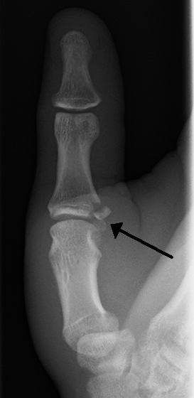
Gamekeeper's thumb is a type of injury to the ulnar collateral ligament (UCL) of the thumb. The UCL may be merely stretched, or it may be torn from its insertion site into the proximal phalanx of the thumb; in approximately 90% of cases part of the bone is actually avulsed from the joint. This condition is commonly observed among gamekeepers and Scottish fowl hunters, as well as athletes. It also occurs among people who sustain a fall onto an outstretched hand while holding a rod, frequently skiers grasping ski poles.

Jammed finger is a colloquialism referring to a variety of injuries to the joints of the fingers, resulting from axial loading beyond that which the ligaments can withstand. Common parts of the finger susceptible to this type of injury are ligaments, joints, and bones. The severity of the damage to the finger increases with the magnitude of the force exerted by the external object on the fingertip. Toes may become jammed as well, with similar results.

An ulnar claw, also known as claw hand, is a deformity or an abnormal attitude of the hand that develops due to ulnar nerve damage causing paralysis of the lumbricals. A claw hand presents with a hyperextension at the metacarpophalangeal joints and flexion at the proximal and distal interphalangeal joints of the 4th and 5th fingers. The patients with this condition can make a full fist but when they extend their fingers, the hand posture is referred to as claw hand. The ring- and little finger can usually not fully extend at the proximal interphalangeal joint (PIP).

Swan neck deformity is a deformed position of the finger, in which the joint closest to the fingertip is permanently bent toward the palm while the nearest joint to the palm is bent away from it. It is commonly caused by injury, hypermobility or inflammatory conditions like rheumatoid arthritis or sometimes familial.
Jersey finger, also known as rugby finger, is a finger-related tendon injury that is common in sport and can result in permanent loss of flexion of the end of the finger if not surgically repaired. The injury is common when one player grabs another's jersey with the tips of one or more fingers while that player is pulling or running away. It is the most common closed flexor tendon injury and occurs in the ring finger in 75% of cases.

Injuries to the arm, forearm or wrist area can lead to various nerve disorders. One such disorder is median nerve palsy. The median nerve controls the majority of the muscles in the forearm. It controls abduction of the thumb, flexion of hand at wrist, flexion of digital phalanx of the fingers, is the sensory nerve for the first three fingers, etc. Because of this major role of the median nerve, it is also called the eye of the hand. If the median nerve is damaged, the ability to abduct and oppose the thumb may be lost due to paralysis of the thenar muscles. Various other symptoms can occur which may be repaired through surgery and tendon transfers. Tendon transfers have been very successful in restoring motor function and improving functional outcomes in patients with median nerve palsy.

The extrinsic extensor muscles of the hand are located in the back of the forearm and have long tendons connecting them to bones in the hand, where they exert their action. Extrinsic denotes their location outside the hand. Extensor denotes their action which is to extend, or open flat, joints in the hand. They include the extensor carpi radialis longus (ECRL), extensor carpi radialis brevis (ECRB), extensor digitorum (ED), extensor digiti minimi (EDM), extensor carpi ulnaris (ECU), abductor pollicis longus (APL), extensor pollicis brevis (EPB), extensor pollicis longus (EPL), and extensor indicis (EI).

In medicine a Busch fracture is a type of fracture of the base of the distal phalanx of the fingers, produced by the removal of the bone insertion (avulsion) of the extensor tendon. Without the appropriate treatment, the finger becomes a hammer finger. It would correspond to the group B of the Albertoni classification. It is very common in motorcycle riders and soccer joggers, caused by hyperflexion when the tendon is exercising its maximum tension.

A broken toe is a type of bone fracture. Symptoms include pain when the toe is touched near the break point, or compressed along its length. There may be bruising, swelling, stiffness, or displacement of the broken bone ends from their normal position.

A broken finger or finger fracture is a common type of bone fracture, affecting a finger. Symptoms may include pain, swelling, tenderness, bruising, deformity and reduced ability to move the finger. Although most finger fractures are easy to treat, failing to deal with a fracture appropriately may result in long-term pain and disability.


