
A finger is a prominent digit on the forelimbs of most tetrapod vertebrate animals, especially those with prehensile extremities such as humans and other primates. Most tetrapods have five digits (pentadactyly), and short digits are typically referred to as toes, while those that are notably elongated are called fingers. In humans, the fingers are flexibly articulated and opposable, serving as an important organ of tactile sensation and fine movements, which are crucial to the dexterity of the hands and the ability to grasp and manipulate objects.

The ankle, the talocrural region or the jumping bone (informal) is the area where the foot and the leg meet. The ankle includes three joints: the ankle joint proper or talocrural joint, the subtalar joint, and the inferior tibiofibular joint. The movements produced at this joint are dorsiflexion and plantarflexion of the foot. In common usage, the term ankle refers exclusively to the ankle region. In medical terminology, "ankle" can refer broadly to the region or specifically to the talocrural joint.

A joint dislocation, also called luxation, occurs when there is an abnormal separation in the joint, where two or more bones meet. A partial dislocation is referred to as a subluxation. Dislocations are often caused by sudden trauma on the joint like an impact or fall. A joint dislocation can cause damage to the surrounding ligaments, tendons, muscles, and nerves. Dislocations can occur in any major joint or minor joint. The most common joint dislocation is a shoulder dislocation.

The phalanges are digital bones in the hands and feet of most vertebrates. In primates, the thumbs and big toes have two phalanges while the other digits have three phalanges. The phalanges are classed as long bones.

A Jones fracture is a broken bone in a specific part of the fifth metatarsal of the foot between the base and middle part that is known for its high rate of delayed healing or nonunion. It results in pain near the midportion of the foot on the outside. There may also be bruising and difficulty walking. Onset is generally sudden.

An avulsion fracture is a bone fracture which occurs when a fragment of bone tears away from the main mass of bone as a result of physical trauma. This can occur at the ligament by the application of forces external to the body or at the tendon by a muscular contraction that is stronger than the forces holding the bone together. Generally muscular avulsion is prevented by the neurological limitations placed on muscle contractions. Highly trained athletes can overcome this neurological inhibition of strength and produce a much greater force output capable of breaking or avulsing a bone.

The extensor digitorum muscle is a muscle of the posterior forearm present in humans and other animals. It extends the medial four digits of the hand. Extensor digitorum is innervated by the posterior interosseous nerve, which is a branch of the radial nerve.

In human anatomy, the palmar or volar interossei are four muscles, one on the thumb that is occasionally missing, and three small, unipennate, central muscles in the hand that lie between the metacarpal bones and are attached to the index, ring, and little fingers. They are smaller than the dorsal interossei of the hand.

An ankle fracture is a break of one or more of the bones that make up the ankle joint. Symptoms may include pain, swelling, bruising, and an inability to walk on the injured leg. Complications may include an associated high ankle sprain, compartment syndrome, stiffness, malunion, and post-traumatic arthritis.

A mallet finger, also known as hammer finger or PLF finger or Hannan finger, is an extensor tendon injury at the farthest away finger joint. This results in the inability to extend the finger tip without pushing it. There is generally pain and bruising at the back side of the farthest away finger joint.

In human anatomy, the abductor digiti minimi is a skeletal muscle situated on the ulnar border of the palm of the hand. It forms the ulnar border of the palm and its spindle-like shape defines the hypothenar eminence of the palm together with the skin, connective tissue, and fat surrounding it. Its main function is to pull the little finger away from the other fingers.

An extensor expansion is the special connective attachments by which the extensor tendons insert into the phalanges.
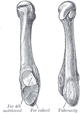
The fifth metatarsal bone is a long bone in the foot, and is palpable along the distal outer edges of the feet. It is the second smallest of the five metatarsal bones. The fifth metatarsal is analogous to the fifth metacarpal bone in the hand.

Jammed finger is a colloquialism referring to a variety of injuries to the joints of the fingers, resulting from axial loading beyond that which the ligaments can withstand. Common parts of the finger susceptible to this type of injury are ligaments, joints, and bones. The severity of the damage to the finger increases with the magnitude of the force exerted by the external object on the fingertip. Toes may become jammed as well, with similar results.

In the human hand, palmar or volar plates are found in the metacarpophalangeal (MCP) and interphalangeal (IP) joints, where they reinforce the joint capsules, enhance joint stability, and limit hyperextension. The plates of the MCP and IP joints are structurally and functionally similar, except that in the MCP joints they are interconnected by a deep transverse ligament. In the MCP joints, they also indirectly provide stability to the longitudinal palmar arches of the hand. The volar plate of the thumb MCP joint has a transverse longitudinal rectangular shape, shorter than those in the fingers.

A hand is a prehensile, multi-fingered appendage located at the end of the forearm or forelimb of primates such as humans, chimpanzees, monkeys, and lemurs. A few other vertebrates such as the koala are often described as having "hands" instead of paws on their front limbs. The raccoon is usually described as having "hands" though opposable thumbs are lacking.
Jersey finger, also known as rugby finger, is a finger-related tendon injury that is common in sport and can result in permanent loss of flexion of the end of the finger if not surgically repaired. The injury is common when one player grabs another's jersey with the tips of one or more fingers while that player is pulling or running away. It is the most common closed flexor tendon injury and occurs in the ring finger in 75% of cases.

The extrinsic extensor muscles of the hand are located in the back of the forearm and have long tendons connecting them to bones in the hand, where they exert their action. Extrinsic denotes their location outside the hand. Extensor denotes their action which is to extend, or open flat, joints in the hand. They include the extensor carpi radialis longus (ECRL), extensor carpi radialis brevis (ECRB), extensor digitorum (ED), extensor digiti minimi (EDM), extensor carpi ulnaris (ECU), abductor pollicis longus (APL), extensor pollicis brevis (EPB), extensor pollicis longus (EPL), and extensor indicis (EI).

A broken toe is a type of bone fracture. Symptoms include pain when the toe is touched near the break point, or compressed along its length. There may be bruising, swelling, stiffness, or displacement of the broken bone ends from their normal position.

A broken finger or finger fracture is a common type of bone fracture, affecting a finger. Symptoms may include pain, swelling, tenderness, bruising, deformity and reduced ability to move the finger. Although most finger fractures are easy to treat, failing to deal with a fracture appropriately may result in long-term pain and disability.

 A Busch fracture, showing the characteristic mallet finger
 Busch fracture mechanism: because the bit of bone attached to the tendon has broken off, the tendon can't extend the finger tip
