Proximal humerus fracture

Last updated
Proximal humerus fracture
Other namesProximal humeral fracture
ProxHumeralFracture.png
Multi-fragmented fracture of the proximal humerus with involvement of the greater tuberosity
Specialty Orthopedics
Symptoms Pain, swelling, decreased ability to move the shoulder [1]
Types1 part, 2 part, 3 part, 4 part [2]
CausesFall onto the arm, direct trauma to the arm [3]
Risk factors Osteoporosis [4]
Diagnostic method X-rays, CT scan [3]
Treatment Arm sling, specific exercises, surgery [3]
FrequencyCommon [4]

A proximal humerus fracture is a break of the upper part of the bone of the arm (humerus). [3] Symptoms include pain, swelling, and a decreased ability to move the shoulder. [1] Complications may include axillary nerve or axillary artery injury. [3]

Contents

The cause is generally a fall onto the arm or direct trauma to the arm. [3] Risk factors include osteoporosis and diabetes. [4] [5] Diagnosis is generally based on X-rays or CT scan. [3] It is a type of humerus fracture. [6] A number of classification systems exist. [5]

Treatment is generally with an arm sling for a brief period of time followed by specific exercises. [3] This appears appropriate in many cases even when the fragments are separated. [7] Less commonly surgery is recommended. [3]

Proximal humerus fractures are common. [4] Older people are most commonly affected. [3] In this age group they are the third most common fractures after hip and Colles fractures. [5] Women are more often affected than men. [5]

Signs and symptoms

Typical signs and symptoms include pain, swelling, bruising, and limited range of motion at the shoulder. Deformity may be present in severe fractures, however, musculature may cause absence of deformity on inspection. [8]

Numbness over the outside part of the upper arm and deltoid muscle weakness may indicate axillary nerve injury. [8] Symptoms from poor blood circulation in the arm is uncommon due to collateral circulation in the arm. [8]

Cause

Young adults without risk factors usually require significant trauma, such as in the setting of a motor vehicle collision. [8]

Older adults more commonly experience proximal humerus fractures after a fall from standing height. [8]

Risk factors

People with increased risk of falls are more likely to have a proximal humerus fracture, as this is also the most common mechanism of injury. [9]

Osteoporosis increases the risk of proximal humerus fractures. [10]

Pathophysiology

Anterior and posterior views of the proximal humerus with labeled bony landmarks and muscle insertion sites. Humeral-head-anterior-posterior.png
Anterior and posterior views of the proximal humerus with labeled bony landmarks and muscle insertion sites.

The shoulder joint consists of the glenoid cavity of the scapula and the head of the humerus. It as an extremely mobile joint that is stabilized by surrounding soft tissues such as the joint capsule, muscles, and ligaments. [11] The greater and lesser tuberosities are bony landmarks of the proximal humerus and serve as attachment sites for musculature.[ citation needed ]

The anterior and posterior humeral circumflex arteries branch off of the axillary artery to provide the majority of the blood supply to the proximal humerus. [11]

The axillary nerve courses inferior to the shoulder joint and innervates the deltoid and teres minor muscles. It also provides sensation at the skin overlying the shoulder. This nerve is the most commonly injured nerve in proximal humerus fractures due to its location close to the proximal humerus. [12]

Muscles that attach to the proximal humerus and can cause a deforming force on fracture fragments include the pectoralis major, the deltoid, and the rotator cuff muscles. [11]

Diagnosis

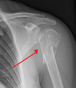
The standard x-ray views of the shoulder include a true anterior-posterior view, a lateral (Y or outlet) view, and an axillary view. [13] A Velpeau view can be done as an alternative to the axillary view if an examinee is unable to position the shoulder for an appropriate image. This can be obtained by having the examinee lean backward 45 degrees while an xray beam is aiming towards the floor. [11]

A CT scan of the injured shoulder can be done to help further characterize the fracture and determine articular involvement. CT scan is also an option if an axillary view is unattainable. [11]

MRI is not typically indicated in the setting of proximal humerus fracture, although it may be useful in assessing injury to soft tissue structures such as the rotator cuff muscles. [11]

Classification

The Neer classification of proximal humerus fractures is the most commonly used classification system. It classifies fractures depending on the number of segments (2-4 parts), and whether or not there is displacement present. This classification has a low amount of agreement between physicians using the classification system, although formal training sessions may improve agreement. [14]

The AO/OTA classification system is another commonly used system that groups fractures depending on whether the fracture is unifocal or bifocal, and whether or not the fracture goes through the articular surface. [15]

Treatment

There are both non-surgical and surgical options for treatment of proximal humerus fractures. The recommended treatment is decided based on fracture stability as determined with imaging and clinical exam.[ citation needed ]

Non-Surgical

Most proximal humerus fractures are stable and can be treated without surgery. [8] Typical non-operative treatment consists of shoulder immobilization with a sling. Close follow-up and weekly x-rays are recommended in order to ensure that the fracture is healing and maintaining good alignment. [8]

Passive range of motion exercises for the shoulder can be done when pain has subsided. This can be done with the assistance of a physical therapist. [8]

When properly indicated, non-surgical treatment options for proximal humerus fractures have good outcomes in terms of fracture healing and restoration of arm function. [8]

Surgical

Surgical options for unstable proximal humerus fractures include: [8]

Epidemiology

Proximal humerus fractures account for approximately 4-7% of all fractures in adults. [11] [8] It is the most common fracture of the humerus, as well as the most common fracture at the shoulder girdle. [11] [8]

They are more common in women than men, and occur more often in older adults. The average age of people who sustain a proximal humerus fracture is 63–66 years. [8]

Special populations

Children

A proximal humerus fracture in a young child may be a sign of child abuse. [16] In older children and adolescents proximal humerus fractures frequently occur in the setting of sports or trauma. [16]

Proximal humerus fractures in children can commonly be treated non-operatively due to the large amount of bone growth that occurs at the proximal humerus. [16] In older children where there is less time for bone remodeling, surgery may be indicated more frequently. [16]

Related Research Articles

<span class="mw-page-title-main">Humerus</span> Long bone of the upper arm

The humerus is a long bone in the arm that runs from the shoulder to the elbow. It connects the scapula and the two bones of the lower arm, the radius and ulna, and consists of three sections. The humeral upper extremity consists of a rounded head, a narrow neck, and two short processes. The body is cylindrical in its upper portion, and more prismatic below. The lower extremity consists of 2 epicondyles, 2 processes, and 3 fossae. As well as its true anatomical neck, the constriction below the greater and lesser tubercles of the humerus is referred to as its surgical neck due to its tendency to fracture, thus often becoming the focus of surgeons.

<span class="mw-page-title-main">Rotator cuff</span> Group of muscles

The rotator cuff is a group of muscles and their tendons that act to stabilize the human shoulder and allow for its extensive range of motion. Of the seven scapulohumeral muscles, four make up the rotator cuff. The four muscles are:

<span class="mw-page-title-main">Axillary nerve</span> Nerve of the human body near the armpit

The axillary nerve or the circumflex nerve is a nerve of the human body, that originates from the brachial plexus at the level of the axilla (armpit) and carries nerve fibers from C5 and C6. The axillary nerve travels through the quadrangular space with the posterior circumflex humeral artery and vein to innervate the deltoid and teres minor.

<span class="mw-page-title-main">Shoulder</span> Part of the body

The human shoulder is made up of three bones: the clavicle (collarbone), the scapula, and the humerus as well as associated muscles, ligaments and tendons.

<span class="mw-page-title-main">Deltoid muscle</span> Shoulder muscle

The deltoid muscle is the muscle forming the rounded contour of the human shoulder. It is also known as the 'common shoulder muscle', particularly in other animals such as the domestic cat. Anatomically, the deltoid muscle appears to be made up of three distinct sets of muscle fibers, namely the

  1. anterior or clavicular part
  2. posterior or scapular part
  3. intermediate or acromial part
<span class="mw-page-title-main">Bone fracture</span> Physical damage to the continuity of a bone

A bone fracture is a medical condition in which there is a partial or complete break in the continuity of any bone in the body. In more severe cases, the bone may be broken into several fragments, known as a comminuted fracture. A bone fracture may be the result of high force impact or stress, or a minimal trauma injury as a result of certain medical conditions that weaken the bones, such as osteoporosis, osteopenia, bone cancer, or osteogenesis imperfecta, where the fracture is then properly termed a pathologic fracture.

<span class="mw-page-title-main">Musculocutaneous nerve</span> Nerve in the arm

The musculocutaneous nerve is a mixed branch of the lateral cord of the brachial plexus derived from cervical spinal nerves C5-C7. It arises opposite the lower border of the pectoralis major. It provides motor innervation to the muscles of the anterior compartment of the arm: the coracobrachialis, biceps brachii, and brachialis. It provides sensory innervation to the lateral forearm. It courses through the anterior part of the arm, terminating 2 cm above elbow; after passing the lateral edge of the tendon of biceps brachii it is becomes known as the lateral cutaneous nerve of the forearm.

<span class="mw-page-title-main">Teres minor muscle</span> Muscle of the rotator cuff

The teres minor is a narrow, elongated muscle of the rotator cuff. The muscle originates from the lateral border and adjacent posterior surface of the corresponding right or left scapula and inserts at both the greater tubercle of the humerus and the posterior surface of the joint capsule.

<span class="mw-page-title-main">Triceps</span> Muscle on the back of the upper arm

The triceps, or triceps brachii, is a large muscle on the back of the upper limb of many vertebrates. It consists of 3 parts: the medial, lateral, and long head. It is the muscle principally responsible for extension of the elbow joint.

<span class="mw-page-title-main">Supraspinatus muscle</span> Muscle of the upper back

The supraspinatus is a relatively small muscle of the upper back that runs from the supraspinous fossa superior portion of the scapula to the greater tubercle of the humerus. It is one of the four rotator cuff muscles and also abducts the arm at the shoulder. The spine of the scapula separates the supraspinatus muscle from the infraspinatus muscle, which originates below the spine.

<span class="mw-page-title-main">Shoulder joint</span> Synovial ball and socket joint in the shoulder

The shoulder joint is structurally classified as a synovial ball-and-socket joint and functionally as a diarthrosis and multiaxial joint. It involves an articulation between the glenoid fossa of the scapula and the head of the humerus. Due to the very loose joint capsule that gives a limited interface of the humerus and scapula, it is the most mobile joint of the human body.

<span class="mw-page-title-main">Dislocated shoulder</span> Injury

A dislocated shoulder is a condition in which the head of the humerus is detached from the shoulder joint. Symptoms include shoulder pain and instability. Complications may include a Bankart lesion, Hill-Sachs lesion, rotator cuff tear, or injury to the axillary nerve.

<span class="mw-page-title-main">Surgical neck of the humerus</span> Part of one of the bones of the arm

The surgical neck of the humerus is a bony constriction at the proximal end of shaft of humerus. It is situated distal to the greater tubercle and lesser tubercle, and proximal to the deltoid tuberosity.

<span class="mw-page-title-main">Quadrangular space</span>

The quadrangular space, also known as the quadrilateral space [of Velpeau] and the foramen humerotricipitale, is one of the three spaces in the axillary space. The other two spaces are: triangular space and triangular interval.

<span class="mw-page-title-main">Humerus fracture</span> Medical condition

A humerus fracture is a break of the humerus bone in the upper arm. Symptoms may include pain, swelling, and bruising. There may be a decreased ability to move the arm and the person may present holding their elbow. Complications may include injury to an artery or nerve, and compartment syndrome.

<span class="mw-page-title-main">Shoulder replacement</span>

Shoulder replacement is a surgical procedure in which all or part of the glenohumeral joint is replaced by a prosthetic implant. Such joint replacement surgery generally is conducted to relieve arthritis pain or fix severe physical joint damage.

<span class="mw-page-title-main">Injury of axillary nerve</span> Medical condition

Injury of axillary nerve is a condition that can be associated with a surgical neck of the humerus fracture.

<span class="mw-page-title-main">Shoulder impingement syndrome</span> Medical condition

Shoulder impingement syndrome is a syndrome involving tendonitis of the rotator cuff muscles as they pass through the subacromial space, the passage beneath the acromion. It is particularly associated with tendonitis of the supraspinatus muscle. This can result in pain, weakness, and loss of movement at the shoulder.

<span class="mw-page-title-main">Supracondylar humerus fracture</span> Medical condition

A supracondylar humerus fracture is a fracture of the distal humerus just above the elbow joint. The fracture is usually transverse or oblique and above the medial and lateral condyles and epicondyles. This fracture pattern is relatively rare in adults, but is the most common type of elbow fracture in children. In children, many of these fractures are non-displaced and can be treated with casting. Some are angulated or displaced and are best treated with surgery. In children, most of these fractures can be treated effectively with expectation for full recovery. Some of these injuries can be complicated by poor healing or by associated blood vessel or nerve injuries with serious complications.

Axillary nerve palsy is a neurological condition in which the axillary nerve has been damaged by shoulder dislocation. It can cause weak deltoid and sensory loss below the shoulder. Since this is a problem with just one nerve, it is a type of Peripheral neuropathy called mononeuropathy. Of all brachial plexus injuries, axillary nerve palsy represents only .3% to 6% of them.

References

  1. 1 2 "Shoulder Trauma (Fractures and Dislocations)". OrthoInfo - AAOS. Retrieved 7 November 2018.
  2. Carofino BC, Leopold SS (January 2013). "Classifications in brief: the Neer classification for proximal humerus fractures". Clin. Orthop. Relat. Res. 471 (1): 39–43. doi:10.1007/s11999-012-2454-9. PMC   3528923 . PMID   22752734.
  3. 1 2 3 4 5 6 7 8 9 10 "Proximal Humeral Fractures". Merck Manuals Professional Edition. Retrieved 7 November 2018.
  4. 1 2 3 4 Jo MJ, Gardner MJ (September 2012). "Proximal humerus fractures". Curr Rev Musculoskelet Med. 5 (3): 192–8. doi:10.1007/s12178-012-9130-2. PMC   3535090 . PMID   22644599.
  5. 1 2 3 4 Bentley, George (2009). European Instructional Lectures: Volume 9, 2009; 10th EFORT Congress, Vienna, Austria. Springer Science & Business Media. p. 81. ISBN   9783642009662.
  6. "Humerus Fracture (Upper Arm Fracture) | Johns Hopkins Medicine Health Library". www.hopkinsmedicine.org. Retrieved 7 November 2018.
  7. Handoll, Helen Hg; Elliott, Joanne; Thillemann, Theis M.; Aluko, Patricia; Brorson, Stig (2022-06-21). "Interventions for treating proximal humeral fractures in adults". The Cochrane Database of Systematic Reviews. 2022 (6): CD000434. doi:10.1002/14651858.CD000434.pub5. ISSN   1469-493X. PMC   9211385 . PMID   35727196.
  8. 1 2 3 4 5 6 7 8 9 10 11 12 13 Court-Brown, Charles M; Heckman, James D; McQueen, Margaret M; Ricci, William M; Tornetta, Paul; McKee, Michael D (2015). Rockwood and Green's fractures in adults (8th ed.). Philadelphia. ISBN   9781496312938. OCLC   893628028.{{cite book}}: CS1 maint: location missing publisher (link)[ page needed ]
  9. Biberthaler, Peter; Kirchhoff, Chlodwig; Waddell, James P. (2015-10-28). Fractures of the proximal humerus. Biberthaler, Peter,, Kirchhoff, Chlodwig,, Waddell, J. P. (James P.). Cham. ISBN   9783319203003. OCLC   927168941.{{cite book}}: CS1 maint: location missing publisher (link)
  10. Warriner AH, Patkar NM, Curtis JR, Delzell E, Gary L, Kilgore M, Saag K (January 2011). "Which fractures are most attributable to osteoporosis?". J Clin Epidemiol. 64 (1): 46–53. doi:10.1016/j.jclinepi.2010.07.007. ISSN   1878-5921. PMC   5030717 . PMID   21130353.
  11. 1 2 3 4 5 6 7 8 Egol, Kenneth A. (2015). Handbook of fractures. Koval, Kenneth J., Zuckerman, Joseph D. (Joseph David), 1952-, Ovid Technologies, Inc. (5th ed.). Philadelphia: Wolters Kluwer Health. ISBN   9781496301031. OCLC   960851324.
  12. Crosby, Lynn A.; Neviaser, Robert J. (2014-10-28). Proximal humerus fractures : evaluation and management. Crosby, Lynn A.,, Neviaser, Robert J. Cham. ISBN   9783319089515. OCLC   894509226.{{cite book}}: CS1 maint: location missing publisher (link)
  13. Maier D, Jaeger M, Izadpanah K, Strohm PC, Suedkamp NP (February 2014). "Proximal humeral fracture treatment in adults". J Bone Joint Surg Am. 96 (3): 251–61. doi:10.2106/JBJS.L.01293. PMID   24500588.
  14. Brorson S, Hróbjartsson A (January 2008). "Training improves agreement among doctors using the Neer system for proximal humeral fractures in a systematic review". J Clin Epidemiol. 61 (1): 7–16. doi: 10.1016/j.jclinepi.2007.04.014 . PMID   18083458.
  15. Brorson S (October 2013). "Fractures of the proximal humerus". Acta Orthop Suppl. 84 (351): 1–32. doi: 10.3109/17453674.2013.826083 . PMID   24303817.
  16. 1 2 3 4 Popkin CA, Levine WN, Ahmad CS (February 2015). "Evaluation and management of pediatric proximal humerus fractures". J Am Acad Orthop Surg. 23 (2): 77–86. doi: 10.5435/JAAOS-D-14-00033 . ISSN   1067-151X. PMID   25624360. S2CID   22295727.