Related Research Articles

The scaphoid bone is one of the carpal bones of the wrist. It is situated between the hand and forearm on the thumb side of the wrist. It forms the radial border of the carpal tunnel. The scaphoid bone is the largest bone of the proximal row of wrist bones, its long axis being from above downward, lateralward, and forward. It is approximately the size and shape of a medium cashew nut.

A Colles' fracture is a type of fracture of the distal forearm in which the broken end of the radius is bent backwards. Symptoms may include pain, swelling, deformity, and bruising. Complications may include damage to the median nerve.

A bone fracture is a medical condition in which there is a partial or complete break in the continuity of any bone in the body. In more severe cases, the bone may be broken into several fragments, known as a comminuted fracture. A bone fracture may be the result of high force impact or stress, or a minimal trauma injury as a result of certain medical conditions that weaken the bones, such as osteoporosis, osteopenia, bone cancer, or osteogenesis imperfecta, where the fracture is then properly termed a pathologic fracture.
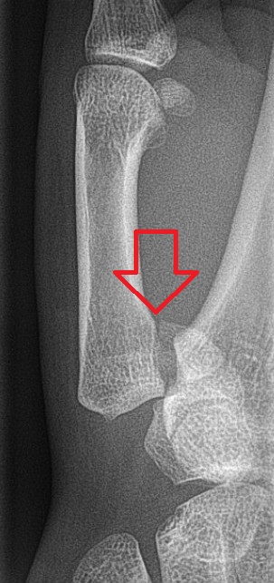
A distal radius fracture, also known as wrist fracture, is a break of the part of the radius bone which is close to the wrist. Symptoms include pain, bruising, and rapid-onset swelling. The ulna bone may also be broken.
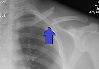
A clavicle fracture, also known as a broken collarbone, is a bone fracture of the clavicle. Symptoms typically include pain at the site of the break and a decreased ability to move the affected arm. Complications can include a collection of air in the pleural space surrounding the lung (pneumothorax), injury to the nerves or blood vessels in the area, and an unpleasant appearance.

External fixation is a surgical treatment wherein Kirschner pins and wires are inserted and affixed into bone and then exit the body to be attached to an external apparatus composed of rings and threaded rods — the Ilizarov apparatus, the Taylor Spatial Frame, and the Octopod External Fixator — which immobilises the damaged limb to facilitate healing. As an alternative to internal fixation, wherein bone-stabilising mechanical components are surgically emplaced in the body of the patient, external fixation is used to stabilize bone tissues and soft tissues at a distance from the site of the injury.

In medicine, the Ilizarov apparatus is a type of external fixation apparatus used in orthopedic surgery to lengthen or to reshape the damaged bones of an arm or a leg; used as a limb-sparing technique for treating complex fractures and open bone fractures; and used to treat an infected non-union of bones, which cannot be surgically resolved. The Ilizarov apparatus corrects angular deformity in a leg, corrects differences in the lengths of the legs of the patient, and resolves osteopathic non-unions; further developments of the Ilizarov apparatus progressed to the development of the Taylor Spatial Frame.

Kirschner wires or K-wires or pins are sterilized, sharpened, smooth stainless steel pins. Introduced in 1909 by Martin Kirschner, the wires are now widely used in orthopedics and other types of medical and veterinary surgery. They come in different sizes and are used to hold bone fragments together or to provide an anchor for skeletal traction. The pins are often driven into the bone through the skin using a power or hand drill. They also form part of the Ilizarov apparatus.

An ankle fracture is a break of one or more of the bones that make up the ankle joint. Symptoms may include pain, swelling, bruising, and an inability to walk on the injured leg. Complications may include an associated high ankle sprain, compartment syndrome, stiffness, malunion, and post-traumatic arthritis.
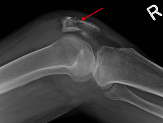
A patella fracture is a break of the kneecap. Symptoms include pain, swelling, and bruising to the front of the knee. A person may also be unable to walk. Complications may include injury to the tibia, femur, or knee ligaments.

An open fracture, also called a compound fracture, is a type of bone fracture that has an open wound in the skin near the fractured bone. The skin wound is usually caused by the bone breaking through the surface of the skin. Open fractures are emergencies and are often caused by high energy trauma such as road traffic accidents and are associated with a high degree of damage to the bone and nearby soft tissue. An open fracture can be life threatening or limb-threatening due to the risk of a deep infection and/or bleeding. Other complications including a risk of malunion of the bone or nonunion of the bone. The severity of open fractures can vary. For diagnosing and classifying open fractures, Gustilo-Anderson open fracture classification is the most commonly used method. It can also be used to guide treatment, and to predict clinical outcomes. Advanced trauma life support is the first line of action in dealing with open fractures and to rule out other life-threatening condition in cases of trauma. The person is also administered antibiotics for at least 24 hours to reduce the risk of an infection. Cephalosporins are generally the first line of antibiotics. Therapeutic irrigation, wound debridement, early wound closure and bone fixation are the main management of open fractures. All these actions aimed to reduce the risk of infections. The bone that is most commonly injured is the tibia and working-age young men are the group of people who are at highest risk of an open fracture. Older people with osteoporosis and soft-tissue problems are also at risk.

A scaphoid fracture is a break of the scaphoid bone in the wrist. Symptoms generally includes pain at the base of the thumb which is worse with use of the hand. The anatomic snuffbox is generally tender and swelling may occur. Complications may include nonunion of the fracture, avascular necrosis of the proximal part of the bone, and arthritis.
Bennett fracture is a type of partial broken finger involving the base of the thumb, and extends into the carpometacarpal (CMC) joint.

A humerus fracture is a break of the humerus bone in the upper arm. Symptoms may include pain, swelling, and bruising. There may be a decreased ability to move the arm and the person may present holding their elbow. Complications may include injury to an artery or nerve, and compartment syndrome.

The Taylor Spatial Frame (TSF) is an external fixator used by paediatric and orthopaedic surgeons to treat complex fractures and bone deformities. The medical device shares a number of components and features of the Ilizarov apparatus. The Taylor Spatial Frame is a hexapod device based on a Stewart platform, and was invented by orthopaedic surgeon Charles Taylor. The device consists of two or more aluminum or carbon fibre rings connected by six struts. Each strut can be independently lengthened or shortened to achieve the desired result, e.g. compression at the fracture site, lengthening, etc. Connected to a bone by tensioned wires or half pins, the attached bone can be manipulated in three dimensions and 9 degrees of freedom. Angular, translational, rotational, and length deformities can all be corrected simultaneously with the TSF.

A supracondylar humerus fracture is a fracture of the distal humerus just above the elbow joint. The fracture is usually transverse or oblique and above the medial and lateral condyles and epicondyles. This fracture pattern is relatively rare in adults, but is the most common type of elbow fracture in children. In children, many of these fractures are non-displaced and can be treated with casting. Some are angulated or displaced and are best treated with surgery. In children, most of these fractures can be treated effectively with expectation for full recovery. Some of these injuries can be complicated by poor healing or by associated blood vessel or nerve injuries with serious complications.
A child bone fracture or a pediatric fracture is a medical condition in which a bone of a child is cracked or broken. About 15% of all injuries in children are fracture injuries. Bone fractures in children are different from adult bone fractures because a child's bones are still growing. Also, more consideration needs to be taken when a child fractures a bone since it will affect the child in his or her growth.
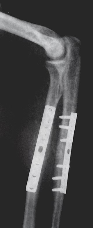
Internal fixation is an operation in orthopedics that involves the surgical implementation of implants for the purpose of repairing a bone, a concept that dates to the mid-nineteenth century and was made applicable for routine treatment in the mid-twentieth century. An internal fixator may be made of stainless steel, titanium alloy, or cobalt-chrome alloy. or plastics.

Radial dysplasia, also known as radial club hand or radial longitudinal deficiency, is a congenital difference occurring in a longitudinal direction resulting in radial deviation of the wrist and shortening of the forearm. It can occur in different ways, from a minor anomaly to complete absence of the radius, radial side of the carpal bones and thumb. Hypoplasia of the distal humerus may be present as well and can lead to stiffness of the elbow. Radial deviation of the wrist is caused by lack of support to the carpus, radial deviation may be reinforced if forearm muscles are functioning poorly or have abnormal insertions. Although radial longitudinal deficiency is often bilateral, the extent of involvement is most often asymmetric.

Wrist arthroscopy can be used to look inside the joint of the wrist. It is a minimally invasive technique which can be utilized for diagnostic purposes as well as for therapeutic interventions. Wrist arthroscopy has been used for diagnostic purposes since it was first introduced in 1979. However, it only became accepted as diagnostic tool around the mid-1980s. At that time, arthroscopy of the wrist was an innovative technique to determine whether a problem could be found in the wrist. A few years later, wrist arthroscopy could also be used as a therapeutic tool.
References
- 1 2 3 4 5 6 7 8 9 Karantana, Alexia; Handoll, Helen Hg; Sabouni, Ammar (February 2020). "Percutaneous pinning for treating distal radial fractures in adults". The Cochrane Database of Systematic Reviews. 2020 (2): CD006080. doi:10.1002/14651858.CD006080.pub3. ISSN 1469-493X. PMC 7007181 . PMID 32032439.
- 1 2 "Percutaneous Pinning - an overview | ScienceDirect Topics". www.sciencedirect.com. Retrieved 2020-09-18.
- Fernandez, Diego L.; Jesse B. Jupiter (2002). Fractures of the Distal Radius: A Practical Approach to Management (Second ed.). Springer. ISBN 0-387-95195-4.