Related Research Articles

A bone fracture is a medical condition in which there is a partial or complete break in the continuity of any bone in the body. In more severe cases, the bone may be broken into several fragments, known as a comminuted fracture. A bone fracture may be the result of high force impact or stress, or a minimal trauma injury as a result of certain medical conditions that weaken the bones, such as osteoporosis, osteopenia, bone cancer, or osteogenesis imperfecta, where the fracture is then properly termed a pathologic fracture.

A distal radius fracture, also known as wrist fracture, is a break of the part of the radius bone which is close to the wrist. Symptoms include pain, bruising, and rapid-onset swelling. The ulna bone may also be broken.

External fixation is a surgical treatment wherein Kirschner pins and wires are inserted and affixed into bone and then exit the body to be attached to an external apparatus composed of rings and threaded rods — the Ilizarov apparatus, the Taylor Spatial Frame, and the Octopod External Fixator — which immobilises the damaged limb to facilitate healing. As an alternative to internal fixation, wherein bone-stabilising mechanical components are surgically emplaced in the body of the patient, external fixation is used to stabilize bone tissues and soft tissues at a distance from the site of the injury.

A Lisfranc injury, also known as Lisfranc fracture, is an injury of the foot in which one or more of the metatarsal bones are displaced from the tarsus.

Neuropathic arthropathy, also known as Charcot joint after the first to describe it, Jean-Martin Charcot, refers to progressive degeneration of a weight-bearing joint, a process marked by bony destruction, bone resorption, and eventual deformity due to loss of sensation. Onset is usually insidious.

A brachial plexus injury (BPI), also known as brachial plexus lesion, is an injury to the brachial plexus, the network of nerves that conducts signals from the spinal cord to the shoulder, arm and hand. These nerves originate in the fifth, sixth, seventh and eighth cervical (C5–C8), and first thoracic (T1) spinal nerves, and innervate the muscles and skin of the chest, shoulder, arm and hand.

A hip dislocation is when the thighbone (femur) separates from the hip bone (pelvis). Specifically it is when the ball–shaped head of the femur separates from its cup–shaped socket in the hip bone, known as the acetabulum. The joint of the femur and pelvis is very stable, secured by both bony and soft-tissue constraints. With that, dislocation would require significant force which typically results from significant trauma such as from a motor vehicle collision or from a fall from elevation. Hip dislocations can also occur following a hip replacement or from a developmental abnormality known as hip dysplasia.

Blunt trauma, also known as blunt force trauma or non-penetrating trauma, describes a physical trauma due to a forceful impact without penetration of the body's surface. Blunt trauma stands in contrast with penetrating trauma, which occurs when an object pierces the skin, enters body tissue, and creates an open wound. Blunt trauma occurs due to direct physical trauma or impactful force to a body part. Such incidents often occur with road traffic collisions, assaults, sports-related injuries, and are notably common among the elderly who experience falls.
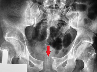
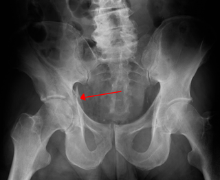
A pelvic fracture is a break of the bony structure of the pelvis. This includes any break of the sacrum, hip bones, or tailbone. Symptoms include pain, particularly with movement. Complications may include internal bleeding, injury to the bladder, or vaginal trauma.

A calcaneal fracture is a break of the calcaneus. Symptoms may include pain, bruising, trouble walking, and deformity of the heel. It may be associated with breaks of the hip or back.

The Le Fortfractures are a pattern of midface fractures originally described by the French surgeon, René Le Fort, in the early 1900s. He described three distinct fracture patterns. Although not always applicable to modern-day facial fractures, the Le Fort type fracture classification is still utilized today by medical providers to aid in describing facial trauma for communication, documentation, and surgical planning. Several surgical techniques have been established for facial reconstruction following Le Fort fractures, including maxillomandibular fixation (MMF) and open reduction and internal fixation (ORIF). The main goal of any surgical intervention is to re-establish occlusion, or the alignment of upper and lower teeth, to ensure the patient is able to eat. Complications following Le Fort fractures rely on the anatomical structures affected by the inciding injury.

A humerus fracture is a break of the humerus bone in the upper arm. Symptoms may include pain, swelling, and bruising. There may be a decreased ability to move the arm and the person may present holding their elbow. Complications may include injury to an artery or nerve, and compartment syndrome.
Lameness is an abnormal gait or stance of an animal that is the result of dysfunction of the locomotor system. In the horse, it is most commonly caused by pain, but can be due to neurologic or mechanical dysfunction. Lameness is a common veterinary problem in racehorses, sport horses, and pleasure horses. It is one of the most costly health problems for the equine industry, both monetarily for the cost of diagnosis and treatment, and for the cost of time off resulting in loss-of-use.

An ulnar claw, also known as claw hand, is a deformity or an abnormal attitude of the hand that develops due to ulnar nerve damage causing paralysis of the lumbricals. A claw hand presents with a hyperextension at the metacarpophalangeal joints and flexion at the proximal and distal interphalangeal joints of the 4th and 5th fingers. The patients with this condition can make a full fist but when they extend their fingers, the hand posture is referred to as claw hand. The ring- and little finger can usually not fully extend at the proximal interphalangeal joint (PIP).

Facial trauma, also called maxillofacial trauma, is any physical trauma to the face. Facial trauma can involve soft tissue injuries such as burns, lacerations and bruises, or fractures of the facial bones such as nasal fractures and fractures of the jaw, as well as trauma such as eye injuries. Symptoms are specific to the type of injury; for example, fractures may involve pain, swelling, loss of function, or changes in the shape of facial structures.

A nasal fracture, commonly referred to as a broken nose, is a fracture of one of the bones of the nose. Symptoms may include bleeding, swelling, bruising, and an inability to breathe through the nose. They may be complicated by other facial fractures or a septal hematoma.

Mandibular fracture, also known as fracture of the jaw, is a break through the mandibular bone. In about 60% of cases the break occurs in two places. It may result in a decreased ability to fully open the mouth. Often the teeth will not feel properly aligned or there may be bleeding of the gums. Mandibular fractures occur most commonly among males in their 30s.
Fractures of the acetabulum occur when the head of the femur is driven into the pelvis. This injury is caused by a blow to either the side or front of the knee and often occurs as a dashboard injury accompanied by a fracture of the femur.

Iliocostal friction syndrome, also known as costoiliac impingement syndrome, is a condition in which the costal margin comes in contact with the iliac crest. The condition presents as low back pain which may radiate to other surrounding areas as a result of irritated nerve, tendon, and muscle structures. It may occur unilaterally due to conditions such as scoliosis, or bilaterally due to conditions such as osteoporosis and hyperkyphosis.

A broken finger or finger fracture is a common type of bone fracture, affecting a finger. Symptoms may include pain, swelling, tenderness, bruising, deformity and reduced ability to move the finger. Although most finger fractures are easy to treat, failing to deal with a fracture appropriately may result in long-term pain and disability.
References
- ↑ "Duverney fracture". Medcyclopaedia. GE.
- 1 2 Amr, SM; Abdel-Meguid, KMS; Kholeif, AM (February 2002). "Neurologic injury caused by fracture of the iliac wing (Duverney's Fracture): Case report". Journal of Trauma-Injury Infection & Critical Care. 52 (2): 370–6. doi:10.1097/00005373-200202000-00027.
- 1 2 3 4 Iliac wing fractures Archived 2012-07-18 at the Wayback Machine at Orthopaedia.com
- ↑ Tile, Marvin; David Helfet; James Kellam (2003). Fractures of the Pelvis and Acetabulum. Lippincott Williams & Wilkins. pp. 263–4. ISBN 978-0-7817-3213-0.