Craniocervical instability

Last updated

Craniocervical instability (CCI) is a medical condition characterized by excessive movement of the vertebra at the atlanto-occipital joint and the atlanto-axial joint located between the skull and the top two vertebra, known as C1 and C2.[ citation needed ] The condition can cause neural injury and compression of nearby structures, including the brain stem, spinal cord, vagus nerve, and vertebral artery, resulting in a constellation of symptoms.

Contents

Craniocervical instability is more common in people with a connective tissue disease, including Ehlers-Danlos syndromes, [1] osteogenesis imperfecta, and rheumatoid arthritis. [2] It is frequently co-morbid with atlanto-axial joint instability, Chiari malformation, [3] or tethered spinal cord syndrome.

The condition can be brought on by physical trauma, including whiplash, laxity of the ligaments surrounding the joint, or other damage to the surrounding connective tissue.

Symptoms and signs

The impact of craniocervical instability can range from minor symptoms to severe disability in which patients are bed-bound. The constellation of symptoms caused by craniocervical instability is known as "cervico-medullary syndrome" [4] and includes: [5] [6] [7]

Symptoms are frequently worsened by a Valsalva maneuver, or by being upright for long periods of time. The reason that being upright is problematic is that gravity allows increased interaction between the brain stem and the top of the spinal column, increasing symptoms.

Lying in the supine position can bring short-term relief. Lying supine eliminates the downward gravitational pull, reducing symptoms to some degree. Lying with the feet somewhat higher and head lower allows gravity can be helpful in symptom reduction.

Diagnosis

Craniocervical instability is usually diagnosed through neuro-anatomical measurement using radiography. Digital Motion X-ray is considered the most accurate method. Upright magnetic resonance imaging, supine magnetic resonance imaging, CT scan, and flexion and extension x-rays may also be used but are far less accurate and have a much higher potential for false negatives.

The measurements to diagnose craniocervical instability are:

Alternatively, craniocervical instability can be diagnosed if a trial of cervical traction, typically using a halo fixation device, results in a significant alleviation of symptoms.

Treatment

Conservative treatment of craniocervical instability includes physical therapy [10] [11] and the use of a cervical collar to keep the neck stable. Prolotherapy, including with stem cells, is a treatment option, but there is no scientific evidence supporting the success of this approach. [12]

Cervical spinal fusion is performed on patients with more severe symptoms.

See also

Related Research Articles

<span class="mw-page-title-main">Ehlers–Danlos syndromes</span> Group of genetic connective tissues disorders

Ehlers–Danlos syndromes (EDS) are a group of 13 genetic connective-tissue disorders in the current classification, with the latest type discovered in 2018. Symptoms often include loose joints, joint pain, stretchy velvety skin, and abnormal scar formation. These may be noticed at birth or in early childhood. Complications may include aortic dissection, joint dislocations, scoliosis, chronic pain, or early osteoarthritis.

<span class="mw-page-title-main">Chiari malformation</span> Medical condition

Chiari malformation (CM) is a structural defect in the cerebellum, characterized by a downward displacement of one or both cerebellar tonsils through the foramen magnum. CMs can cause headaches, difficulty swallowing, vomiting, dizziness, neck pain, unsteady gait, poor hand coordination, numbness and tingling of the hands and feet, and speech problems. Less often, people may experience ringing or buzzing in the ears, weakness, slow heart rhythm, or fast heart rhythm, curvature of the spine (scoliosis) related to spinal cord impairment, abnormal breathing, such as central sleep apnea, characterized by periods of breathing cessation during sleep, and, in severe cases, paralysis.

<span class="mw-page-title-main">Cervical spine disorder</span> Medical condition

Cervical spine disorders are illnesses that affect the cervical spine, which is made up of the upper first seven vertebrae, encasing and shielding the spinal cord. This fragment of the spine starts from the region above the shoulder blades and ends by supporting and connecting the skull.

<span class="mw-page-title-main">Cervical vertebrae</span> Vertebrae of the neck

In tetrapods, cervical vertebrae are the vertebrae of the neck, immediately below the skull. Truncal vertebrae lie caudal of cervical vertebrae. In sauropsid species, the cervical vertebrae bear cervical ribs. In lizards and saurischian dinosaurs, the cervical ribs are large; in birds, they are small and completely fused to the vertebrae. The vertebral transverse processes of mammals are homologous to the cervical ribs of other amniotes. Most mammals have seven cervical vertebrae, with the only three known exceptions being the manatee with six, the two-toed sloth with five or six, and the three-toed sloth with nine.

<span class="mw-page-title-main">Klippel–Feil syndrome</span> Congenital condition characterised by fusion of two or more vertebrae in the neck

Klippel–Feil syndrome (KFS), also known as cervical vertebral fusion syndrome, is a rare congenital condition characterized by the abnormal fusion of any two of the seven bones in the neck. It results in a limited ability to move the neck and shortness of the neck, resulting in the appearance of a low hairline.

A connective tissue disease (collagenosis) is any disease that has the connective tissues of the body as a target of pathology. Connective tissue is any type of biological tissue with an extensive extracellular matrix that supports, binds together, and protects organs. These tissues form a framework, or matrix, for the body, and are composed of two major structural protein molecules: collagen and elastin. There are many different types of collagen protein in each of the body's tissues. Elastin has the capability of stretching and returning to its original length—like a spring or rubber band. Elastin is the major component of ligaments and skin. In patients with connective tissue disease, it is common for collagen and elastin to become injured by inflammation (ICT). Many connective tissue diseases feature abnormal immune system activity with inflammation in tissues as a result of an immune system that is directed against one's own body tissues (autoimmunity).

<span class="mw-page-title-main">Ligamentous laxity</span> Looseness of the ligaments

Ligamentous laxity, or ligament laxity, is a cause of chronic body pain characterized by loose ligaments. When this condition affects joints in the entire body, it is called generalized joint hypermobility, which occurs in about ten percent of the population, and may be genetic. Loose ligaments can appear in a variety of ways and levels of severity. It also does not always affect the entire body. One could have loose ligaments of the feet, but not of the arms.

<span class="mw-page-title-main">Rectus capitis posterior minor muscle</span> Tendon

The rectus capitis posterior minor is a muscle in the upper back part of the neck. It is one of the suboccipital muscles. Its inferior attachment is at the posterior arch of atlas; its superior attachment is onto the occipital bone at and below the inferior nuchal line. The muscle is innervated by the suboccipital nerve. The muscle acts as a weak extensor of the head.

<span class="mw-page-title-main">Hypermobility (joints)</span> Human condition: joints that stretch further than normal

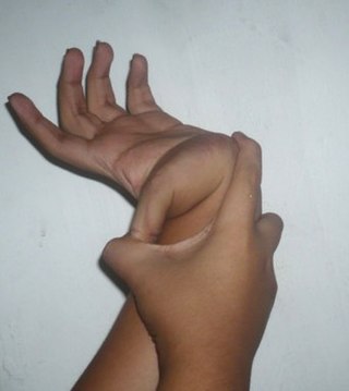
Hypermobility, also known as double-jointedness, describes joints that stretch farther than normal. For example, some hypermobile people can bend their thumbs backwards to their wrists and bend their knee joints backwards, put their leg behind the head or perform other contortionist "tricks". It can affect one or more joints throughout the body.

<span class="mw-page-title-main">Hypermobility spectrum disorder</span> Heritable connective tissue disorder

Hypermobility spectrum disorder (HSD), related to earlier diagnoses such as hypermobility syndrome (HMS), and joint hypermobility syndrome (JHS) is a heritable connective tissue disorder that affects joints and ligaments. Different forms and sub-types have been distinguished, but it does not include asymptomatic joint hypermobility, sometimes known as double-jointedness.

<span class="mw-page-title-main">Vertebral artery dissection</span> Tear of the inner lining of the vertebral artery

Vertebral artery dissection (VAD) is a flap-like tear of the inner lining of the vertebral artery, which is located in the neck and supplies blood to the brain. After the tear, blood enters the arterial wall and forms a blood clot, thickening the artery wall and often impeding blood flow. The symptoms of vertebral artery dissection include head and neck pain and intermittent or permanent stroke symptoms such as difficulty speaking, impaired coordination, and visual loss. It is usually diagnosed with a contrast-enhanced CT or MRI scan.

<span class="mw-page-title-main">Occipital condyles</span> Undersurface protuberances of the occipital bone in vertebrates

The occipital condyles are undersurface protuberances of the occipital bone in vertebrates, which function in articulation with the superior facets of the atlas vertebra.

<span class="mw-page-title-main">Dural ectasia</span> Medical condition

Dural ectasia is widening or ballooning of the dural sac surrounding the spinal cord. This usually occurs in the lumbosacral region, as this is where the cerebrospinal fluid pressure is greatest, but the spinal canal can be affected in any plane.

Atlanto-occipital dislocation, orthopedic decapitation, or internal decapitation describes ligamentous separation of the spinal column from the skull base. It is possible for a human to survive such an injury; however, 70% of cases result in immediate death. It should not be confused with atlanto-axial dislocation, which describes ligamentous separation between the first and second cervical vertebra.

<span class="mw-page-title-main">Cruciate ligament of atlas</span> Ligament forming part of theatlanto-axial joint

The cruciate ligament of the atlas is a cross-shaped ligament in the neck forming part of the atlanto-axial joint. It consists of the transverse ligament of atlas, a superior longitudinal band, and an inferior longitudinal band.

<span class="mw-page-title-main">Cerebrospinal fluid leak</span> Medical condition

A cerebrospinal fluid leak is a medical condition where the cerebrospinal fluid (CSF) surrounding the brain or spinal cord leaks out of one or more holes or tears in the dura mater. A cerebrospinal fluid leak can be either cranial or spinal, and these are two different disorders. A spinal CSF leak can be caused by one or more meningeal diverticula or CSF-venous fistulas not associated with an epidural leak.

Basilar invagination is invagination (infolding) of the base of the skull that occurs when the top of the C2 vertebra migrates upward. It can cause narrowing of the foramen magnum. It also may press on the lower brainstem.

The Chiari Institute is a medical institution that focuses on the treatment of Arnold–Chiari malformation and syringomyelia. It was established in 2001 by the North Shore-LIJ Health System, and is located in Great Neck, New York. The institute was founded by Thomas H. Milhorat, MD shortly after he was appointed the Chairman of Neurosurgery at the North Shore-Long Island Jewish Health System, and Paolo Bolognese, MD. It is now led by Raj Narayan, MD. Paolo Bolognese left TCI on September, 1st, 2014

<span class="mw-page-title-main">Cervicocranial syndrome</span> Medical condition

Cervicocranial syndrome or is a neurological illness. It is a combination of symptoms that are caused by an abnormality in the neck. The bones of the neck that are affected are cervical vertebrae. This syndrome can be identified by confirming cervical bone shifts, collapsed cervical bones or misalignment of the cervical bone leading to improper functioning of cervical spinal nerves.Greenberg Regenerative Medicine | Bryn Mawr, Pennsylvania Cervicocranial syndrome is either congenital or acquired. Some examples of diseases that could result in cervicocranial syndrome are Chiari disease, Klippel-Feil malformation osteoarthritis, and trauma. Treatment options include neck braces, pain medication and surgery. The quality of life for individuals suffering from CCJ syndrome can improve through surgery.

<span class="mw-page-title-main">Rheumatoid disease of the spine</span> Medical condition

Rheumatoid disease of the spine is a morbid consequence of untreated longstanding severe cervical spinal rheumatoid arthritis (RA)–an inflammatory autoimmune disease that attacks the ligaments, joints, and bones of the neck. Although the anterior subluxation of the atlantoaxial joint is the most common manifestation of the disorder, subluxation can also occur with posterior or vertical movement, and subaxial joints can also be involved.

References

  1. Henderson, Fraser C.; Austin, Claudiu; Benzel, Edward; Bolognese, Paolo; Ellenbogen, Richard; Francomano, Clair A.; Ireton, Candace; Klinge, Petra; Koby, Myles; Long, Donlin; Patel, Sunil; Singman, Eric L.; Voermans, Nicol C. (2017). "Neurological and spinal manifestations of the Ehlers-Danlos syndromes". American Journal of Medical Genetics Part C: Seminars in Medical Genetics. 175 (1): 195–211. doi: 10.1002/ajmg.c.31549 . PMID   28220607.
  2. Henderson, F. C.; Geddes, J. F.; Crockard, H. A. (1993). "Neuropathology of the brainstem and spinal cord in end stage rheumatoid arthritis: Implications for treatment". Annals of the Rheumatic Diseases. 52 (9): 629–637. doi: 10.1136/ard.52.9.629 . PMC   1005138 . PMID   8239756.
  3. Nishikawa, Misao; h. Milhorat, Thomas; a. Bolognese, Paolo; b. Mcdonnell, Nazli; a. Francomano, Clair (2009). "Occipito-atlanto-axial Hypermobility : Clinical Features and Dynamic Analysis of Cranial Settling and Posterior Gliding of Occipital Condyle. Part 1 : Findings in Patients with Hereditary Disorders of Connective Tissue and Ehlers-Danlos Syndrome". Spinal Surgery. 23 (2): 168–175. doi: 10.2531/spinalsurg.23.168 .
  4. Batzdorf U, Henderson F, Rigamonti D 2015. "Consensus statement on Cervico-Medullary Syndrome." In Co-morbidities that complicate the treatment and outcomes of Chiari malformation. Ulrich Batzdorf.
  5. Flanagan, Michael F. (2015). "The Role of the Craniocervical Junction in Craniospinal Hydrodynamics and Neurodegenerative Conditions". Neurology Research International. 2015: 1–20. doi: 10.1155/2015/794829 . PMC   4681798 . PMID   26770824.
  6. Martin, Vincent T.; Neilson, Derek (2014). "Joint Hypermobility and Headache: The Glue That Binds the Two Together - Part 2". Headache: The Journal of Head and Face Pain. 54 (8): 1403–1411. doi:10.1111/head.12417. PMID   24958300. S2CID   40251988.
  7. Rozen, TD; Roth, JM; Denenberg, N. (2006). "Cervical Spine Joint Hypermobility: A Possible Predisposing Factor for New Daily Persistent Headache". Cephalalgia. 26 (10): 1182–1185. doi:10.1111/j.1468-2982.2006.01187.x. PMID   16961783. S2CID   25434393.
  8. Franck, Joel Ira; Perrin, Pamela (2015). "The Cranial Cervical Syndrome Defined: New Hope for Postwhiplash Migraine Headache Patients - Cervical Digital Motion X-Ray, FONAR Upright® Weight-Bearing Multi-Position™ MRI and Minimally Invasive C1-C2 Transarticular Lag Screw Fixation Fusion". The Craniocervical Syndrome and MRI. pp. 9–21. doi:10.1159/000365467. ISBN   978-3-318-02696-2.
  9. Henderson, Fraser C.; Austin, Claudiu; Benzel, Edward; Bolognese, Paolo; Ellenbogen, Richard; Francomano, Clair A.; Ireton, Candace; Klinge, Petra; Koby, Myles; Long, Donlin; Patel, Sunil; Singman, Eric L.; Voermans, Nicol C. (2017). "Neurological and spinal manifestations of the Ehlers-Danlos syndromes". American Journal of Medical Genetics Part C: Seminars in Medical Genetics. 175 (1): 195–211. doi: 10.1002/ajmg.c.31549 . PMID   28220607.
  10. Chu, ECP (November 2021). "Cervicogenic Dizziness Associated With Craniocervical Instability: A Case Report". Journal of Medical Cases. 12 (11): 451–454. doi:10.14740/jmc3792. PMC   8577610 . PMID   34804305.
  11. Chu, ECP (April 2021). "Craniocervical instability associated with rheumatoid arthritis: a case report and brief review". AME Case Reports. 5 (12): 12. doi: 10.21037/acr-20-131 . PMC   8060151 . PMID   33912801.
  12. Steilen, Danielle; Hauser, Ross; Woldin, Barbara; Sawyer, Sarah (2014). "Chronic Neck Pain: Making the Connection Between Capsular Ligament Laxity and Cervical Instability". The Open Orthopaedics Journal. 8: 326–345. doi: 10.2174/1874325001408010326 . PMC   4200875 . PMID   25328557.