Bone malrotation

Last updated
Bone malrotation
Other namesBone malalignment
Specialty Orthopedics
Complications Osteoarthritis
TypesMain occurrences are femoral and tibial malrotation
CausesSurgical Complication after fracture
TreatmentCompensatory methods or Osteotomy

Bone malrotation refers to the situation that results when a bone heals out of rotational alignment from another bone, or part of bone. It often occurs as the result of a surgical complication after a fracture where intramedullary nailing (IMN) occurs, [1] especially in the femur and tibial bones, but can also occur genetically at birth. The severity of this complication is often neglected due to its complexity to detect and treat, [1] yet if left untreated, bone malrotation can significantly impact regular bodily functioning, and even lead to severe arthritis. Detection throughout history has become more advanced and accurate, ranging from clinical assessment to ultrasounds to CT (computed tomography) scans. Treatment can include an osteotomy, a major surgical procedure where bones are cut and realigned correctly, or compensatory methods, where individuals learn to externally or internally rotate their limb to compensate for the rotation. Further research is currently being examined in this area to reduce occurrences of malrotation, including detailed computer navigation to improve visual accuracy during surgery. [2]

Contents

Cause

Bone malrotation predominantly occurs after an injury where a bone is fractured, however malrotation can genetically occur during foetal development. It usually occurs during a surgery which involves intramedullary nailing, which is the insertion of metal rods and nails to stabilise bones. Nailing is used as it requires minimal surgical dissection, less disruption of the fracture hematoma and allows faster functionality to a patient post-surgery. [1] [3] However, due to the semi-closed nature of IM, it is impossible to correct under direct vision, so there is less rotational control compared to traditional open methods such as plate fixation. [3] As a result, in many cases of intramedullary nailing, the bone is misaligned which causes malrotated regrowth. This torsional error is a major problem for femoral and tibial fractures, and occurs in 17 to 35% of patients who receive these surgeries, [4] and up to 40% of femur fracture patients. [1]

An x-ray of intramedullary nailing in a tibial fracture X ray internal fixation leg fracture.jpg
An x-ray of intramedullary nailing in a tibial fracture

Femoral malrotation is the most significant bone malrotation issue, and these errors cause cosmetic problems but can also cause drastic physical problems. [4]

Historically, bone malrotation occurred due to a lack of adequate treatment measures, where fixation methods such as traction, casting and non-locked nails provided poor torsional stability. Currently, the utilisation of locked intramedullary nailing, has reduced the occurrence of rotational malalignment during fracture healing, yet femoral malrotation continues to remain very prevalent due to surgeon's inability to reliably restore the pre-injury alignment during operation. [1] A high attention to detail is necessary intra-operatively to avoid this complication when locking the intramedullary device. [5] The utilisation of both radiological and clinical assessment techniques to compare with an injured limb would also help prevent bone malrotation, however these can be difficult and inexact methods of assessment, which is why complications are so common. [1]

Diagnosis

It is difficult to assess and diagnose the rotational malalignment of a bone after an operation, and many methodologies have been developed.

Clinical assessment

Clinical assessment and observation is one methodology, however it is unreliable, and can lead to complications. For femoral or tibial malrotation, many surgeons use the patient's ankle or patella to symmetrically align them with the injured side or to the floor but this method does not consider the position of the proximal fragment and could be moved during reduction attempts. [5] In order to measure femoral malalignment, many doctors will compare the internal and external rotation of both hips, while the patient is supine or prone, and a change in a patient's range of movement indicates malrotation. However, while clinical assessment can indicate the direction of malrotation, it often reports inaccurate measurements of the degree of malalignment, so it is unreliable in determining the potential impacts and necessary treatment methods. [3]

Radiography

Another diagnosis methodology sometimes utilised are radiographs ; however, they are unreliable as they require difficult patient positioning to quantify the rotational deformity. [5] This difficulty is due to a patient's restriction in movement, as they may exhibit post-traumatic deformities and severe pain. To determine the level of femoral malrotation, with this method, two radiographs of the pelvis and upper legs must be made. Firstly, an anteroposterior (AP) perspective which shows the degree of difference between the femoral neck and femur, and another view where the hips and knees are both flexed to a right angle, which determines antetorsion. Both of these radiographs are utilised to calculate the angle of anteversion of the femoral neck. [3]

Ultrasounds

Ultrasounds can also be used to measure bone malrotation, and are considered highly reliable. The main issue with utilising ultrasound measurement is that it relies largely on the skills of an ultrasound technician, and consequently is not widely used. When measuring femoral malrotation with ultrasounds, a patient's thighs are fixed symmetrically while the degree of rotation of the femoral shaft is measured. This methodology requires exact positioning of a patient to correctly measure any deformity. [3]

MRI scans

MRI scans can be utilised and are proven to be reliable and effective at determining malrotation of bones. However, they are not as readily available as CT scanners, and are also time consuming and very expensive. [5]

CT scans

A person getting a CT scan. Contrast CT.jpg
A person getting a CT scan.

The standard bone malrotation detection practice utilised are computed tomography scans, which are able to exactly quantify the amount of rotational malalignment. [5] It is also considered highly reliable and utilised because of its reproducibility. For femoral malrotation, the scan involves doctors measuring the angle between a line through the axis of the femoral neck and a line tangential to the femoral condyles. The angle difference between the uninjured and injured sides determines the degree of malrotation. An increase in the anteversion of the femoral neck of the injured side denotes internal rotation, and a decrease means external rotation has occurred. Another advantage of CT scans is that patient positioning does not impact the measurement accuracy of femoral torsion, which is unlike radiographs and ultrasounds. However, inaccurate measurements of malrotation can still occur in CT scans, but are mostly related difficulty in drawing clear and accurate lines along the femoral neck within the image. To avoid this inaccuracy, there needs to be improved accuracy of the line drawn, and this can be fixed by the utilisation of multi-image superimposed projection of the CT images so a more accurate measurement can be drawn. [3]

Impacts

Whilst malrotation of a bone can be tolerated for most cases, it can still cause severe impacts on functional outcome for some patients. Whilst tibial fractures are the most common long bone fractures, it is malrotation of the femur which can cause the most significant impacts on regular functioning. [6]

If untreated, femoral malrotation can cause considerable gait disturbance and abnormal hip joint pressures. [2] Malrotation of the femur in the setting of a mid-shaft fracture has an impact on the axis of the entire leg, which shifts the centre of force in the knee away from its neutral position. Consequently, femoral malrotation has significant impacts on the mechanical axis and force vectors within the knee. This can cause pain in the hip and knee, and patients may be limited in their movement, which can impair their function, especially in physically demanding activities such as walking up stairs and running. [3] This can impact a person's satisfaction in life, as they may be physically unable to do things they want and need to, such as for laborious work, leisure activities, or raising a family, which can have significant impacts on their psychological health. Femoral malrotation can also cause an abnormally rotated foot, as the angle of a person's foot is directly related to the angle of the femur. [7]

Another significant long term consequence of femoral malrotation if untreated is degenerative arthritis of the hip and knee. [3] This joint arthrosis occurs as there is a proven correlation between rotational error, axis deviation and arthrosis of the knee and hip joints. However, the severity of the arthrosis is dependent on the percentage of malrotation. [4]

Whilst most malrotation problems occurred during surgery, patients with 10° or less difference of malrotation compared with the uninjured side rarely complained of any issues. [8] However, patients with differences greater than 15° found noticeable issues, and above 30° malrotation difference resulted in serious complaints. [5] External femoral malrotation is usually much better tolerated than internal malrotation. [1] However, older studies have found that external rotational deformities are more poorly tolerated, [9] which shows the need for more research in this area.

Treatment

There are two main treatment methods; corrective surgery to fix the malrotation, or compensatory methods where patients learn to compensate for any malrotation when walking.

Compensatory methods

Many people with bone malrotation, such as femoral malrotation are able to functionally tolerate and learn compensatory methods, such as externally or internally adjusting their limb. Patients will compensate for even considerable rotational malalignment when they are active. However, femoral malrotation is more difficult to compensate for and maintain with non-operative treatment methods. [5] Patients with femoral malrotation of less than 15 degrees give less complaints than those with higher amounts of malalignment, yet many patients are able to tolerate the deformity well. [3]

Osteotomy

In extreme cases, the malalignment can be treated by means of a derotational osteotomy which is a major surgical procedure. [3] It can be conducted around the existing intramedullary fixtures, but it usually requires a subtrochanteric osteotomy, which is an invasive surgical approach where bone is cut and realigned. As this is a major procedure, it is beneficial if discovered early, before callus has set, and consequently, patients should be assessed for malrotation in the early post-operative period and a CT scan should occur if abnormalities are found. [5] Correct measurement of bone malalignment with a CT scan is vital when considering an osteotomies in fixing rotational deformities, as torsional differences below 15 degrees are often easily compensated for with non surgical treatment. [3] Revision surgery should be avoided, if not necessary, as implementing femoral nails for a second time leads to higher rates of problems such as infection, nonunion, or nail destabilisation due to overlapping holes for the interlocking screws. [10]

Further research

There is currently limited clinical attention in the area of bone malrotation and research into more accurate methods, likely due to the complexity of the issue. [1] Whilst computed tomography scans are useful in determining the degree of bone malrotation after a surgery has occurred, research is being conducted into how to prevent this deformity occurring at all during surgery. CT scans are impractical intraoperatively due to high cost, lack of equipment portability and increased exposure to radiation. [10]

Consequently, one area being researched is that of computer navigation, which could be utilised during these surgeries to avoid malalignment issues. Computer assisted surgery (CAS) matches a patient's anatomy with pre or intraoperatively generated fluoroscopic image data, by using camera detected infrared signals. The computer then calculates the positioning of each component and creates a visual of their position on the monitor. As a result, the surgeon can monitor in real time the position of the surgical instruments in relation to the patient's anatomy and conducts the procedure accordingly. [11] The surgeon can plan the torsional adjustment and control the fracture reduction and nail insertion on a touchscreen of the navigation module.

Currently, this navigation may add extra time during surgery, so it is sometimes considered unnecessary in an urgent surgery. However, without computer navigation, the risk of bone malrotation is significantly increased. The benefit of computer navigation means that surgeons are able to view more clearly and restore axial alignment, but also ensure the injured limb and matches the alignment and length of the uninjured limb. [2] CAS provides increased surgical accuracy and safety for both patients and surgeons, radiation time is reduced, and it utilises minimally invasive techniques which means less tissue damage, improved wound healing, and reduced infections and scarring for patients. [11]

Related Research Articles

<span class="mw-page-title-main">Knee</span> Region around the kneecap

In humans and other primates, the knee joins the thigh with the leg and consists of two joints: one between the femur and tibia, and one between the femur and patella. It is the largest joint in the human body. The knee is a modified hinge joint, which permits flexion and extension as well as slight internal and external rotation. The knee is vulnerable to injury and to the development of osteoarthritis.

An osteotomy is a surgical operation whereby a bone is cut to shorten or lengthen it or to change its alignment. It is sometimes performed to correct a hallux valgus, or to straighten a bone that has healed crookedly following a fracture. It is also used to correct a coxa vara, genu valgum, and genu varum. The operation is done under a general anaesthetic.

<span class="mw-page-title-main">Coxa vara</span> Deformity of the hip

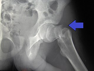
Coxa vara is a deformity of the hip, whereby the angle between the head and the shaft of the femur is reduced to less than 120 degrees. This results in the leg being shortened and the development of a limp. It may be congenital and is commonly caused by injury, such as a fracture. It can also occur when the bone tissue in the neck of the femur is softer than normal, causing it to bend under the weight of the body. This may either be congenital or the result of a bone disorder. The most common cause of coxa vara is either congenital or developmental. Other common causes include metabolic bone diseases, post-Perthes deformity, osteomyelitis, and post traumatic. Shepherd's Crook deformity is a severe form of coxa vara where the proximal femur is severely deformed with a reduction in the neck shaft angle beyond 90 degrees. It is most commonly a sequela of osteogenesis imperfecta, Paget's disease, osteomyelitis, tumour and tumour-like conditions.

<span class="mw-page-title-main">Genu varum</span> Varus deformity marked by (outward) bowing at the knee

Genu varum is a varus deformity marked by (outward) bowing at the knee, which means that the lower leg is angled inward (medially) in relation to the thigh's axis, giving the limb overall the appearance of an archer's bow. Usually medial angulation of both lower limb bones is involved.

<span class="mw-page-title-main">Hip fracture</span> Broken bone in hip joint region

A hip fracture is a break that occurs in the upper part of the femur, at the femoral neck or (rarely) the femoral head. Symptoms may include pain around the hip, particularly with movement, and shortening of the leg. Usually the person cannot walk.

<span class="mw-page-title-main">Hip dislocation</span> Orthopedic injury

A hip dislocation is when the thighbone (femur) separates from the hip bone (pelvis). Specifically it is when the ball–shaped head of the femur separates from its cup–shaped socket in the hip bone, known as the acetabulum. The joint of the femur and pelvis is very stable, secured by both bony and soft-tissue constraints. With that, dislocation would require significant force which typically results from significant trauma such as from a motor vehicle collision or from a fall from elevation. Hip dislocations can also occur following a hip replacement or from a developmental abnormality known as hip dysplasia.

<span class="mw-page-title-main">Osteochondritis dissecans</span> Ischemic bone disease

Osteochondritis dissecans is a joint disorder primarily of the subchondral bone in which cracks form in the articular cartilage and the underlying subchondral bone. OCD usually causes pain during and after sports. In later stages of the disorder there will be swelling of the affected joint which catches and locks during movement. Physical examination in the early stages does only show pain as symptom, in later stages there could be an effusion, tenderness, and a crackling sound with joint movement.

<span class="mw-page-title-main">Cruciate ligament</span> Type of ligament shaped like an X

Cruciate ligaments are pairs of ligaments arranged like a letter X. They occur in several joints of the body, such as the knee joint, wrist joint and the atlanto-axial joint. In a fashion similar to the cords in a toy Jacob's ladder, the crossed ligaments stabilize the joint while allowing a very large range of motion.

<span class="mw-page-title-main">Taylor Spatial Frame</span>

The Taylor Spatial Frame (TSF) is an external fixator used by paediatric and orthopaedic surgeons to treat complex fractures and bone deformities. The medical device shares a number of components and features of the Ilizarov apparatus. The Taylor Spatial Frame is a hexapod device based on a Stewart platform, and was invented by orthopaedic surgeon Charles Taylor. The device consists of two or more aluminum or carbon fibre rings connected by six struts. Each strut can be independently lengthened or shortened to achieve the desired result, e.g. compression at the fracture site, lengthening, etc. Connected to a bone by tensioned wires or half pins, the attached bone can be manipulated in three dimensions and 9 degrees of freedom. Angular, translational, rotational, and length deformities can all be corrected simultaneously with the TSF.

Bone segment navigation is a surgical method used to find the anatomical position of displaced bone fragments in fractures, or to position surgically created fragments in craniofacial surgery. Such fragments are later fixed in position by osteosynthesis. It has been developed for use in craniofacial and oral and maxillofacial surgery.

<span class="mw-page-title-main">Pigeon toe</span> Medical condition affecting the feet

Pigeon toe, also known as in-toeing, is a condition which causes the toes to point inward when walking. It is most common in infants and children under two years of age and, when not the result of simple muscle weakness, normally arises from underlying conditions, such as a twisted shin bone or an excessive anteversion resulting in the twisting of the thigh bone when the front part of a person's foot is turned in.

<span class="mw-page-title-main">Intramedullary rod</span> Metal rod inserted to treat fractures

An intramedullary rod, also known as an intramedullary nail or inter-locking nail or Küntscher nail, is a metal rod forced into the medullary cavity of a bone. IM nails have long been used to treat fractures of long bones of the body. Gerhard Küntscher is credited with the first use of this device in 1939, during World War II, for soldiers with fractures of the femur. Prior to that, treatment of such fractures was limited to traction or plaster, both of which required long periods of inactivity. IM nails resulted in earlier return to activity for the soldiers, sometimes even within a span of a few weeks, since they share the load with the bone, rather than entirely supporting the bone.

<span class="mw-page-title-main">Dror Paley</span> Canadian-trained orthopedic surgeon (born 1956)

Dror Paley is a Canadian-trained orthopedic surgeon, who specializes in limb lengthening and deformity correction procedures.

<span class="mw-page-title-main">Femoral fracture</span> Broken femur, at shaft or distally

A femoral fracture is a bone fracture that involves the femur. They are typically sustained in high-impact trauma, such as car crashes, due to the large amount of force needed to break the bone. Fractures of the diaphysis, or middle of the femur, are managed differently from those at the head, neck, and trochanter; those are conventionally called hip fractures. Thus, mentions of femoral fracture in medicine usually refer implicitly to femoral fractures at the shaft or distally.

Posterolateral corner injuries of the knee are injuries to a complex area formed by the interaction of multiple structures. Injuries to the posterolateral corner can be debilitating to the person and require recognition and treatment to avoid long term consequences. Injuries to the PLC often occur in combination with other ligamentous injuries to the knee; most commonly the anterior cruciate ligament (ACL) and posterior cruciate ligament (PCL). As with any injury, an understanding of the anatomy and functional interactions of the posterolateral corner is important to diagnosing and treating the injury.

<span class="mw-page-title-main">Acetabular fracture</span> Broken bone in acetabular portion of hip bone

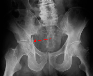
Fractures of the acetabulum occur when the head of the femur is driven into the pelvis. This injury is caused by a blow to either the side or front of the knee and often occurs as a dashboard injury accompanied by a fracture of the femur.

<span class="mw-page-title-main">Medial knee injuries</span> Medical condition

Medial knee injuries are the most common type of knee injury. The medial ligament complex of the knee consists of:

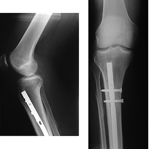
High tibial osteotomy is an orthopaedic surgical procedure which aims to correct a varus deformation with compartmental osteoarthritis. Since the inception of the procedure, advancements to technique, fixation devices, and a better understanding of patient selection has allowed HTO to become more popular in younger, more active patients hoping to combat arthritis. The idea behind the procedure is to realign the weight-bearing line of the knee. By realigning the knee, the force produced from weight-bearing is shifted from the arthritic, medial compartment to the healthy, lateral compartment. This decrease in force or load in the diseased part of the knee joint decreases knee pain and can delay the development or progression of osteoarthritis in the medial compartment.

<span class="mw-page-title-main">Index of trauma and orthopaedics articles</span>

Orthopedic surgery is the branch of surgery concerned with conditions involving the musculoskeletal system. Orthopedic surgeons use both surgical and nonsurgical means to treat musculoskeletal injuries, sports injuries, degenerative diseases, infections, bone tumours, and congenital limb deformities. Trauma surgery and traumatology is a sub-specialty dealing with the operative management of fractures, major trauma and the multiply-injured patient.

References

  1. 1 2 3 4 5 6 7 8 Gugala, Z., Qaisi, Y., Hipp, J., & Lindsey, R. (2011). "Long-term functional implications of the iatrogenic rotational malalignment of healed diaphyseal femur fractures following intramedullary nailing". Clinical Biomechanics, 26(3), 274–277.
  2. 1 2 3 Kahler, D. (2017). "Computer-assisted surgery: The use of stored intraoperative images for accurate restoration of femoral length and rotational alignment after fracture". Injury, 48(s1), S35–S40.
  3. 1 2 3 4 5 6 7 8 9 10 11 Jaarsma, R, van Kampen, A. (2004). "Rotational malalignment after fractures of the femur". The Journal of Bone and Joint Surgery, 86(8), 1100–1104.
  4. 1 2 3 Bretin, P., O'Loughlin, P., Suero, E., Kendoff, D., Ostermeier, S., Hüfner, T., Krettek, C & Citak, M. (2011). "Influence of femoral malrotation on knee joint alignment and intra-articular contract pressures". Archives of Orthopaedic and Trauma Surgery, 131(8), 1115–1120.
  5. 1 2 3 4 5 6 7 8 Kent, E, Arora, A, Owen, J & Khanduja, V. (2010). "Assessment and correction of femoral malrotation following intramedullary nailing of the femur". Acta Orthopaedica. 76, 580–584.
  6. Theriault, B, Turgeon, A & Pelet, S. (2012). "Functional Impact of Tibial Malrotation Following Intramedullary Nailing of Tibial Shaft Fractures". The Journal of Bone and Joint Surgery. 94, 2033–2039.
  7. Staheli LT, Corbett M, Wyss C, King H. (1985). "Lower-extremity rotational problems in children: normal values to guide management". Journal of Bone and Joint Surgery. 67-A,39–47.
  8. Yves Vetter S, Swartman B, Beisemann N, Schnetzke M, Keil H, et al. (2016). "Malrotation After Closed Reduction and Intramedullary Nailing of Femoral Shaft Fractures", Journal of Orthopedic and Spine Trauma. 2, 2.
  9. Johnson KD, Greenberg M. (1987). "Comminuted femoral shaft fractures". Orthopaedics Clinic of North America. 18:133–47.
  10. 1 2 Lieberman, G, Jansen, K, Mast, L, Brady, J, Yoo, B. (2018). "Comparison of fluoroscopic techniques for assessment of femoral rotational alignment". Orthopaedic Trauma Association International. 1, 1, 4.
  11. 1 2 Dirhold, B, Citak, M, Al-Khateeb, H, Haasper, C, Kendoff, D, Krettek, C, Citak, M. (2012). "Current state of computer-assisted trauma surgery". Musculoskeletal Medical Journal. 5,3, 184–191.