Chondromalacia patellae

Last updated
Chondromalacia patella
Other namesCMP
Knee MRI, T1T2PD 08.jpg
Specialty Orthopedics
Diagnostic method MRI

Chondromalacia patellae (also known as CMP) is an inflammation of the underside of the patella and softening of the cartilage.

Contents

The cartilage under the kneecap is a natural shock absorber, and overuse, injury, and many other factors can cause increased deterioration and breakdown of the cartilage. The cartilage is no longer smooth and therefore movement and use is very painful. [1] While it often affects young individuals engaged in active sports, it also afflicts older adults who overwork their knees. [2] [3]

Chondromalacia patellae is sometimes used synonymously with patellofemoral pain syndrome. [4] However, there is general consensus that patellofemoral pain syndrome applies only to individuals without cartilage damage. [4] [5] This condition is also known as Chondrosis. [6] [7] [8] [9] The term literally translates to softening (malakia) of cartilage (chondros) behind patella in Greek. [10]

Cause

The condition may result from acute injury to the patella or chronic friction between the patella and a groove in the femur through which it passes during knee flexion. [11] Possible causes include a tight iliotibial band, neuromas, bursitis, overuse, malalignment, core instability, and patellar maltracking.[ citation needed ]

Pain at the front or inner side of the knee is common in adults of all ages especially when engaging in soccer, gymnastics, cycling, rowing, tennis, ballet, basketball, horseback riding, volleyball, running, combat sports, figure skating, snowboarding, skateboarding and even swimming. The pain is typically felt after prolonged sitting. [12] Skateboarders most commonly experience this injury in their non-dominant foot due to the constant kicking and twisting required of it.[ citation needed ] Swimmers acquire it doing the breaststroke, which demands an unusual motion of the knee. People who are involved in an active lifestyle with high impact on the knees are at greatest risk. Proper management of physical activity may help prevent worsening of the condition. Athletes are advised to talk to a physician for further medical diagnosis, as symptoms may be similar to more serious problems within the knee. Tests are not necessarily needed for diagnosis, but in some situations they may confirm diagnosis or rule out other causes for pain. Commonly used tests are blood tests, MRI scans, and arthroscopy. [13]

While the term chondromalacia sometimes refers to abnormal-appearing cartilage anywhere in the body, [14] it most commonly denotes irritation of the underside of the kneecap (or "patella"). The patella's posterior surface is covered with a layer of smooth cartilage, which the base of the femur normally glides smoothly against when the knee is bent. However, in some individuals the kneecap tends to rub against one side of the knee joint, irritating the cartilage and causing knee pain. [15]

Diagnosis

When investigating a possible diagnosis of chondromalacia, a physician will carry out a complete examination of the affected knee. The physician will palpate the patella and surrounding tissue, feel the joint to observe when and how the distress manifests and obtains a list of symptoms and clinical history. The following tests or procedures may be used to refine the diagnosis:

Treatment

In the absence of cartilage damage, pain at the front of the knee due to overuse can be managed with a combination of RICE (rest, ice, compression, elevation), anti-inflammatory medications, and physiotherapy. [17]

Usually chondromalacia develops without swelling or bruising and most individuals benefit from rest and adherence to an appropriate physical therapy program. Allowing inflammation to subside while avoiding irritating activities for several weeks is followed by a gradual resumption. Cross-training activities such as swimming – using strokes other than the breaststroke – can help to maintain general fitness and body composition. This is beneficial until a physical therapy program emphasizing strengthening and flexibility of the hip and thigh muscles can be undertaken. Use of nonsteroidal anti-inflammatory medication is also helpful to minimize the swelling amplifying patellar pain. Treatment with surgery is declining in popularity due to positive non-surgical outcomes and the relative ineffectiveness of surgical intervention. [15]

See also

Related Research Articles

Iliotibial band syndrome (ITBS) is the second most common knee injury, and is caused by inflammation located on the lateral aspect of the knee due to friction between the iliotibial band and the lateral epicondyle of the femur. Pain is felt most commonly on the lateral aspect of the knee and is most intensive at 30 degrees of knee flexion. Risk factors in women include increased hip adduction and knee internal rotation. Risk factors seen in men are increased hip internal rotation and knee adduction. ITB syndrome is most associated with long-distance running, cycling, weight-lifting, and with military training.

<span class="mw-page-title-main">Knee</span> Leg joint in primates

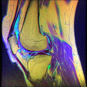
In humans and other primates, the knee joins the thigh with the leg and consists of two joints: one between the femur and tibia, and one between the femur and patella. It is the largest joint in the human body. The knee is a modified hinge joint, which permits flexion and extension as well as slight internal and external rotation. The knee is vulnerable to injury and to the development of osteoarthritis.

<span class="mw-page-title-main">Patella</span> Kneecap, bone covering knee joint

The patella, also known as the kneecap, is a flat, rounded triangular bone which articulates with the femur and covers and protects the anterior articular surface of the knee joint. The patella is found in many tetrapods, such as mice, cats, birds and dogs, but not in whales, or most reptiles.

<span class="mw-page-title-main">Luxating patella</span> Medical condition

A luxating patella, sometimes called a trick knee, is a condition in which the patella, or kneecap, dislocates or moves out of its normal location. It can be associated with damage to the anterior cruciate ligament.

<span class="mw-page-title-main">Osgood–Schlatter disease</span> Inflammation of the patellar ligament

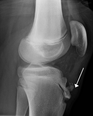
Osgood–Schlatter disease (OSD) is inflammation of the patellar ligament at the tibial tuberosity (apophysitis). It is characterized by a painful bump just below the knee that is worse with activity and better with rest. Episodes of pain typically last a few weeks to months. One or both knees may be affected and flares may recur.

Runner's knee may refer to a number of overuse injuries involving pain around the kneecap (patella), such as:

<span class="mw-page-title-main">Patellar tendinitis</span> Human disease

Patellar tendinitis, also known as jumper's knee, is an overuse injury of the tendon that straightens the knee. Symptoms include pain in the front of the knee. Typically the pain and tenderness is at the lower part of the kneecap, though the upper part may also be affected. Generally there is no pain when the person is at rest. Complications may include patellar tendon rupture.

<span class="mw-page-title-main">Osteochondritis dissecans</span> Ischemic bone disease

Osteochondritis dissecans is a joint disorder primarily of the subchondral bone in which cracks form in the articular cartilage and the underlying subchondral bone. OCD usually causes pain during and after sports. In later stages of the disorder there will be swelling of the affected joint which catches and locks during movement. Physical examination in the early stages does only show pain as symptom, in later stages there could be an effusion, tenderness, and a crackling sound with joint movement.

<span class="mw-page-title-main">Snapping hip syndrome</span> Medical condition

Snapping hip syndrome, also referred to as dancer's hip, is a medical condition characterized by a snapping sensation felt when the hip is flexed and extended. This may be accompanied by a snapping or popping noise and pain or discomfort. Pain often decreases with rest and diminished activity. Snapping hip syndrome is commonly classified by the location of the snapping as either extra-articular or intra-articular.

The knee examination, in medicine and physiotherapy, is performed as part of a physical examination, or when a patient presents with knee pain or a history that suggests a pathology of the knee joint.

<span class="mw-page-title-main">Nail–patella syndrome</span> Medical condition

Nail–patella syndrome is a genetic disorder that results in small, poorly developed nails and kneecaps, but can also affect many other areas of the body, such as the elbows, chest, and hips. The name "nail–patella" can be very misleading because the syndrome often affects many other areas of the body, including even the production of certain proteins. The severity of these effects varies depending on the individual. It is also referred to as iliac horn syndrome, hereditary onychoosteodysplasia, Fong disease or Turner–Kieser syndrome.

Plica syndrome is a condition that occurs when a plica becomes irritated, enlarged, or inflamed.

<span class="mw-page-title-main">Patellofemoral pain syndrome</span> Medical condition

Patellofemoral pain syndrome is knee pain as a result of problems between the kneecap and the femur. The pain is generally in the front of the knee and comes on gradually. Pain may worsen with sitting, excessive use, or climbing and descending stairs.

A lateral release is a surgical procedure to release tight capsular structures on the outer aspect of the kneecap (patella). This is usually performed because of knee pain related to the kneecap being pulled over to the outer (lateral) side and not being able to run properly in the centre of the groove of the femur bone as the knee bends and straightens. The procedure is also known as a 'lateral retinacular release'.

Patellar subluxation syndrome, is an injury that is concerned with the kneecap. Patellar subluxation is more common than patellar dislocation and is just as disabling.

<span class="mw-page-title-main">Patellar dislocation</span> Medical condition

A patellar dislocation is a knee injury in which the patella (kneecap) slips out of its normal position. Often the knee is partly bent, painful and swollen. The patella is also often felt and seen out of place. Complications may include a patella fracture or arthritis.

<span class="mw-page-title-main">Knee pain</span> Medical condition

Knee pain is pain in or around the knee.

Anterior interval release (AIR) is a type of arthroscopic knee surgery performed to alleviate pain and associated symptoms caused by scar tissue accumulation in the anterior region of the knee, behind and under the knee cap, in a condition called arthrofibrosis. In normal, asymptomatic knees, this anterior compartment of the knee comprises mobile, scar-free tissues such as the infrapatellar (Hoffa's) fat pad. With progression, scar tissue leads to closure of the anterior interval, tethering the patella tendon and causing pain, loss of range of motion, damage to knee cartilage, and/or pain, among other symptoms.

<span class="mw-page-title-main">Clarke's test</span>

In medicine, Clarke's test is a component of knee examination which may be used to test for patellofemoral pain syndrome, chondromalacia patellae, patellofemoral arthritis, or anterior knee pain. It is not a standard part of the knee examination but is used to diagnose anterior knee pain where the history indicates this as the likely pathology. The patient is asked to actively contract the quadriceps muscle while the examiner's hand exerts pressure on the superior pole of the patella, so trying to prevent the proximal movement of the patella. While it can produce some discomfort even in normal people, the reproduction of the symptoms suggest pain of patellofemoral origin.

Running injuries affect about half of runners annually. The frequencies of various RRI depend on the type of running, such as speed and mileage. Some injuries are acute, caused by sudden overstress, such as side stitch, strains, and sprains. Many of the common injuries that affect runners are chronic, developing over longer periods as the result of overuse. Common overuse injuries include shin splints, stress fractures, Achilles tendinitis, Iliotibial band syndrome, Patellofemoral pain, and plantar fasciitis.

References

  1. "Chondromalicia patella". Mayo Clinic. Mayo Foundation for Medial Education and Research (MFMER). Retrieved 9 December 2013.
  2. Grelsamer, Ronald P (2005). "Patellar Nomenclature". Clinical Orthopaedics and Related Research (436): 60–5. doi:10.1097/01.blo.0000171545.38095.3e. PMID   15995421.
  3. "Isolated patellofemoral arthritis often overlooked". Academy News. The American Academy of Orthopaedic Surgeons. February 6, 1999.
  4. 1 2 Heintjes, E; Berger, MY; Bierma-Zeinstra, SM; Bernsen, RM; Verhaar, JA; Koes, BW (2004). "Pharmacotherapy for patellofemoral pain syndrome". The Cochrane Database of Systematic Reviews. 2008 (3): CD003470. doi:10.1002/14651858.CD003470.pub2. PMC   8276350 . PMID   15266488.
  5. Dixit, S; DiFiori, JP; Burton, M; Mines, B (Jan 15, 2007). "Management of patellofemoral pain syndrome". American Family Physician. 75 (2): 194–202. PMID   17263214.
  6. "Disorders of the Patellofemoral Joint Chapter 11". www.patellofemoral.org. Archived from the original on 2021-07-14. Retrieved 2021-07-14.
  7. Skalski, Matt. "Chondromalacia grading | Radiology Case | Radiopaedia.org". Radiopaedia. Retrieved 2021-07-14.
  8. "Chondral Injuries - Causes, Symptoms, Treatment & Prevention". Orthosports Medical Center. Retrieved 2021-07-14.
  9. "Treatment of Chondral (Cartilage) Lesions | Knee Arthroscopy | Knee Osteoarthritis Treatment". www.drkharrazi.com. Retrieved 2021-07-14.
  10. "Chondromalacia Patellae". Physiopedia. Retrieved 2021-07-14.
  11. Shiel, William C.; Cunha, John P. (June 27, 2012). "Chondromalacia Patella". MedicineNet. Retrieved May 19, 2013.
  12. Gauresh. "Knee Cap Pain".[ unreliable medical source? ]
  13. "Chondromalacia patellae". Health Information Patient.info. Egton Medical Information Systems Ltd. Retrieved 9 December 2013.
  14. Schindler, Oliver S. (2004). "Synovial plicae of the knee". Current Orthopaedics. 18 (3): 210–9. doi:10.1016/j.cuor.2004.03.005.
  15. 1 2 Cluett, Jonathan (June 14, 2011). "Chondromalacia". About.com. Archived from the original on April 13, 2016. Retrieved June 26, 2010.
  16. "Knee Pain (Chondromalacia Patella): Causes, Symptoms & Treatment". Cleveland Clinic. Retrieved 2021-04-01.
  17. Jenkins, Mark A.; Caryn Honig (2005-06-02). "Patello-Femoral Syndrome". Archived from the original on 2008-10-09. Retrieved 2008-10-06.[ unreliable medical source? ]