The ulna is a long bone found in the forearm that stretches from the elbow to the wrist, and when in anatomical position, is found on the medial side of the forearm. That is, the ulna is on the same side of the forearm as the little finger. It runs parallel to the radius, the other long bone in the forearm. The ulna is usually slightly longer than the radius, but the radius is thicker. Therefore, the radius is considered to be the larger of the two.

The radial nerve is a nerve in the human body that supplies the posterior portion of the upper limb. It innervates the medial and lateral heads of the triceps brachii muscle of the arm, as well as all 12 muscles in the posterior osteofascial compartment of the forearm and the associated joints and overlying skin.
The forearm is the region of the upper limb between the elbow and the wrist. The term forearm is used in anatomy to distinguish it from the arm, a word which is used to describe the entire appendage of the upper limb, but which in anatomy, technically, means only the region of the upper arm, whereas the lower "arm" is called the forearm. It is homologous to the region of the leg that lies between the knee and the ankle joints, the crus.
Wrist drop is a medical condition in which the wrist and the fingers cannot extend at the metacarpophalangeal joints. The wrist remains partially flexed due to an opposing action of flexor muscles of the forearm. As a result, the extensor muscles in the posterior compartment remain paralyzed.

The radius or radial bone is one of the two large bones of the forearm, the other being the ulna. It extends from the lateral side of the elbow to the thumb side of the wrist and runs parallel to the ulna. The ulna is usually slightly longer than the radius, but the radius is thicker. Therefore the radius is considered to be the larger of the two. It is a long bone, prism-shaped and slightly curved longitudinally.

The upper limbs or upper extremities are the forelimbs of an upright-postured tetrapod vertebrate, extending from the scapulae and clavicles down to and including the digits, including all the musculatures and ligaments involved with the shoulder, elbow, wrist and knuckle joints. In humans, each upper limb is divided into the arm, forearm and hand, and is primarily used for climbing, lifting and manipulating objects.

The tibial nerve is a branch of the sciatic nerve. The tibial nerve passes through the popliteal fossa to pass below the arch of soleus.

In human anatomy, the supinator is a broad muscle in the posterior compartment of the forearm, curved around the upper third of the radius. Its function is to supinate the forearm.
In human anatomy, the abductor pollicis longus (APL) is one of the extrinsic muscles of the hand. Its major function is to abduct the thumb at the wrist. Its tendon forms the anterior border of the anatomical snuffbox.
The posterior interosseous nerve is a nerve in the forearm. It is the continuation of the deep branch of the radial nerve, after this has crossed the supinator muscle. It is considerably diminished in size compared to the deep branch of the radial nerve. The nerve fibers originate from cervical segments C7 and C8 in the spinal column.

The posterior interosseous artery is an artery of the forearm. It is a branch of the common interosseous artery, which is a branch of the ulnar artery.
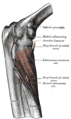
The radial nerve divides into a superficial (sensory) and deep (motor) branch at the cubital fossa. The deep branch of the radial nerve winds to the back of the forearm around the lateral side of the radius between the two planes of fibers of the Supinator, and is prolonged downward between the superficial and deep layers of muscles, to the middle of the forearm. The deep branch provides motor function to the muscles in the posterior compartment of the forearm, which is mostly the extensor muscles of the hand.
The posterior interosseous recurrent artery branches off the posterior interosseous artery near its origin. It ascends between the lateral epicondyle and olecranon, beneath the anconeus, upon or through the substance of the supinator muscle. It anastomoses with the middle collateral artery, posterior ulnar recurrent artery, and ulnar collateral artery. The artery is sometimes absent.

The anterior interosseous nerve is a branch of the median nerve that supplies the deep muscles on the anterior of the forearm, except the ulnar (medial) half of the flexor digitorum profundus. Its nerve roots come from C8 and T1.
The interosseous membrane of the forearm is a fibrous sheet that connects the interosseous margins of the radius and the ulna. It is the main part of the radio-ulnar syndesmosis, a fibrous joint between the two bones.
Radial tunnel syndrome (RTS) is caused by increased pressure on the radial nerve as it travels from the upper arm to the hand and wrist.
Anterior interosseous syndrome is a medical condition in which damage to the anterior interosseous nerve (AIN), a distal motor and sensory branch of the median nerve, classically with severe weakness of the pincer movement of the thumb and index finger, and can cause transient pain in the wrist.

Pronator teres syndrome is a compression neuropathy of the median nerve at the elbow. It is rare compared to compression at the wrist or isolated injury of the anterior interosseous branch of the median nerve.
Nerve compression syndrome, or compression neuropathy, or nerve entrapment syndrome, is a medical condition caused by chronic, direct pressure on a peripheral nerve. It is known colloquially as a trapped nerve, though this may also refer to nerve root compression. Its symptoms include pain, tingling, numbness and muscle weakness. The symptoms affect just one particular part of the body, depending on which nerve is affected. The diagnosis is largely clinical and can be confirmed with diagnostic nerve blocks. Occasionally imaging and electrophysiology studies aid in the diagnosis. Timely diagnosis is important as untreated chronic nerve compression may cause permanent damage. A surgical nerve decompression can relieve pressure on the nerve but cannot always reverse the physiological changes that occurred before treatment. Nerve injury by a single episode of physical trauma is in one sense an acute compression neuropathy but is not usually included under this heading, as chronic compression takes a unique pathophysiological course.
Radial nerve dysfunction is a problem associated with the radial nerve resulting from injury consisting of acute trauma to the radial nerve. The damage has sensory consequences, as it interferes with the radial nerve's innervation of the skin of the posterior forearm, lateral three digits, and the dorsal surface of the lateral side of the palm. The damage also has motor consequences, as it interferes with the radial nerve's innervation of the muscles associated with the extension at the elbow, wrist, and fingers, as well the supination of the forearm. This type of injury can be difficult to localize, but relatively common, as many ordinary occurrences can lead to the injury and resulting mononeuropathy. One out of every ten patients with radial nerve dysfunction do so because of a fractured humerus.