The carpal bones are the eight small bones that make up the wrist that connects the hand to the forearm. The term "carpus" is derived from the Latin carpus and the Greek καρπός (karpós), meaning "wrist". In human anatomy, the main role of the wrist is to facilitate effective positioning of the hand and powerful use of the extensors and flexors of the forearm, and the mobility of individual carpal bones increase the freedom of movements at the wrist.

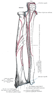
The extensor carpi radialis longus is one of the five main muscles that control movements at the wrist. This muscle is quite long, starting on the lateral side of the humerus, and attaching to the base of the second metacarpal bone.
In human anatomy, extensor carpi radialis brevis is a muscle in the forearm that acts to extend and abduct the wrist. It is shorter and thicker than its namesake extensor carpi radialis longus which can be found above the proximal end of the extensor carpi radialis brevis.
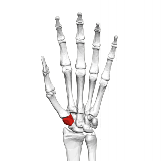
The trapezium bone is a carpal bone in the hand. It forms the radial border of the carpal tunnel.

In human anatomy, the metacarpal bones or metacarpus, form the intermediate part of the skeletal hand located between the phalanges of the fingers and the carpal bones of the wrist which forms the connection to the forearm. The metacarpal bones are equivalent to the metatarsal bones in the foot.

The capitate bone is found in the center of the carpal bone region, colloquially known as the wrist, which is at the distal end of the radius and ulna bones. It articulates with the third metacarpal bone and forms the third carpometacarpal joint. The capitate bone is the largest of the carpal bones in the human hand. It presents, above, a rounded portion or head, which is received into the concavity formed by the scaphoid and lunate bones; a constricted portion or neck; and below this, the body. The bone is also found in many other mammals, and is homologous with the "third distal carpal" of reptiles and amphibians.

The hamate bone or unciform bone is a bone in the human wrist readily distinguishable by its wedge shape and a hook-like process ("hamulus") projecting from its palmar surface.

The upper limb or upper extremity is the region in a vertebrate animal extending from the deltoid region up to and including the hand, including the arm, axilla and shoulder.

A boxer's fracture is the break of the 5th metacarpal bones of the hand near the knuckle. Occasionally it is used to refer to fractures of the 4th metacarpal as well. Symptoms include pain and a depressed knuckle.

The flexor carpi ulnaris muscle is a muscle of the human forearm that acts to flex and adduct the hand.

In human anatomy, the palmar or volar interossei are three small, unipennate muscles in the hand that lie between the metacarpal bones and are attached to the index, ring, and little fingers. They are smaller than the dorsal interossei of the hand.

The carpometacarpal (CMC) joints are five joints in the wrist that articulate the distal row of carpal bones and the proximal bases of the five metacarpal bones.

The deep transverse metacarpal ligament is a narrow fibrous band which runs across the palmar surfaces of the heads of the second, third, fourth and fifth metacarpal bones, connecting them together.

The intercarpal joints can be subdivided into three sets of joints : Those of the proximal row of carpal bones, those of the distal row of carpal bones, and those of the two rows with each other.
The midcarpal joint is formed by the scaphoid, lunate, and triquetral bones in the proximal row, and the trapezium, trapezoid, capitate, and hamate bones in the distal row. The distal pole of the scaphoid articulates with two trapezial bones as a gliding type of joint. The proximal end of the scaphoid combines with the lunate and triquetrum to form a deep concavity that articulates with the convexity of the combined capitate and hamate in a form of diarthrodial, almost condyloid joint.
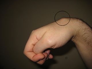
Carpometacarpal bossing is a small, immovable mass of bone on the back of the wrist. The mass occurs in one of the joints between the carpus and metacarpus of the hand, called the carpometacarpal joints, where a small immovable protuberance occurs when this joint becomes swollen or bossed.

In human anatomy, the annular ligaments of the fingers, often referred to as A pulleys, are the annular part of the fibrous sheathes of the fingers. Four or five such annular pulleys, together with three cruciate pulleys, form a fibro-osseous tunnel on the palmar aspect of the hand through which passes the deep and superficial flexor tendons. The annular and cruciate ligaments serve to govern the flexor mechanism of the hand and wrist, providing critical constraints to the flexor tendons to prevent bowstringing upon contraction and excursion of extrinsic flexor musculo-tendinous units.
The extrinsic extensor muscles of the hand are located in the back of the forearm and have long tendons connecting them to bones in the hand, where they exert their action. Extrinsic denotes their location outside the hand. Extensor denotes their action which is to extend, or open flat, joints in the hand. They include the extensor carpi radialis longus (ECRL), extensor carpi radialis brevis (ECRB), extensor digitorum (ED), extensor digiti minimi (EDM), extensor carpi ulnaris (ECU), abductor pollicis longus (APL), extensor pollicis brevis (EPB), extensor pollicis longus (EPL), and extensor indicis (EI).