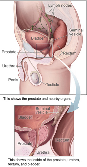
The prostate is both an accessory gland of the male reproductive system and a muscle-driven mechanical switch between urination and ejaculation. It is found in all male mammals. It differs between species anatomically, chemically, and physiologically. Anatomically, the prostate is found below the bladder, with the urethra passing through it. It is described in gross anatomy as consisting of lobes and in microanatomy by zone. It is surrounded by an elastic, fibromuscular capsule and contains glandular tissue, as well as connective tissue.

A lymph node, or lymph gland, is a kidney-shaped organ of the lymphatic system and the adaptive immune system. A large number of lymph nodes are linked throughout the body by the lymphatic vessels. They are major sites of lymphocytes that include B and T cells. Lymph nodes are important for the proper functioning of the immune system, acting as filters for foreign particles including cancer cells, but have no detoxification function.
Articles related to anatomy include:

The femoral triangle is an anatomical region of the upper third of the thigh. It is a subfascial space which appears as a triangular depression below the inguinal ligament when the thigh is flexed, abducted and laterally rotated.

Inguinal lymph nodes are lymph nodes in the groin. They are situated in the femoral triangle of the inguinal region. They are subdivided into two groups: the superficial inguinal lymph nodes and deep inguinal lymph nodes.

The periaortic lymph nodes are a group of lymph nodes that lie in front of the lumbar vertebrae near the aorta. These lymph nodes receive drainage from the gastrointestinal tract and the abdominal organs.

The femoral sheath is a funnel-shaped downward extension of abdominal fascia within which the femoral artery and femoral vein pass between the abdomen and the thigh. The femoral sheath is subdivided by two vertical partitions to form three compartments ; the medial compartment is known as the femoral canal and contains lymphatic vessels and a lymph node, whereas the intermediate canal and the lateral canal accommodate the femoral vein and the femoral artery (respectively). Some neurovascular structures perforate the femoral sheath. Topographically, the femoral sheath is contained within the femoral triangle.

The axillary lymph nodes or armpit lymph nodes are lymph nodes in the human armpit. Between 20 and 49 in number, they drain lymph vessels from the lateral quadrants of the breast, the superficial lymph vessels from thin walls of the chest and the abdomen above the level of the navel, and the vessels from the upper limb. They are divided in several groups according to their location in the armpit. These lymph nodes are clinically significant in breast cancer, and metastases from the breast to the axillary lymph nodes are considered in the staging of the disease.

The internal iliac lymph nodes surround the internal iliac artery and its branches, and receive the lymphatics corresponding to the distribution of the branches of it, i. e., they receive lymphatics from all the pelvic viscera, from the deeper parts of the perineum, including the membranous and cavernous portions of the urethra, and from the buttock and back of the thigh. The internal iliac lymph nodes also drain the superior half of the rectum, above the pectinate line.

The common iliac lymph nodes, four to six in number, are grouped behind and on the sides of the common iliac artery, one or two being placed below the bifurcation of the aorta, in front of the fifth lumbar vertebra.
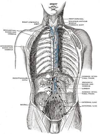
The lumbar trunks are formed by the union of the efferent vessels from the lateral aortic lymph nodes.

The popliteal lymph nodes, small in size and some six or seven in number, are embedded in the fat contained in the popliteal fossa, sometimes referred to as the 'knee pit'. One lies immediately beneath the popliteal fascia, near the terminal part of the small saphenous vein, and drains the region from which this vein derives its tributaries, such as superficial regions of the posterolateral aspect of the leg and the plantar aspect of the foot.

The deep cervical lymph nodes are a group of cervical lymph nodes in the neck that form a chain along the internal jugular vein within the carotid sheath.
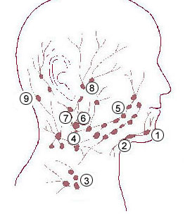
The submental lymph nodes are 2-3 lymph nodes situated in the submental triangle, between the anterior bellies of the digastric muscle and the hyoid bone.
The submandibular lymph nodes, are some 3-6 lymph nodes situated at the inferior border of the ramus of mandible.
The intercostal lymph nodes occupy the posterior parts of the intercostal spaces, in relation to the intercostal vessels.

The following outline is provided as an overview of and topical guide to human anatomy:
The superficial lateral cervical lymph nodes are found along the course of the external jugular vein, between the inferior aspect of the parotid gland and the supraclavicular nodes. The nodes are intercalated along the course of the vessels draining the parotid nodes and the infraauricular nodes. These nodes drain into the supraclavicular nodes, and on to the jugular trunk, followed by the thoracic duct on the left or the right lymphatic duct.
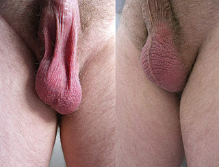
In most terrestrial mammals, the scrotum or scrotal sac is a part of the external male genitalia located at the base of the penis. It consists of a sac of skin containing the external spermatic fascia, testicles, epididymides, and vasa deferentia. The scrotum will usually tighten when exposed to cold temperatures.


