
The lungs are the most important organs of the respiratory system in humans and most other animals, including some snails and a small number of fish. In mammals and most other vertebrates, two lungs are located near the backbone on either side of the heart. Their function in the respiratory system is to extract oxygen from the air and transfer it into the bloodstream, and to release carbon dioxide from the bloodstream into the atmosphere, in a process of gas exchange. The pleurae, which are thin, smooth, and moist, serve to reduce friction between the lungs and chest wall during breathing, allowing for easy and effortless movements of the lungs.

Veins are blood vessels in the circulatory system of humans and most other animals that carry blood towards the heart. Most veins carry deoxygenated blood from the tissues back to the heart; exceptions are those of the pulmonary and fetal circulations which carry oxygenated blood to the heart. In the systemic circulation, arteries carry oxygenated blood away from the heart, and veins return deoxygenated blood to the heart, in the deep veins.

The thoracic cavity is the chamber of the body of vertebrates that is protected by the thoracic wall. The central compartment of the thoracic cavity is the mediastinum. There are two openings of the thoracic cavity, a superior thoracic aperture known as the thoracic inlet and a lower inferior thoracic aperture known as the thoracic outlet.

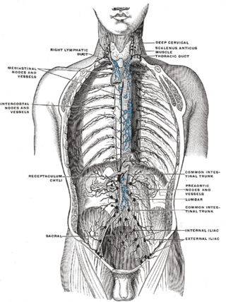
A lymph node, or lymph gland, is a kidney-shaped organ of the lymphatic system and the adaptive immune system. A large number of lymph nodes are linked throughout the body by the lymphatic vessels. They are major sites of lymphocytes that include B and T cells. Lymph nodes are important for the proper functioning of the immune system, acting as filters for foreign particles including cancer cells, but have no detoxification function.
Articles related to anatomy include:
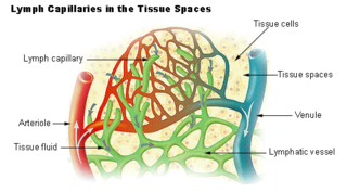
The lymphatic vessels are thin-walled vessels (tubes), structured like blood vessels, that carry lymph. As part of the lymphatic system, lymph vessels are complementary to the cardiovascular system. Lymph vessels are lined by endothelial cells, and have a thin layer of smooth muscle, and adventitia that binds the lymph vessels to the surrounding tissue. Lymph vessels are devoted to the propulsion of the lymph from the lymph capillaries, which are mainly concerned with the absorption of interstitial fluid from the tissues. Lymph capillaries are slightly bigger than their counterpart capillaries of the vascular system. Lymph vessels that carry lymph to a lymph node are called afferent lymph vessels, and those that carry it from a lymph node are called efferent lymph vessels, from where the lymph may travel to another lymph node, may be returned to a vein, or may travel to a larger lymph duct. Lymph ducts drain the lymph into one of the subclavian veins and thus return it to general circulation.

The cardiac plexus is a plexus of nerves situated at the base of the heart that innervates the heart.

The root of the lung is a group of structures that emerge at the hilum of each lung, just above the middle of the mediastinal surface and behind the cardiac impression of the lung. It is nearer to the back than the front. The root of the lung is connected by the structures that form it to the heart and the trachea. The rib cage is separated from the lung by a two-layered membranous coating, the pleura. The hilum is the large triangular depression where the connection between the parietal pleura and the visceral pleura is made, and this marks the meeting point between the mediastinum and the pleural cavities.

In human male anatomy, the dorsal veins of the penis are blood vessels that drain the shaft, the skin and the glans of the human penis. They are typically located in the midline on the dorsal aspect of the penis and they comprise the superficial dorsal veinof the penis, that lies in the subcutaneous tissue of the shaft, and the deep dorsal veinof the penis, that lies beneath the deep fascia.

A bronchopulmonary segment is a portion of lung supplied by a specific segmental bronchus and its vessels. These arteries branch from the pulmonary and bronchial arteries, and run together through the center of the segment. Veins and lymphatic vessels drain along the edges of the segment. The segments are separated from each other by layers of connective tissue that forms them into discrete anatomical and functional units. This separation means that a bronchopulmonary segment can be surgically removed without affecting the function of the others.

The tracheobronchial lymph nodes are lymph nodes that are located around the division of trachea and main bronchi.

The axillary lymph nodes or armpit lymph nodes are lymph nodes in the human armpit. Between 20 and 49 in number, they drain lymph vessels from the lateral quadrants of the breast, the superficial lymph vessels from thin walls of the chest and the abdomen above the level of the navel, and the vessels from the upper limb. They are divided in several groups according to their location in the armpit. These lymph nodes are clinically significant in breast cancer, and metastases from the breast to the axillary lymph nodes are considered in the staging of the disease.

The popliteal lymph nodes, small in size and some six or seven in number, are embedded in the fat contained in the popliteal fossa, sometimes referred to as the 'knee pit'. One lies immediately beneath the popliteal fascia, near the terminal part of the small saphenous vein, and drains the region from which this vein derives its tributaries, such as superficial regions of the posterolateral aspect of the leg and the plantar aspect of the foot.

One or two supratrochlear lymph nodes are placed above the medial epicondyle of the humerus, medial to the basilic vein.

The deep parotid lymph nodes are lymph nodes found below the parotid gland.
The intercostal lymph nodes occupy the posterior parts of the intercostal spaces, in relation to the intercostal vessels.

The following outline is provided as an overview of and topical guide to human anatomy:

In human anatomy, the hilum, sometimes formerly called a hilus, is a depression or fissure where structures such as blood vessels and nerves enter an organ. Examples include:

The pulmonary pleurae are the two opposing layers of serous membrane overlying the lungs, mediastinum and the inside surfaces of the surrounding chest walls.