The right and left paratracheal lymph nodes are lymph nodes in the neck situated lateral to the trachea and esophagus alongside the recurrent laryngeal nerve. They drain to the deep cervical lymph nodes.

The stylopharyngeus muscle is a muscle in the head. It originates from the temporal styloid process. Some of its fibres insert onto the thyroid cartilage, while others end by intermingling with proximal structures. It is innervated by the glossopharyngeal nerve. It acts to elevate the larynx and pharynx, and dilate the pharynx, thus facilitating swallowing.

The carotid sheath is a condensation of the deep cervical fascia enveloping multiple vital neurovascular structures of the neck, including the common and internal carotid arteries, the internal jugular vein, the vagus nerve, and ansa cervicalis. The carotid sheath helps protects the structures contained therein.

The retropharyngeal space is a potential space and deep compartment of the head and neck situated posterior to the pharynx. The RPS is bounded anteriorly by the buccopharyngeal fascia, posteriorly by the alar fascia, and laterally by the carotid sheath. It extends between the base of the skull superiorly, and the mediastinum inferiorly. It contains the retropharyngeal lymph nodes. Its function is to facilitate movements in the superoinferior axis of the larynx, pharynx, and esophagus in relation to the cervical spine.

Cervical lymph nodes are lymph nodes found in the neck. Of the 800 lymph nodes in the human body, 300 are in the neck. Cervical lymph nodes are subject to a number of different pathological conditions including tumours, infection and inflammation.

The occipital lymph nodes, one to three in number, are located on the back of the head close to the margin of the trapezius and resting on the insertion of the semispinalis capitis.

The prevertebral fascia is the layer of deep cervical fascia that surrounds the vertebral column. It is the deepest layer of deep cervical fascia.
The submental lymph nodes are 2-3 lymph nodes situated in the submental triangle, between the anterior bellies of the digastric muscle and the hyoid bone.
Supraclavicular lymph nodes are lymph nodes found above the clavicle, that can be felt in the supraclavicular fossa. The supraclavicular lymph nodes on the left side are called Virchow's nodes. It leads to an appreciable mass that can be recognized clinically, called Troisier sign.
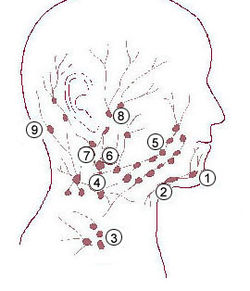
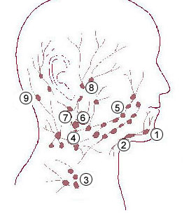
The submandibular lymph nodes, are some 3-6 lymph nodes situated at the inferior border of the ramus of mandible.

The deep parotid lymph nodes are lymph nodes found below the parotid gland.

Retropharyngeal abscess (RPA) is an abscess located in the tissues in the back of the throat behind the posterior pharyngeal wall. Because RPAs typically occur in deep tissue, they are difficult to diagnose by physical examination alone. RPA is a relatively uncommon illness, and therefore may not receive early diagnosis in children presenting with stiff neck, malaise, difficulty swallowing, or other symptoms listed below. Early diagnosis is key, while a delay in diagnosis and treatment may lead to death. Parapharyngeal space communicates with retropharyngeal space and an infection of retropharyngeal space can pass down behind the esophagus into the mediastinum. RPAs can also occur in adults of any age.
Parotid lymph nodes are lymph nodes found near the parotid gland in the immune system.
The superficial parotid lymph nodes are a group of lymph nodes anterior to the ear.
The deep lateral cervical lymph nodes are found near the upper part of the internal jugular vein in the neck, lateral or posterior to the carotid sheath.
The superficial lateral cervical lymph nodes are found along the course of the external jugular vein, between the inferior aspect of the parotid gland and the supraclavicular nodes. The nodes are intercalated along the course of the vessels draining the parotid nodes and the infraauricular nodes. These nodes drain into the supraclavicular nodes, and on to the jugular trunk, followed by the thoracic duct on the left or the right lymphatic duct.
The anterior cervical lymph nodes are a group of nodes found on the anterior part of the neck, in front of the sternocleidomastoid muscle. These can be grouped into a deep and superficial group.
The pretracheal lymph nodes are lymph nodes located anterior to the trachea in the neck.

The parapharyngeal space, is a potential space in the head and the neck. It has clinical importance in otolaryngology due to parapharyngeal space tumours and parapharyngeal abscess developing in this area. It is also a key anatomic landmark for localizing disease processes in the surrounding spaces of the neck; the direction of its displacement indirectly reflects the site of origin for masses or infection in adjacent areas, and consequently their appropriate differential diagnosis.

Guttural pouches are large, auditory-tube diverticula that contain between 300 and 600 ml of air. They are present in odd-toed mammals, some bats, hyraxes, and the American forest mouse. They are paired bilaterally just below the ears, behind the skull and connect to the nasopharynx.