
In human anatomy, the subclavian arteries are paired major arteries of the upper thorax, below the clavicle. They receive blood from the aortic arch. The left subclavian artery supplies blood to the left arm and the right subclavian artery supplies blood to the right arm, with some branches supplying the head and thorax. On the left side of the body, the subclavian comes directly off the aortic arch, while on the right side it arises from the relatively short brachiocephalic artery when it bifurcates into the subclavian and the right common carotid artery.

In human anatomy, the thoracic duct is the larger of the two lymph ducts of the lymphatic system. The thoracic duct usually begins from the upper aspect of the cisterna chyli, passing out of the abdomen through the aortic hiatus into first the posterior mediastinum and then the superior mediastinum, extending as high up as the root of the neck before descending to drain into the systemic (blood) circulation at the venous angle.

The superior thoracic aperture, also known as the thoracic outlet, or thoracic inlet refers to the opening at the top of the thoracic cavity. It is also clinically referred to as the thoracic outlet, in the case of thoracic outlet syndrome. A lower thoracic opening is the inferior thoracic aperture.
The cisterna chyli or receptaculum chyli is a dilated sac at the lower end of the thoracic duct in most mammals into which lymph from the intestinal trunk and two lumbar lymphatic trunks flow. It receives fatty chyle from the intestines and thus acts as a conduit for the lipid products of digestion. It is the most common drainage trunk of most of the body's lymphatics. The cisterna chyli is a retroperitoneal structure.

The internal intercostal muscles are a group of skeletal muscles located between the ribs. They are eleven in number on either side. They commence anteriorly at the sternum, in the intercostal spaces between the cartilages of the true ribs, and at the anterior extremities of the cartilages of the false ribs, and extend backward as far as the angles of the ribs, hence they are continued to the vertebral column by thin aponeuroses, the posterior intercostal membranes. They pull the sternum and ribs upward and inward.

The intercostal space (ICS) is the anatomic space between two ribs. Since there are 12 ribs on each side, there are 11 intercostal spaces, each numbered for the rib superior to it.

The right lymphatic duct is an important lymphatic vessel that drains the right upper quadrant of the human body. It forms various combinations with the right subclavian vein and right internal jugular vein.

The costocervical trunk arises from the upper and back part of the second part of subclavian artery, behind the scalenus anterior on the right side, and medial to that muscle on the left side.

This article describes the anatomy of the head and neck of the human body, including the brain, bones, muscles, blood vessels, nerves, glands, nose, mouth, teeth, tongue, and throat.
The intercostal arteries are a group of arteries passing within an intercostal space. There are 9 anterior and 11 posterior intercostal arteries on each side of the body. The anterior intercostal arteries are branches of the internal thoracic artery and its terminal branch - the musculophrenic artery. The posterior intercostal arteries are branches of the supreme intercostal artery and thoracic aorta.

The axillary lymph nodes or armpit lymph nodes are lymph nodes in the human armpit. Between 20 and 49 in number, they drain lymph vessels from the lateral quadrants of the breast, the superficial lymph vessels from thin walls of the chest and the abdomen above the level of the navel, and the vessels from the upper limb. They are divided in several groups according to their location in the armpit. These lymph nodes are clinically significant in breast cancer, and metastases from the breast to the axillary lymph nodes are considered in the staging of the disease.
An apical group of six to twelve glands is situated partly posterior to the upper portion of the pectoralis minor and partly above the upper border of this muscle.

The jugular trunk is a lymphatic vessel in the neck. It is formed by vessels that emerge from the superior deep cervical lymph nodes and unite to efferents of the inferior deep cervical lymph nodes.
The efferent vessels of the tracheobronchial lymph nodes ascend upon the trachea and unite with efferents of the internal mammary and anterior mediastinal glands to form the right and left bronchomediastinal trunks.
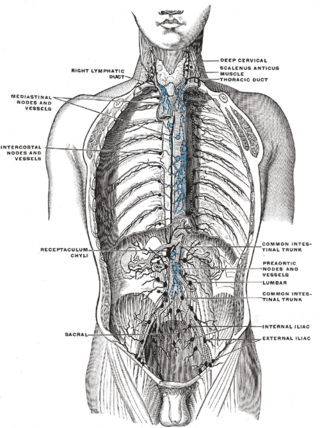
The intercostal lymph nodes occupy the posterior parts of the intercostal spaces, in relation to the intercostal vessels.

The following outline is provided as an overview of and topical guide to human anatomy:

The superior mesenteric lymph nodes may be divided into three principal groups:
Lymph trunk is a collection of lymph vessels that carries lymph, and is formed by confluence of many efferent lymph vessels. It in turn drains into one of the two lymph ducts.
The superior diaphragmatic lymph nodes lie on the thoracic aspect of the diaphragm, and consist of three sets – anterior, middle, and posterior.

