
The left and right brachiocephalic veins are major veins in the upper chest, formed by the union of the ipsilateral internal jugular vein and subclavian vein behind the sternoclavicular joint. The left brachiocephalic vein is more than twice the length of the right brachiocephalic vein.

In human anatomy, the subclavian arteries are paired major arteries of the upper thorax, below the clavicle. They receive blood from the aortic arch. The left subclavian artery supplies blood to the left arm and the right subclavian artery supplies blood to the right arm, with some branches supplying the head and thorax. On the left side of the body, the subclavian comes directly off the aortic arch, while on the right side it arises from the relatively short brachiocephalic artery when it bifurcates into the subclavian and the right common carotid artery.

In human anatomy, the thoracic duct is the larger of the two lymph ducts of the lymphatic system. The thoracic duct usually begins from the upper aspect of the cisterna chyli, passing out of the abdomen through the aortic hiatus into first the posterior mediastinum and then the superior mediastinum, extending as high up as the root of the neck before descending to drain into the systemic (blood) circulation at the venous angle.

The internal jugular vein is a paired jugular vein that collects blood from the brain and the superficial parts of the face and neck. This vein runs in the carotid sheath with the common carotid artery and vagus nerve.

The external jugular vein receives the greater part of the blood from the exterior of the cranium and the deep parts of the face, being formed by the junction of the posterior division of the retromandibular vein with the posterior auricular vein.
In anatomy, the left and right common carotid arteries (carotids) are arteries that supply the head and neck with oxygenated blood; they divide in the neck to form the external and internal carotid arteries.

The right lymphatic duct is an important lymphatic vessel that drains the right upper quadrant of the human body. It forms various combinations with the right subclavian vein and right internal jugular vein.

The subclavian triangle, the smaller division of the posterior triangle, is bounded, above, by the inferior belly of the omohyoideus; below, by the clavicle; its base is formed by the posterior border of the sternocleidomastoideus.

This article describes the anatomy of the head and neck of the human body, including the brain, bones, muscles, blood vessels, nerves, glands, nose, mouth, teeth, tongue, and throat.

A central or intermediate group of three or four large glands is imbedded in the adipose tissue near the base of the axilla.
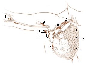
A brachial lymph nodes are group of four to six lymph nodes which lies in relation to the medial and posterior aspects of the axillary vein; the afferents of these glands drain the whole arm with the exception of that portion whose vessels accompany the cephalic vein.
An anterior or pectoral group consists of four or five glands along the lower border of the Pectoralis minor, in relation with the lateral thoracic artery.

The prevertebral fascia is the layer of deep cervical fascia that surrounds the vertebral column. It is the deepest layer of deep cervical fascia.

The jugular trunk is a lymphatic vessel in the neck. It is formed by vessels that emerge from the superior deep cervical lymph nodes and unite to efferents of the inferior deep cervical lymph nodes.

The inferior deep cervical lymph nodes are one of the two groups of the deep cervical lymph nodes.
The efferent vessels of the tracheobronchial lymph nodes ascend upon the trachea and unite with efferents of the internal mammary and anterior mediastinal glands to form the right and left bronchomediastinal trunks.
The parasternal lymph nodes are placed at the anterior ends of the intercostal spaces, by the side of the internal thoracic artery.
The efferent vessels of the subclavicular group unite to form the subclavian trunk, which opens either directly into the junction of the internal jugular and subclavian veins or into the jugular lymphatic trunk; on the left side it may end in the thoracic duct.
Lymph trunk is a collection of lymph vessels that carries lymph, and is formed by confluence of many efferent lymph vessels. It in turn drains into one of the two lymph ducts.


