The prostate is both an accessory gland of the male reproductive system and a muscle-driven mechanical switch between urination and ejaculation. It is found in all male mammals. It differs between species anatomically, chemically, and physiologically. Anatomically, the prostate is found below the bladder, with the urethra passing through it. It is described in gross anatomy as consisting of lobes and in microanatomy by zone. It is surrounded by an elastic, fibromuscular capsule and contains glandular tissue, as well as connective tissue.

The seminal vesicles are a pair of convoluted tubular accessory glands that lie behind the urinary bladder of male mammals. They secrete fluid that partly composes the semen.

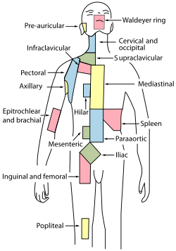
The periaortic lymph nodes are a group of lymph nodes that lie in front of the lumbar vertebrae near the aorta. These lymph nodes receive drainage from the gastrointestinal tract and the abdominal organs.

Sir Astley Paston Cooper, 1st Baronet was a British surgeon and anatomist, who made contributions to otology, vascular surgery, the anatomy and pathology of the mammary glands and testicles, and the pathology and surgery of hernia.

In human anatomy, the common iliac veins are formed by the external iliac veins and internal iliac veins. The left and right common iliac veins come together in the abdomen at the level of the fifth lumbar vertebra, forming the inferior vena cava. They drain blood from the pelvis and lower limbs.
The external iliac veins are large veins that connect the femoral veins to the common iliac veins. Their origin is at the inferior margin of the inguinal ligaments and they terminate when they join the internal iliac veins.

The external iliac arteries are two major arteries which bifurcate off the common iliac arteries anterior to the sacroiliac joint of the pelvis.

The common iliac artery is a large artery of the abdomen paired on each side. It originates from the aortic bifurcation at the level of the 4th lumbar vertebra. It ends in front of the sacroiliac joint, one on either side, and each bifurcates into the external and internal iliac arteries.
The internal iliac artery is the main artery of the pelvis.

The median sacral artery is a small artery that arises posterior to the abdominal aorta and superior to its bifurcation.

The external iliac lymph nodes are lymph nodes, from eight to ten in number, that lie along the external iliac vessels.

The internal iliac vein begins near the upper part of the greater sciatic foramen, passes upward behind and slightly medial to the internal iliac artery and, at the brim of the pelvis, joins with the external iliac vein to form the common iliac vein.
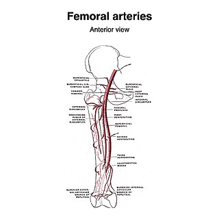
The superficial iliac circumflex artery, the smallest of the cutaneous branches of the femoral artery, arises close to the superficial epigastric artery, and, piercing the fascia lata, runs lateralward, parallel with the inguinal ligament, as far as the crest of the ilium.

The urogenital triangle is the anterior part of the perineum. In female mammals, it contains the vulva, while in male mammals, it contains the penis and scrotum.

The median sacral vein is a vein of the abdomen. It accompanies the median sacral artery along the front of the sacrum. It ends in the left common iliac vein. Sometimes, it ends in the angle of junction of the two common iliac veins.
The lateral sacral veins accompany the lateral sacral arteries on the anterior surface of the sacrum. They drain into the internal iliac vein. They communicate with each other via the sacral venous plexus.

The vesical venous plexus is a venous plexus situated at the fundus of the urinary bladder. It collects venous blood from the urinary bladder in both sexes, from the accessory sex glands in males, and from the corpora cavernosa of clitoris in females. It drains into the internal iliac veins via several vesical veins.

The following outline is provided as an overview of and topical guide to human anatomy:

The human abdomen is divided into quadrants and regions by anatomists and physicians for the purposes of study, diagnosis, and treatment. The division into four quadrants allows the localisation of pain and tenderness, scars, lumps, and other items of interest, narrowing in on which organs and tissues may be involved. The quadrants are referred to as the left lower quadrant, left upper quadrant, right upper quadrant and right lower quadrant. These terms are not used in comparative anatomy, since most other animals do not stand erect.

This glossary of medical terms is a list of definitions about medicine, its sub-disciplines, and related fields.