Related Research Articles

The stomach is a muscular, hollow organ in the upper gastrointestinal tract of humans and many other animals, including several invertebrates. The stomach has a dilated structure and functions as a vital organ in the digestive system. The stomach is involved in the gastric phase of digestion, following the cephalic phase in which the sight and smell of food and the act of chewing are stimuli. In the stomach a chemical breakdown of food takes place by means of secreted digestive enzymes and gastric acid. It also plays a role in regulating gut microbiota, influencing digestion and overall health.

The gastrointestinal tract is the tract or passageway of the digestive system that leads from the mouth to the anus. The GI tract contains all the major organs of the digestive system, in humans and other animals, including the esophagus, stomach, and intestines. Food taken in through the mouth is digested to extract nutrients and absorb energy, and the waste expelled at the anus as feces. Gastrointestinal is an adjective meaning of or pertaining to the stomach and intestines.
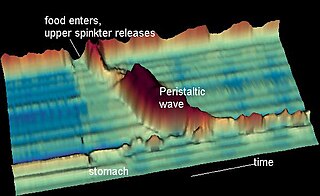
Peristalsis is a type of intestinal motility, characterized by radially symmetrical contraction and relaxation of muscles that propagate in a wave down a tube, in an anterograde direction. Peristalsis is progression of coordinated contraction of involuntary circular muscles, which is preceded by a simultaneous contraction of the longitudinal muscle and relaxation of the circular muscle in the lining of the gut.
Digestion is the breakdown of large insoluble food compounds into small water-soluble components so that they can be absorbed into the blood plasma. In certain organisms, these smaller substances are absorbed through the small intestine into the blood stream. Digestion is a form of catabolism that is often divided into two processes based on how food is broken down: mechanical and chemical digestion. The term mechanical digestion refers to the physical breakdown of large pieces of food into smaller pieces which can subsequently be accessed by digestive enzymes. Mechanical digestion takes place in the mouth through mastication and in the small intestine through segmentation contractions. In chemical digestion, enzymes break down food into the small compounds that the body can use.

The enteric nervous system (ENS) is one of the three divisions of the autonomic nervous system (ANS), the others being the sympathetic nervous system (SNS) and parasympathetic nervous system (PSNS). It consists of a mesh-like system of neurons that governs the function of the gastrointestinal tract. The ENS is nicknamed the "second brain". It is derived from neural crest cells.

The cardiac pacemaker is the heart's natural rhythm generator. It employs pacemaker cells that produce electrical impulses, known as cardiac action potentials, which control the rate of contraction of the cardiac muscle, that is, the heart rate. In most humans, these cells are concentrated in the sinoatrial (SA) node, the primary pacemaker, which regulates the heart’s sinus rhythm.

Cardiac muscle is one of three types of vertebrate muscle tissues, the others being skeletal muscle and smooth muscle. It is an involuntary, striated muscle that constitutes the main tissue of the wall of the heart. The cardiac muscle (myocardium) forms a thick middle layer between the outer layer of the heart wall and the inner layer, with blood supplied via the coronary circulation. It is composed of individual cardiac muscle cells joined by intercalated discs, and encased by collagen fibers and other substances that form the extracellular matrix.

The cardiac conduction system transmits the signals generated by the sinoatrial node – the heart's pacemaker, to cause the heart muscle to contract, and pump blood through the body's circulatory system. The pacemaking signal travels through the right atrium to the atrioventricular node, along the bundle of His, and through the bundle branches to Purkinje fibers in the walls of the ventricles. The Purkinje fibers transmit the signals more rapidly to stimulate contraction of the ventricles.

Unlike the action potential in skeletal muscle cells, the cardiac action potential is not initiated by nervous activity. Instead, it arises from a group of specialized cells known as pacemaker cells, that have automatic action potential generation capability. In healthy hearts, these cells form the cardiac pacemaker and are found in the sinoatrial node in the right atrium. They produce roughly 60–100 action potentials every minute. The action potential passes along the cell membrane causing the cell to contract, therefore the activity of the sinoatrial node results in a resting heart rate of roughly 60–100 beats per minute. All cardiac muscle cells are electrically linked to one another, by intercalated discs which allow the action potential to pass from one cell to the next. This means that all atrial cells can contract together, and then all ventricular cells.

Muscle contraction is the activation of tension-generating sites within muscle cells. In physiology, muscle contraction does not necessarily mean muscle shortening because muscle tension can be produced without changes in muscle length, such as when holding something heavy in the same position. The termination of muscle contraction is followed by muscle relaxation, which is a return of the muscle fibers to their low tension-generating state.

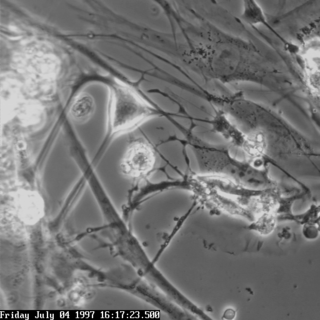
Enterochromaffin (EC) cells are a type of enteroendocrine cell, and neuroendocrine cell. They reside alongside the epithelium lining the lumen of the digestive tract and play a crucial role in gastrointestinal regulation, particularly intestinal motility and secretion. They were discovered by Nikolai Kulchitsky.
An electrogastrogram (EGG) is a computer generated graphic produced by electrogastrography, which detects, analyzes and records the myoelectrical signal generated by the movement of the smooth muscle of the stomach, intestines and other smooth muscle containing organs. An electrogastroenterogram or electroviscerogram is a similar display of the recording of myoelectrical activity of gastrointestinal or other organs which are able to generate myoelectrical activity.
Interstitial cells of Cajal (ICC) are interstitial cells found in the gastrointestinal tract. There are different types of ICC with different functions. ICC and another type of interstitial cell, known as platelet-derived growth factor receptor alpha (PDGFRα) cells, are electrically coupled to smooth muscle cells via gap junctions, that work together as an SIP functional syncytium. Myenteric interstitial cells of Cajal (ICC-MY) serve as pacemaker cells that generate the bioelectrical events known as slow waves. Slow waves conduct to smooth muscle cells and cause phasic contractions.
Vagovagal reflex refers to gastrointestinal tract reflex circuits where afferent and efferent fibers of the vagus nerve coordinate responses to gut stimuli via the dorsal vagal complex in the brain. The vagovagal reflex controls contraction of the gastrointestinal muscle layers in response to distension of the tract by food. This reflex also allows for the accommodation of large amounts of food in the gastrointestinal tracts.
Gastrointestinal physiology is the branch of human physiology that addresses the physical function of the gastrointestinal (GI) tract. The function of the GI tract is to process ingested food by mechanical and chemical means, extract nutrients and excrete waste products. The GI tract is composed of the alimentary canal, that runs from the mouth to the anus, as well as the associated glands, chemicals, hormones, and enzymes that assist in digestion. The major processes that occur in the GI tract are: motility, secretion, regulation, digestion and circulation. The proper function and coordination of these processes are vital for maintaining good health by providing for the effective digestion and uptake of nutrients.

Gastroparesis is a medical disorder of ineffective neuromuscular contractions (peristalsis) of the stomach, resulting in food and liquid remaining in the stomach for a prolonged period of time. Stomach contents thus exit more slowly into the duodenum of the digestive tract, a medical sign called delayed gastric emptying. The opposite of this, where stomach contents exit quickly into the duodenum, is called dumping syndrome.
A slow-wave potential is a rhythmic electrophysiological event in the gastrointestinal tract. The normal conduction of slow waves is one of the key regulators of gastrointestinal motility. Slow waves are generated and propagated by a class of pacemaker cells called the interstitial cells of Cajal, which also act as intermediates between nerves and smooth muscle cells. Slow waves generated in interstitial cells of Cajal spread to the surrounding smooth muscle cells and control motility.
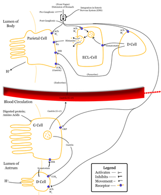
The nervous system, and endocrine system collaborate in the digestive system to control gastric secretions, and motility associated with the movement of food throughout the gastrointestinal tract, including peristalsis, and segmentation contractions.

The gastrointestinal wall of the gastrointestinal tract is made up of four layers of specialised tissue. From the inner cavity of the gut outwards, these are the mucosa, the submucosa, the muscular layer and the serosa or adventitia.

The human digestive system consists of the gastrointestinal tract plus the accessory organs of digestion. Digestion involves the breakdown of food into smaller and smaller components, until they can be absorbed and assimilated into the body. The process of digestion has three stages: the cephalic phase, the gastric phase, and the intestinal phase.
References
- 1 2 3 4 5 6 Rhoades, Rodney A.; Bell, David R., eds. (2022). "Gastrointestinal Physiology". Medical Physiology: Principles for Clinical Medicine. Lippincott Williams & Wilkins. pp. 556–638. ISBN 978-1-9751-6045-6. OCLC 1313900098.
- 1 2 3 4 5 6 7 8 9 10 Widmaier, Raff & Strang 2016, p. [ page needed ].
- ↑ Thomson, Lars; Robinson, Tim L.; Lee, Jonathan C.F.; Farraway, Laura A.; Hughes, Martin J.G.; Andrews, David W.; Huizinga, Jan D. (July 1998). "Interstitial cells of Cajal generate a rhythmic pacemaker current". Nature Medicine. 4 (7): 848–851. doi:10.1038/nm0798-848. PMID 9662380.
- 1 2 3 Ju, Yue-Kun; Woodcock, Elizabeth A.; Allen, David G.; Cannell, Mark B. (September 2012). "Inositol 1,4,5-trisphosphate receptors and pacemaker rhythms". Journal of Molecular and Cellular Cardiology. 53 (3): 375–381. doi:10.1016/j.yjmcc.2012.06.004. PMID 22713798.
- 1 2 3 4 5 6 Wu, Tongzhi; Rayner, Christopher K; Young, Richard L; Horowitz, Michael (December 2013). "Gut motility and enteroendocrine secretion". Current Opinion in Pharmacology. 13 (6): 928–934. doi:10.1016/j.coph.2013.09.002. PMID 24060702.