Renal papillary necrosis

Last updated
Renal papillary necrosis
Other namesRenal medullary necrosis [1]
Illu kidney2.jpg
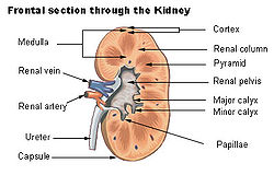
Frontal section through the kidney
Specialty Urology, nephrology   OOjs UI icon edit-ltr-progressive.svg
Symptoms Back pain, cloudy urine [1]
CausesDiabetic nephropathy, Kidney infection [1]
Diagnostic method Blood and urine test [1]
TreatmentDepends on cause [1]

Renal papillary necrosis is a form of nephropathy involving the necrosis of the renal papilla. [1] Lesions that characterize renal papillary necrosis come from an impairment of the blood supply and from subsequent ischemic necrosis that is diffuse. [2]

Contents

Signs and symptoms

Symptoms (and signs) consistent with renal papillary necrosis are: [1]

Causes

In terms of cause, almost any condition that involves ischemia can lead to renal papillary necrosis. A mnemonic for the causes of renal papillary necrosis is POSTCARDS: pyelonephritis, obstruction of the urogenital tract, sickle cell disease, tuberculosis, cirrhosis of the liver, analgesia/alcohol use disorder, renal vein thrombosis, diabetes mellitus, and systemic vasculitis. [3] Often, a patient with renal papillary necrosis will have numerous conditions acting synergistically to bring about the disease. [4]

Analgesic nephropathy is a common cause of renal papillary necrosis. The risk is higher for phenacetin (which was withdrawn from market in the United States) and paracetamol (acetaminophen) compared to aspirin and other NSAIDs.[ medical citation needed ]

Pathophysiology

This condition is due to ischemia of the renal papillae, the portion of the kidney that collects urine from the nephron. The papillae are vulnerable to ischemia as they are supplied by small caliber arteries which are liable to obstruction. All of the underlying causes of papillary necrosis cause diminished flow through these arteries, either through direct mechanical obstruction (sickle cell), obstruction secondary to inflammation (vasculitides), or vasoconstriction (NSAIDs). [5] Papillary necrosis is more likely to develop when multiple of these underlying factors are present. [6] Ultimately, necrosis of the papillae results in sloughing into the lumen, causing hematuria. If the degree of necrosis is substantial post-renal failure may occur, though this is uncommon. [7]

Diagnosis

Cystoscope Cystoscope-med-20050425.jpg
Cystoscope

Individuals with renal papillary necrosis due to excess use of analgesic have an elevated risk of epithelial tumors, hence a urine cytology exam is useful. [8] In terms of imaging this condition can be identified by retrograde pyelography (RGP). [9] The diagnosis of renal papillary necrosis is therefore done via: [10]

Treatment

Treatment of renal papillary necrosis is supportive, any obstruction (ureteral) can be dealt with via stenting. This condition is not linked to a higher possibility of kidney failure. [11] Control of infection is important, thus antimicrobial treatment is begun, so as to avert surgery (should the infection not respond). [12]

Related Research Articles

<span class="mw-page-title-main">Kidney failure</span> Disease where the kidneys fail to adequately filter waste products from the blood

Kidney failure, also known as end-stage kidney disease, is a medical condition in which the kidneys can no longer adequately filter waste products from the blood, functioning at less than 15% of normal levels. Kidney failure is classified as either acute kidney failure, which develops rapidly and may resolve; and chronic kidney failure, which develops slowly and can often be irreversible. Symptoms may include leg swelling, feeling tired, vomiting, loss of appetite, and confusion. Complications of acute and chronic failure include uremia, hyperkalaemia, and volume overload. Complications of chronic failure also include heart disease, high blood pressure, and anaemia.

<span class="mw-page-title-main">Nephritis</span> Inflammation of the kidneys

Nephritis is inflammation of the kidneys and may involve the glomeruli, tubules, or interstitial tissue surrounding the glomeruli and tubules. It is one of several different types of nephropathy.

<span class="mw-page-title-main">Renal medulla</span> Innermost part of the kidney

The renal medulla is the innermost part of the kidney. The renal medulla is split up into a number of sections, known as the renal pyramids. Blood enters into the kidney via the renal artery, which then splits up to form the segmental arteries which then branch to form interlobar arteries. The interlobar arteries each in turn branch into arcuate arteries, which in turn branch to form interlobular arteries, and these finally reach the glomeruli. At the glomerulus the blood reaches a highly disfavourable pressure gradient and a large exchange surface area, which forces the serum portion of the blood out of the vessel and into the renal tubules. Flow continues through the renal tubules, including the proximal tubule, the loop of Henle, through the distal tubule and finally leaves the kidney by means of the collecting duct, leading to the renal pelvis, the dilated portion of the ureter.

<span class="mw-page-title-main">Kidney disease</span> Damage to or disease of a kidney

Kidney disease, or renal disease, technically referred to as nephropathy, is damage to or disease of a kidney. Nephritis is an inflammatory kidney disease and has several types according to the location of the inflammation. Inflammation can be diagnosed by blood tests. Nephrosis is non-inflammatory kidney disease. Nephritis and nephrosis can give rise to nephritic syndrome and nephrotic syndrome respectively. Kidney disease usually causes a loss of kidney function to some degree and can result in kidney failure, the complete loss of kidney function. Kidney failure is known as the end-stage of kidney disease, where dialysis or a kidney transplant is the only treatment option.

<span class="mw-page-title-main">Acute kidney injury</span> Medical condition

Acute kidney injury (AKI), previously called acute renal failure (ARF), is a sudden decrease in kidney function that develops within 7 days, as shown by an increase in serum creatinine or a decrease in urine output, or both.

<span class="mw-page-title-main">Pyelonephritis</span> Medical condition

Pyelonephritis is inflammation of the kidney, typically due to a bacterial infection. Symptoms most often include fever and flank tenderness. Other symptoms may include nausea, burning with urination, and frequent urination. Complications may include pus around the kidney, sepsis, or kidney failure.

<span class="mw-page-title-main">Hypertensive kidney disease</span> Medical condition

Hypertensive kidney disease is a medical condition referring to damage to the kidney due to chronic high blood pressure. It manifests as hypertensive nephrosclerosis. It should be distinguished from renovascular hypertension, which is a form of secondary hypertension, and thus has opposite direction of causation.

<span class="mw-page-title-main">Hydronephrosis</span> Medical condition

Hydronephrosis describes hydrostatic dilation of the renal pelvis and calyces as a result of obstruction to urine flow downstream. Alternatively, hydroureter describes the dilation of the ureter, and hydronephroureter describes the dilation of the entire upper urinary tract.

<span class="mw-page-title-main">Renal colic</span> Medical condition

Renal colic, also known as ureteric colic, is a type of abdominal pain commonly caused by obstruction of ureter from dislodged kidney stones. The most frequent site of obstruction is the vesico-ureteric junction (VUJ), the narrowest point of the upper urinary tract. Acute obstruction and the resultant urinary stasis can distend the ureter (hydroureter) and cause a reflexive peristaltic smooth muscle spasm, which leads to a very intense visceral pain transmitted via the ureteric plexus.

<span class="mw-page-title-main">Interstitial nephritis</span> Medical condition

Interstitial nephritis, also known as tubulointerstitial nephritis, is inflammation of the area of the kidney known as the renal interstitium, which consists of a collection of cells, extracellular matrix, and fluid surrounding the renal tubules. It is also known as intestinal nephritis because the clinical picture may include mesenteric lymphadenitis in some cases of acute pyelonephritis. More specifically, in case of recurrent urinary tract infection, secondary infection can spread to adjacent intestine. In addition to providing a scaffolding support for the tubular architecture, the interstitium has been shown to participate in the fluid and electrolyte exchange as well as endocrine functions of the kidney.

Acute uric acid nephropathy is a rapidly worsening (decreasing) kidney function that is caused by high levels of uric acid in the urine (hyperuricosuria).

Phosphate nephropathy or nephrocalcinosis is an adverse renal condition that arises with a formation of phosphate crystals within the kidney's tubules. This renal insufficiency is associated with the use of oral sodium phosphate (OSP) such as C.B. Fleet's Phospho soda and Salix's Visocol, for bowel cleansing prior to a colonoscopy.   

<span class="mw-page-title-main">Analgesic nephropathy</span> Medical condition

Analgesic nephropathy is injury to the kidneys caused by analgesic medications such as aspirin, bucetin, phenacetin, and paracetamol. The term usually refers to damage induced by excessive use of combinations of these medications, especially combinations that include phenacetin. It may also be used to describe kidney injury from any single analgesic medication.

Pyelogram is a form of imaging of the renal pelvis and ureter.

<span class="mw-page-title-main">Nephrocalcinosis</span> Medical condition caused by the deposition of calcium salts in the kidneys

Nephrocalcinosis, once known as Albright's calcinosis after Fuller Albright, is a term originally used to describe the deposition of poorly soluble calcium salts in the renal parenchyma due to hyperparathyroidism. The term nephrocalcinosis is used to describe the deposition of both calcium oxalate and calcium phosphate. It may cause acute kidney injury. It is now more commonly used to describe diffuse, fine, renal parenchymal calcification in radiology. It is caused by multiple different conditions and is determined by progressive kidney dysfunction. These outlines eventually come together to form a dense mass. During its early stages, nephrocalcinosis is visible on x-ray, and appears as a fine granular mottling over the renal outlines. It is most commonly seen as an incidental finding with medullary sponge kidney on an abdominal x-ray. It may be severe enough to cause renal tubular acidosis or even end stage kidney disease, due to disruption of the kidney tissue by the deposited calcium salts.

<span class="mw-page-title-main">Medullary sponge kidney</span> Congenital disorder of urinary system

Medullary sponge kidney is a congenital disorder of the kidneys characterized by cystic dilatation of the collecting tubules in one or both kidneys. Individuals with medullary sponge kidney are at increased risk for kidney stones and urinary tract infection (UTI). Patients with MSK typically pass twice as many stones per year as do other stone formers without MSK. While having a low morbidity rate, as many as 10% of patients with MSK have an increased risk of morbidity associated with frequent stones and UTIs. While many patients report increased chronic kidney pain, the source of the pain, when a UTI or blockage is not present, is unclear at this time. Renal colic is present in 55% of patients. Women with MSK experience more stones, UTIs, and complications than men. MSK was previously believed not to be hereditary but there is more evidence coming forth that may indicate otherwise.

<span class="mw-page-title-main">Loin pain hematuria syndrome</span> Medical condition

Loin pain hematuria syndrome (LPHS) is the combination of debilitating unilateral or bilateral flank pain and microscopic or macroscopic amounts of blood in the urine that is otherwise unexplained.

Urologic diseases or conditions include urinary tract infections, kidney stones, bladder control problems, and prostate problems, among others. Some urologic conditions do not affect a person for that long and some are lifetime conditions. Kidney diseases are normally investigated and treated by nephrologists, while the specialty of urology deals with problems in the other organs. Gynecologists may deal with problems of incontinence in women.

Sickle cell nephropathy is a type of nephropathy associated with sickle cell disease which causes kidney complications as a result of sickling of red blood cells in the small blood vessels. The hypertonic and relatively hypoxic environment of the renal medulla, coupled with the slow blood flow in the vasa recta, favors sickling of red blood cells, with resultant local infarction. Functional tubule defects in patients with sickle cell disease are likely the result of partial ischemic injury to the renal tubules.

Renal cortical necrosis (RCN) is a rare cause of acute kidney failure. The condition is "usually caused by significantly diminished arterial perfusion of the kidneys due to spasms of the feeding arteries, microvascular injury, or disseminated intravascular coagulation" and is the pathological progression of acute tubular necrosis. It is frequently associated with obstetric catastrophes such as abruptio placentae and septic shock, and is three times more common in developing nations versus industrialized nations.

References

  1. 1 2 3 4 5 6 7 "Renal Papillary Necrosis". Medline. NIH. Retrieved 15 October 2015.
  2. Jung, Dae Chul; Kim, Seung Hyup; Jung, Sung Il; Hwang, Sung Il; Kim, Sun Ho (November 2006). "Renal Papillary Necrosis: Review and Comparison of Findings at Multi–Detector Row CT and Intravenous Urography1". RadioGraphics. 26 (6): 1827–1836. PMID   17102053.
  3. Kim, Seung Hyup (2011-11-19). Radiology Illustrated: Uroradiology. Springer Science & Business Media. p. 471. ISBN   9783642053221.
  4. Powell, Christopher. "Papillary Necrosis". Medscape Reference. Retrieved 10 Nov 2011.
  5. "Analgesic Nephropathy. Chronic kidney disease information. Patient | Patient". Patient. Retrieved 2015-10-15.
  6. Greenberg, Arthur; Cheung, Alfred K. (2005-01-01). Primer on Kidney Diseases. Elsevier Health Sciences. p. 387. ISBN   978-1416023128.
  7. Powell, Christopher (Jan 12, 2017). "Papillary Necrosis". Medscape.
  8. Wein, Alan J.; Kavoussi, Louis R.; Novick, Andrew C.; Partin, Alan W.; Peters, Craig A. (2011-09-28). Campbell-Walsh Urology. Elsevier Health Sciences. p. 269. ISBN   978-1455722983.
  9. Kim, Seung Hyup (2011-11-19). Radiology Illustrated: Uroradiology. Springer Science & Business Media. p. 472. ISBN   9783642053221.
  10. "Papillary Necrosis Workup: Laboratory Studies, Imaging Studies, Diagnostic Procedures". emedicine.medscape.com. Retrieved 2015-10-15.
  11. Schrier, Robert W. (2007-01-01). Diseases of the Kidney and Urinary Tract. Lippincott Williams & Wilkins. p. 2008. ISBN   9780781793070.
  12. Schlossberg, David (2015-04-23). Clinical Infectious Disease. Cambridge University Press. p. 438. ISBN   9781107038912.

Further reading