Nephritic syndrome

Last updated
Nephritic syndrome
Other namesAcute nephritic syndrome [1]
Blausen 0593 KidneyAnatomy 02.png
A graphic representation of the kidney.
Specialty Nephrology   OOjs UI icon edit-ltr-progressive.svg
Symptoms Oliguria [2]
CausesInfectious, autoimmune, or thrombotic [3]
Diagnostic method Urinalysis, kidney biopsy [4]
TreatmentAntihypertensives [5]

Nephritic syndrome is a syndrome comprising signs of nephritis, which is kidney disease involving inflammation. It often occurs in the glomerulus, where it is called glomerulonephritis. Glomerulonephritis is characterized by inflammation and thinning of the glomerular basement membrane and the occurrence of small pores in the podocytes of the glomerulus. These pores become large enough to permit both proteins and red blood cells to pass into the urine (yielding proteinuria and hematuria, respectively). By contrast, nephrotic syndrome is characterized by proteinuria and a constellation of other symptoms that specifically do not include hematuria. [6] Nephritic syndrome, like nephrotic syndrome, may involve low level of albumin in the blood due to the protein albumin moving from the blood to the urine. [7]

Contents

Signs and symptoms

Hematuria (one of the symptoms of Nephritic syndrome MicroHematuria.JPG
Hematuria (one of the symptoms of Nephritic syndrome

Historically, nephritic syndrome has been characterized by blood in the urine (hematuria), high blood pressure (hypertension), decreased urine output <400 ml/day (oliguria), red blood cell casts, pyuria, and mild to moderate proteinuria. [8] [9] If the condition is allowed to progress without treatment, it can eventually lead to azotemia and uremic symptoms. [9] This constellation of symptoms contrasts with the classical presentation of nephrotic syndrome (excessive proteinuria >3.5 g/day, low plasma albumin levels (hypoalbuminemia) <3 g/L, generalized edema, and hyperlipidemia). [8] [10]

Signs and symptoms that are consistent with nephritic syndrome include:

Causes

Purpura Purpura.jpg
Purpura

Nephritic syndrome is caused by extensive inflammatory damage to the glomerulus capillaries, which is associated with a variety of medical conditions that we will discuss. Furthermore, the cause of this inflammation can be infectious, autoimmune, or thrombotic. [3] The causative conditions can be divided conveniently between age groups as follows, though it is important to note that many of the conditions listed in children/adolescents can also occur in adults with lower frequency, and vice versa: [4]

Children/adolescents

Adults

Pathophysiology

The pathophysiology of nephritic syndrome is dependent on the underlying disease process, which can vary depending on what condition the nephritic syndrome is secondary to. More specifically, different diseases (many of which are mentioned above in the Causes section) affect different segments of the glomerulus and cause disease-specific segments of the glomerulus to become inflamed. Most often, it is dependent on what part of the glomerulus is damaged by antibody-antigen complex (immune complex) deposition. [9] In all cases, however, the inflammatory processes in the glomerulus cause the capillaries to swell and the pores between podocytes become large enough that inappropriate contents in the blood plasma (i.e. red blood cells, protein, etc.) will begin to spill into the urine. This causes a decrease in glomerular filtration rate (GFR) and, if left untreated over time, will eventually produce uremic symptoms and retention of sodium and water in the body, leading to both edema and hypertension. [9]

Diagnosis

The diagnostic approach to nephritic syndrome includes evaluating the patient for any suspected underlying pathology that could cause a nephritic syndrome.[ citation needed ]

Physical examination

If the person in the office is being examined by a physician, some physical exam findings consistent with nephritic syndrome include the following:

Laboratory testing

If the physician is suspicious of a possible nephritic syndrome, then he/she may order some common lab tests including:

If nephritic syndrome is identified and diagnosed, then it is important for the physician to determine the underlying cause. To do this, he/she may order any of a large variety of relevant lab tests, some of which are included here:

Invasive testing

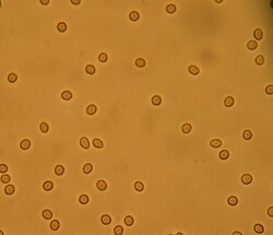
A kidney biopsy will provide a fully definitive diagnosis of nephritic syndrome and may also reveal the underlying cause of the nephritic syndrome depending on the underlying pathological process. On biopsy, a patient with nephritic syndrome would show inflammation of numerous glomeruli. [37]

Treatment

When a patient is confirmed to have nephritic syndrome, the main goal of treatment (regardless of the underlying cause) is to control elevated blood pressures and reduce active inflammation in the kidney itself. [4] Most often, the patient will need to be admitted to the hospital for close monitoring to ensure the efficacy of treatment and make adjustments as needed. Some treatment modalities commonly used to meet these goals include:

Once the acute phase of the nephritic syndrome is controlled, it is crucial to determine the underlying pathology that caused the onset of the acute nephritic syndrome and to treat that condition. If the underlying cause is not determined and treated appropriately, it increases the risk of a recurrence of nephritic syndrome or chronic kidney disease (CKD) in the future. [4]

Prognosis

Because nephritic syndrome is a syndrome and not a disease, the prognosis depends on the underlying cause. Generally, the prognosis of nephritic syndrome in children is better than it is in adults. [5]

Epidemiology

According to the CDC, nephritis/nephrosis/nephritic syndrome was the 9th leading cause of death in the United States in 2017. [45] It was listed as the cause of death for 50,633 out of the total 2,813,503 deaths reported in 2017. [45]

Geography

The southeast region of the United States reported a significantly higher death rate due to kidney disease than any other region in 2017. Mississippi reported the highest death rate due to kidney disease (21.7), followed by Louisiana (20.6) and Arkansas (19.7). [46] Although Vermont reported the lowest death rate due to kidney disease (3.3), the western United States reported the lowest regional average death rate due to kidney disease in 2017. [46]

Gender

Out of the 1,374,392 female deaths reported in the US in 2017, kidney disease was listed as the cause of death for 24,889 women and was reported as the 9th overall cause of death for women in 2017. [45]

Out of the 1,439,111 male deaths reported in the US in 2017, kidney disease was not listed in the top 10 causes of death. [45]

Race and ethnicity

Out of the 2,378,385 deaths reported in individuals who identified as White, kidney disease was ranked 10th overall (39,105 deaths) in causes of death in the US in 2017. [45]

Out of the 340,644 deaths reported in individuals who identified as Black or African American, kidney disease was ranked 8th overall (9,609 deaths) in causes of death in the US in 2017. [45]

Out of the 74,094 deaths reported in individuals who identified as Asian or Pacific Islander, kidney disease was ranked 9th overall (1,563 deaths) in causes of death in the US in 2017. [45]

Out of the 197,249 deaths reported in individuals who identified as Hispanic or Latino, kidney disease was ranked 10th overall (3,928 deaths) in causes of death in the US 2017. [45]

Other countries of world

In a review of Romanian cases, a 10-year review yielded that upon biopsy, nephritic syndrome was the second most common clinical syndrome at 21.9% (nephrotic syndrome was 52.3%) [47]

Related Research Articles

Albuminuria is a pathological condition wherein the protein albumin is abnormally present in the urine. It is a type of proteinuria. Albumin is a major plasma protein ; in healthy people, only trace amounts of it are present in urine, whereas larger amounts occur in the urine of patients with kidney disease. For a number of reasons, clinical terminology is changing to focus on albuminuria more than proteinuria.

<span class="mw-page-title-main">Proteinuria</span> Presence of an excess of serum proteins in the urine

Proteinuria is the presence of excess proteins in the urine. In healthy persons, urine contains very little protein, less than 150 mg/day; an excess is suggestive of illness. Excess protein in the urine often causes the urine to become foamy. Severe proteinuria can cause nephrotic syndrome in which there is worsening swelling of the body.

<span class="mw-page-title-main">Nephrotic syndrome</span> Collection of symptoms due to kidney damage

Nephrotic syndrome is a collection of symptoms due to kidney damage. This includes protein in the urine, low blood albumin levels, high blood lipids, and significant swelling. Other symptoms may include weight gain, feeling tired, and foamy urine. Complications may include blood clots, infections, and high blood pressure.

<span class="mw-page-title-main">Nephritis</span> Inflammation of the kidneys

Nephritis is inflammation of the kidneys and may involve the glomeruli, tubules, or interstitial tissue surrounding the glomeruli and tubules. It is one of several different types of nephropathy.

<span class="mw-page-title-main">Hematuria</span> Medical condition

Hematuria or haematuria is defined as the presence of blood or red blood cells in the urine. "Gross hematuria" occurs when urine appears red, brown, or tea-colored due to the presence of blood. Hematuria may also be subtle and only detectable with a microscope or laboratory test. Blood that enters and mixes with the urine can come from any location within the urinary system, including the kidney, ureter, urinary bladder, urethra, and in men, the prostate. Common causes of hematuria include urinary tract infection (UTI), kidney stones, viral illness, trauma, bladder cancer, and exercise. These causes are grouped into glomerular and non-glomerular causes, depending on the involvement of the glomerulus of the kidney. But not all red urine is hematuria. Other substances such as certain medications and foods can cause urine to appear red. Menstruation in women may also cause the appearance of hematuria and may result in a positive urine dipstick test for hematuria. A urine dipstick test may also give an incorrect positive result for hematuria if there are other substances in the urine such as myoglobin, a protein excreted into urine during rhabdomyolysis. A positive urine dipstick test should be confirmed with microscopy, where hematuria is defined by three or more red blood cells per high power field. When hematuria is detected, a thorough history and physical examination with appropriate further evaluation can help determine the underlying cause.

<span class="mw-page-title-main">Assessment of kidney function</span> Ways of assessing the function of the kidneys

Assessment of kidney function occurs in different ways, using the presence of symptoms and signs, as well as measurements using urine tests, blood tests, and medical imaging.

<span class="mw-page-title-main">Kidney disease</span> Damage to or disease of a kidney

Kidney disease, or renal disease, technically referred to as nephropathy, is damage to or disease of a kidney. Nephritis is an inflammatory kidney disease and has several types according to the location of the inflammation. Inflammation can be diagnosed by blood tests. Nephrosis is non-inflammatory kidney disease. Nephritis and nephrosis can give rise to nephritic syndrome and nephrotic syndrome respectively. Kidney disease usually causes a loss of kidney function to some degree and can result in kidney failure, the complete loss of kidney function. Kidney failure is known as the end-stage of kidney disease, where dialysis or a kidney transplant is the only treatment option.

<span class="mw-page-title-main">IgA nephropathy</span> Disease of the kidney

IgA nephropathy (IgAN), also known as Berger's disease, or synpharyngitic glomerulonephritis, is a disease of the kidney and the immune system; specifically it is a form of glomerulonephritis or an inflammation of the glomeruli of the kidney. Aggressive Berger's disease can attack other major organs, such as the liver, skin and heart.

<span class="mw-page-title-main">Henoch–Schönlein purpura</span> Medical condition

Henoch–Schönlein purpura (HSP), also known as IgA vasculitis, is a disease of the skin, mucous membranes, and sometimes other organs that most commonly affects children. In the skin, the disease causes palpable purpura, often with joint pain and abdominal pain. With kidney involvement, there may be a loss of small amounts of blood and protein in the urine, but this usually goes unnoticed; in a small proportion of cases, the kidney involvement proceeds to chronic kidney disease. HSP is often preceded by an infection, such as a throat infection.

<span class="mw-page-title-main">Glomerulonephritis</span> Term for several kidney diseases

Glomerulonephritis (GN) is a term used to refer to several kidney diseases. Many of the diseases are characterised by inflammation either of the glomeruli or of the small blood vessels in the kidneys, hence the name, but not all diseases necessarily have an inflammatory component.

<span class="mw-page-title-main">Diabetic nephropathy</span> Chronic loss of kidney function

Diabetic nephropathy, also known as diabetic kidney disease, is the chronic loss of kidney function occurring in those with diabetes mellitus. Diabetic nephropathy is the leading causes of chronic kidney disease (CKD) and end-stage renal disease (ESRD) globally. The triad of protein leaking into the urine, rising blood pressure with hypertension and then falling renal function is common to many forms of CKD. Protein loss in the urine due to damage of the glomeruli may become massive, and cause a low serum albumin with resulting generalized body swelling (edema) so called nephrotic syndrome. Likewise, the estimated glomerular filtration rate (eGFR) may progressively fall from a normal of over 90 ml/min/1.73m2 to less than 15, at which point the patient is said to have end-stage renal disease. It usually is slowly progressive over years.

<span class="mw-page-title-main">Membranous glomerulonephritis</span> Medical condition

Membranous glomerulonephritis (MGN) is a slowly progressive disease of the kidney affecting mostly people between ages of 30 and 50 years, usually white people.

<span class="mw-page-title-main">Focal segmental glomerulosclerosis</span> Kidney disease

Focal segmental glomerulosclerosis (FSGS) is a histopathologic finding of scarring (sclerosis) of glomeruli and damage to renal podocytes. This process damages the filtration function of the kidney, resulting in protein presence in the urine due to protein loss. FSGS is a leading cause of excess protein loss—nephrotic syndrome—in children and adults. Signs and symptoms include proteinuria and edema. Kidney failure is a common long-term complication of the disease. FSGS can be classified as primary, secondary, or genetic, depending on whether a particular toxic or pathologic stressor or genetic predisposition can be identified as the cause. Diagnosis is established by renal biopsy, and treatment consists of glucocorticoids and other immune-modulatory drugs. Response to therapy is variable, with a significant portion of patients progressing to end-stage kidney failure. An American epidemiological study 20 years ago demonstrated that FSGS is estimated to occur in 7 persons per million, with males and African-Americans at higher risk.

<span class="mw-page-title-main">Acute proliferative glomerulonephritis</span> Medical condition

Acute proliferative glomerulonephritis is a disorder of the small blood vessels of the kidney. It is a common complication of bacterial infections, typically skin infection by Streptococcus bacteria types 12, 4 and 1 (impetigo) but also after streptococcal pharyngitis, for which it is also known as postinfectious glomerulonephritis (PIGN) or poststreptococcal glomerulonephritis (PSGN). It can be a risk factor for future albuminuria. In adults, the signs and symptoms of infection may still be present at the time when the kidney problems develop, and the terms infection-related glomerulonephritis or bacterial infection-related glomerulonephritis are also used. Acute glomerulonephritis resulted in 19,000 deaths in 2013, down from 24,000 deaths in 1990 worldwide.

<span class="mw-page-title-main">Rapidly progressive glomerulonephritis</span> Medical condition

Rapidly progressive glomerulonephritis (RPGN) is a syndrome of the kidney that is characterized by a rapid loss of kidney function, with glomerular crescent formation seen in at least 50% or 75% of glomeruli seen on kidney biopsies. If left untreated, it rapidly progresses into acute kidney failure and death within months. In 50% of cases, RPGN is associated with an underlying disease such as Goodpasture syndrome, systemic lupus erythematosus or granulomatosis with polyangiitis; the remaining cases are idiopathic. Regardless of the underlying cause, RPGN involves severe injury to the kidneys' glomeruli, with many of the glomeruli containing characteristic glomerular crescents.

<span class="mw-page-title-main">Mesangial proliferative glomerulonephritis</span> Medical condition

Mesangial proliferative glomerulonephritis (MesPGN) is a morphological pattern characterized by a numerical increase in mesangial cells and expansion of the extracellular matrix within the mesangium of the glomerulus. The increase in the number of mesangial cells can be diffuse or local and immunoglobulin and/or complement deposition can also occur. MesPGN is associated with a variety of disease processes affecting the glomerulus, though can be idiopathic. The clinical presentation of MesPGN usually consists of hematuria or nephrotic syndrome. Treatment is often consistent with the histologic pattern of and/or disease process contributing to mesangial proliferative glomerulonephritis, and usually involves some form of immunosuppressant.

Glomerulonephrosis is a non-inflammatory disease of the kidney (nephrosis) presenting primarily in the glomerulus as nephrotic syndrome. The nephron is the functional unit of the kidney and it contains the glomerulus, which acts as a filter for blood to retain proteins and blood lipids. Damage to these filtration units results in important blood contents being released as waste in urine. This disease can be characterized by symptoms such as fatigue, swelling, and foamy urine, and can lead to chronic kidney disease and ultimately end-stage renal disease, as well as cardiovascular diseases. Glomerulonephrosis can present as either primary glomerulonephrosis or secondary glomerulonephrosis.

Diffuse proliferative glomerulonephritis (DPGN) is a type of glomerulonephritis that is the most serious form of renal lesions in SLE and is also the most common, occurring in 35% to 60% of patients. In absence of SLE, DPGN pathology looks more like Membranoproliferative glomerulonephritis

Sickle cell nephropathy is a type of nephropathy associated with sickle cell disease which causes kidney complications as a result of sickling of red blood cells in the small blood vessels. The hypertonic and relatively hypoxic environment of the renal medulla, coupled with the slow blood flow in the vasa recta, favors sickling of red blood cells, with resultant local infarction. Functional tubule defects in patients with sickle cell disease are likely the result of partial ischemic injury to the renal tubules.

<span class="mw-page-title-main">IgM nephropathy</span> Medical condition

IgM nephropathy or immunoglobulin M nephropathy (IgMN) is a kind of idiopathic glomerulonephritis that is marked by IgM diffuse deposits in the glomerular mesangium. IgM nephropathy was initially documented in 1978 by two separate teams of researchers.

References

  1. "acute nephritic syndrome""at Dorland's Medical Dictionary
  2. 1 2 3 Madara, Bernadette; Pomarico-Denino, Vanessa (2008-07-08). Quick Look Nursing: Pathophysiology. Jones & Bartlett Learning. p. 484. ISBN   9780763749323.
  3. 1 2 Schrier, Robert W. (2008-10-01). Manual of Nephrology. Lippincott Williams & Wilkins. pp. 144–145. ISBN   9780781796194.
  4. 1 2 3 4 5 "Acute nephritic syndrome: MedlinePlus Medical Encyclopedia". www.nlm.nih.gov. Retrieved 2015-10-27.
  5. 1 2 "Acute Nephritis; Nephrosis; Nephritic syndrome information. Patient | Patient". Patient. Retrieved 2015-10-27.
  6. Ferri, Fred F. (2017-05-25). Ferri's clinical advisor 2018 : 5 books in 1. Preceded by: Ferri, Fred F. Philadelphia, PA. p. 889. ISBN   978-0-323-52957-0. OCLC   989151714.{{cite book}}: CS1 maint: location missing publisher (link)
  7. 1 2 Schrier, Robert W. (2010). Renal and Electrolyte Disorders. Lippincott Williams & Wilkins. pp. 559–560. ISBN   978-1-60831-072-2.
  8. 1 2 Kibble, Jonathan David (2009). Medical physiology : the big picture. Halsey, Colby Ray. New York: McGraw-Hill. p. 221. ISBN   978-0-07-164302-3. OCLC   469141953.
  9. 1 2 3 4 5 6 7 8 9 10 Harrison's principles of internal medicine. Longo, Dan L. (Dan Louis), 1949-, Fauci, Anthony S., 1940-, Kasper, Dennis L., Hauser, Stephen L., Jameson, J. Larry., Loscalzo, Joseph. (18th ed.). New York: McGraw-Hill. 2012. pp. 2334–2345. ISBN   978-0-07-174890-2. OCLC   747712285.{{cite book}}: CS1 maint: others (link)
  10. Floege, Jürgen; Johnson, Richard J.; Feehally, John (2010). Comprehensive Clinical Nephrology (Fourth ed.). Mosby. p. 198. doi:10.1016/B978-0-323-05876-6.00015-0. ISBN   9780323058766.
  11. Schrier, Robert W. (2010-01-01). Renal and Electrolyte Disorders. Lippincott Williams & Wilkins. p. 560. ISBN   9781608310722.
  12. Floege, Jurgen; Johnson, Richard J.; Feehally, John (2010-11-08). Comprehensive Clinical Nephrology E-Book. Elsevier Health Sciences. pp. 202–203. ISBN   978-0-323-08133-7.
  13. Krueger, Gerhard R. F.; Buja, L. Maximilian (2013-03-12). Atlas of Anatomic Pathology with Imaging: A Correlative Diagnostic Companion. Springer Science & Business Media. p. 287. ISBN   9781447128465.
  14. Whelton, Paul K.; Carey, Robert M.; Aronow, Wilbert S.; Casey, Donald E.; Collins, Karen J.; Dennison Himmelfarb, Cheryl; DePalma, Sondra M.; Gidding, Samuel; Jamerson, Kenneth A.; Jones, Daniel W.; MacLaughlin, Eric J. (June 2018). "2017 ACC/AHA/AAPA/ABC/ACPM/AGS/APhA/ASH/ASPC/NMA/PCNA Guideline for the Prevention, Detection, Evaluation, and Management of High Blood Pressure in Adults: A Report of the American College of Cardiology/American Heart Association Task Force on Clinical Practice Guidelines". Hypertension. 71 (6): e13–e115. doi: 10.1161/HYP.0000000000000065 . ISSN   1524-4563. PMID   29133356.
  15. 1 2 "Subtopic 3: Microscopic Examination of Urine Sediment". Texas Collaborative. November 19, 2004. Archived from the original on 2004-11-19. Retrieved January 22, 2020.
  16. Davin, J. C.; Ten Berge, I. J.; Weening, J. J. (March 2001). "What is the difference between IgA nephropathy and Henoch–Schönlein purpura nephritis?". Kidney International. 59 (3): 823–834. doi: 10.1046/j.1523-1755.2001.059003823.x . ISSN   0085-2538. PMID   11231337.
  17. "Acute Poststreptococcal Glomerulonephritis: Background, Pathophysiology, Epidemiology". 2018-12-05.{{cite journal}}: Cite journal requires |journal= (help)
  18. Comprehensive pediatric nephrology . Geary, Denis F., Schaefer, Franz. Philadelphia, PA: Mosby/Elsevier. 2008. pp.  309–317. ISBN   978-0-323-07096-6. OCLC   324995599.{{cite book}}: CS1 maint: others (link)
  19. Kraft, D. M.; Mckee, D.; Scott, C. (August 1998). "Henoch–Schönlein purpura: a review". American Family Physician. 58 (2): 405–408, 411. ISSN   0002-838X. PMID   9713395.
  20. Cody, Ellen M.; Dixon, Bradley P. (February 2019). "Hemolytic Uremic Syndrome". Pediatric Clinics of North America. 66 (1): 235–246. doi:10.1016/j.pcl.2018.09.011. ISSN   1557-8240. PMID   30454746. S2CID   53875876.
  21. Salama, A. D.; Levy, J. B.; Lightstone, L.; Pusey, C. D. (2001-09-15). "Goodpasture's disease". Lancet. 358 (9285): 917–920. doi:10.1016/S0140-6736(01)06077-9. ISSN   0140-6736. PMID   11567730. S2CID   40175400.
  22. Ward, M. M. (2000-11-13). "Changes in the incidence of end-stage renal disease due to lupus nephritis, 1982-1995". Archives of Internal Medicine. 160 (20): 3136–3140. doi:10.1001/archinte.160.20.3136. ISSN   0003-9926. PMID   11074743.
  23. Lohr, James W. (January 10, 2018). "Rapidly Progressive Glomerulonephritis". Medscape. Retrieved January 21, 2020.
  24. Bhowmik, Dipankar (January 2011). "Clinical Approach to Rapidly Progressive Renal Failure" (PDF). Journal of the Association of Physicians of India. 59: 38–44. PMID   21751663.
  25. Robbins basic pathology. Kumar, Vinay, 1944-, Robbins, Stanley L. (Stanley Leonard), 1915-2003. (8th ed.). Philadelphia, PA: Saunders/Elsevier. 2007. pp. 406–408. ISBN   978-1-4160-2973-1. OCLC   69672074.{{cite book}}: CS1 maint: others (link)
  26. Retamozo, Soledad; Brito-Zerón, Pilar; Bosch, Xavier; Stone, John H.; Ramos-Casals, Manuel (November 2013). "Cryoglobulinemic disease". Oncology (Williston Park, N.Y.). 27 (11): 1098–1105, 1110–1116. ISSN   0890-9091. PMID   24575538.
  27. Habib, R.; Gubler, M. C.; Loirat, C.; Mäiz, H. B.; Levy, M. (April 1975). "Dense deposit disease: a variant of membranoproliferative glomerulonephritis". Kidney International. 7 (4): 204–215. doi: 10.1038/ki.1975.32 . ISSN   0085-2538. PMID   1095806.
  28. "Tips for having your blood pressure taken". National Heart, Lung, and Blood Institute. July 2, 2014. Archived from the original on July 2, 2014.
  29. "Techniques: Heart Sounds & Murmurs". University of Washington Department of Medicine. Retrieved January 22, 2020.
  30. "Electrolytes". MedlinePlus. November 20, 2017. Retrieved January 22, 2020.
  31. "Creatinine Blood Test". MedlinePlus. July 15, 2017. Retrieved January 22, 2020.
  32. "BUN - blood test". MedlinePlus. April 29, 2019. Retrieved January 22, 2020.
  33. "Urinalysis". MedlinePlus. February 7, 2019. Retrieved January 22, 2020.
  34. Lee, Andrew; Mirrett, Stanley; Reller, L. Barth; Weinstein, Melvin P. (November 2007). "Detection of bloodstream infections in adults: how many blood cultures are needed?". Journal of Clinical Microbiology. 45 (11): 3546–3548. doi:10.1128/JCM.01555-07. ISSN   0095-1137. PMC   2168497 . PMID   17881544.
  35. Kavanaugh, A.; Tomar, R.; Reveille, J.; Solomon, D. H.; Homburger, H. A. (January 2000). "Guidelines for clinical use of the antinuclear antibody test and tests for specific autoantibodies to nuclear antigens". Archives of Pathology & Laboratory Medicine. 124 (1): 71–81. doi:10.5858/2000-124-0071-GFCUOT. ISSN   0003-9985. PMID   10629135.
  36. Bradwell, A. R. (1999). Advanced atlas of autoantibody patterns. Mead, G. P., Stokes, R. P., Binding Site Limited. Birmingham: The Binding Site. ISBN   0-7044-8510-9. OCLC   41258931.
  37. "Acute nephritic syndrome". MedlinePlus. July 16, 2019. Retrieved January 22, 2020.
  38. Allen, C.; Glasziou, P.; Del Mar, C. (1999-10-09). "Bed rest: a potentially harmful treatment needing more careful evaluation". Lancet. 354 (9186): 1229–1233. doi:10.1016/s0140-6736(98)10063-6. ISSN   0140-6736. PMID   10520630. S2CID   12196831.
  39. "Fluid Restricted Diet". Intermountain Healthcare. 2016. Retrieved January 23, 2020.
  40. Mahtani, Kamal R.; Heneghan, Carl; Onakpoya, Igho; Tierney, Stephanie; Aronson, Jeffrey K.; Roberts, Nia; Hobbs, F. D. Richard; Nunan, David (December 1, 2018). "Reduced Salt Intake for Heart Failure: A Systematic Review". JAMA Internal Medicine. 178 (12): 1693–1700. doi:10.1001/jamainternmed.2018.4673. ISSN   2168-6114. PMC   6422065 . PMID   30398532. S2CID   53241717.
  41. Ali, Syed Salman; Sharma, Pramod Kumar; Garg, Vipin Kumar; Singh, Avnesh Kumar; Mondal, Sambhu Charan (April 2012). "The target-specific transporter and current status of diuretics as antihypertensive". Fundamental & Clinical Pharmacology. 26 (2): 175–179. doi:10.1111/j.1472-8206.2011.01012.x. ISSN   1472-8206. PMID   22145583. S2CID   43171023.
  42. Wright, Jackson T.; Bakris, George; Greene, Tom; Agodoa, Larry Y.; Appel, Lawrence J.; Charleston, Jeanne; Cheek, DeAnna; Douglas-Baltimore, Janice G.; Gassman, Jennifer; Glassock, Richard; Hebert, Lee (2002-11-20). "Effect of blood pressure lowering and antihypertensive drug class on progression of hypertensive kidney disease: results from the AASK trial". JAMA. 288 (19): 2421–2431. doi:10.1001/jama.288.19.2421. ISSN   0098-7484. PMID   12435255.
  43. Rhen, Turk; Cidlowski, John A. (2005-10-20). "Antiinflammatory action of glucocorticoids--new mechanisms for old drugs". The New England Journal of Medicine. 353 (16): 1711–1723. doi:10.1056/NEJMra050541. ISSN   1533-4406. PMID   16236742. S2CID   5744727.
  44. Tattersall, James; Dekker, Friedo; Heimbürger, Olof; Jager, Kitty J.; Lameire, Norbert; Lindley, Elizabeth; Van Biesen, Wim; Vanholder, Raymond; Zoccali, Carmine; ERBP Advisory Board (July 2011). "When to start dialysis: updated guidance following publication of the Initiating Dialysis Early and Late (IDEAL) study". Nephrology, Dialysis, Transplantation. 26 (7): 2082–2086. doi:10.1093/ndt/gfr168. ISSN   1460-2385. PMID   21551086.
  45. 1 2 3 4 5 6 7 8 "Leading causes of death and numbers of deaths, by sex, race, and Hispanic origin: United States, 1980 and 2017" (PDF). CDC. 2018. Retrieved January 21, 2020.
  46. 1 2 "Kidney Disease Mortality by State". CDC. 2017. Retrieved January 21, 2020.
  47. Covic, Adrian; Schiller, Adalbert; Volovat, Carmen; Gluhovschi, Gheorghe; Gusbeth-Tatomir, Paul; Petrica, Ligia; Caruntu, Irina-Draga; Bozdog, Gheorghe; Velciov, Silvia; Trandafirescu, Virginia; Bob, Flaviu; Gluhovschi, Cristina (1 February 2006). "Epidemiology of renal disease in Romania: a 10 year review of two regional renal biopsy databases". Nephrology Dialysis Transplantation. 21 (2): 419–424. doi: 10.1093/ndt/gfi207 . ISSN   0931-0509. PMID   16249204 . Retrieved 1 May 2020.

Further reading