Pachygyria

Last updated
Pachygyria
Specialty Medical genetics, neurology   OOjs UI icon edit-ltr-progressive.svg

Pachygyria (from the Greek pachy meaning "thick" or "fat" gyri) is a congenital malformation of the cerebral hemisphere. It results in unusually thick convolutions of the cerebral cortex. Typically, children have developmental delay and seizures, the onset and severity depending on the severity of the cortical malformation. Infantile spasms are common in affected children, as is intractable epilepsy.[ citation needed ]

Contents

Presentation

The term 'pachygyria' does not directly relate to a specific malformation but rather is used to generally describe physical characteristics of the brain in association with several neuronal migration disorders; most commonly disorders relating to varied degrees of lissencephaly. Lissencephaly is present in 1 of 85,470 births and the life span of those affected is short as only a few survive past the age of 20. [1] Pachygyria is a condition identified by a type of cortical genetic malformation. Clinicians will subjectively determine the malformation based on the degree of malposition and the extent of thickened abnormal grey differentiation present. [2]

Connections to epilepsy, lissencephaly, and subcortical band heterotopia

Various degrees of intensity and locations of epilepsy are associated with malformations of cortical development. Researchers suggest that approximately 40% of children diagnosed with drug-resistant epilepsy have some degree of cortical malformation. [1] [2]

Lissencephaly (to which pachygyria is most closely linked) is associated with severe mental retardation, epilepsy, and motor disability. Two characteristics of lissencephaly include its absence of convolutions (agyria) and decreased presence of convolutions (pachygyria). [2] The types of seizures associated with lissencephaly include:

Other possible symptoms of lissencephaly include telecanthus, estropia, hypertelorism, varying levels of mental retardation, cerebellar hypoplasia, corpus callosum aplasia, and decreased muscle tone and tendon reflexes. [3] Over 90% of children affected with lissencephaly have seizures. [2]

Patients with subcortical band heterotopia (another disorder associated with pachygyria) typically have milder symptoms and their cognitive function is closely linked to the thickness of the subcortical band and the degree of pachygyria present. [2]

Causes

Pachygyria is caused by a breakdown in the fetal neuronal migration process due to genetic or possibly environmental influences. The affected cerebral cortex will typically have only four developed layers instead of the normal six. One of the best known and most common types of neuronal migration disorders is lissencephaly, a diffuse cortical malformation relating directly to agyria and pachygyria. [4] Incomplete neuronal migration during the early fetal brain development is the precursor to lissencephaly. [5] Should neurons follow an abnormal migration during development possible cortical malformations include classical lissencephaly (as stated above) and subcortical band heterotopia with an agyria-pachygyria band spectrum. [2]

Normal neuronal migration

Normal neuronal migration involves the development of six cortical layers, each one performing distinct functions. [2]

Normal cerebral development occurs in three dynamic and overlapping stages:[ citation needed ]

Most types of incomplete neuronal migration to the cortex occur during the third and fourth gestational months. [4] The abnormal migration of the neurons causes them to not reach their proper final destinations, which results in failure of the sulci and gyri to form. [2]

The stage of cortical development at which migration is arrested is directly related to the level of structural malposition. [1]

One of the most critical stages in brain development is when the post-mitotic neurons migrate from the ventricular zone to form the cortical plate. [6] Migration arrested toward the latter part of development usually restricts the abnormal cell position to the cortex level. [1]

Neuronal migration disorder caused by genetic mutations

Several genetic mutations have been isolated and linked to specific malformations of the cerebral cortex. [1] Genes shown to cause lissencephaly include both autosomal and X-linked genes. [3] Below, the mutations of LIS1 or DCX genes are discussed as they are most commonly linked to neuronal migration disorders including lissencephaly-pachygyria and subcortical band heterotopia. [2]

LIS1

LIS1 is responsible for the autosomal form of lissencephaly. [2] Mutations of the LIS1 gene are associated with about 80% of those affected with lissencephaly. [5] LIS1 was the first human neuronal migration gene to be cloned. It is responsible for encoding the alpha subunit of the intracellular Ib isoform of platelet-activating factor acetylhydrolase. It is located on chromosome 17p13.3 and has 11 exons with a coding region of 1233bp. LIS1 protein appears to interact with tubulin to suppress microtubule dynamics. The protein is highly conserved and studies have shown that it participates in cytoplasmic dynein-mediated nucleokinesis, somal translocation, cell motility, mitosis, and chromosome segregation. [6] LIS1 encodes for a 45kDa protein called PAFAH1B1 that contains seven WD40 repeats required for proper neuronal migration. [5] The LIS1 gene encodes for a protein similar to the β subunit of G proteins responsible for degrading bioactive lipid platelet-activating factor (PAF). [2] This leads to theories that LIS1 might exert its effect on migration through microtubules. Specific concentrations of PAF may be necessary for optimal neuronal migration by influencing cell morphology adhesion properties. Studies have shown that addition of PAF or inhibition of platelet-activating factor acetylhydrolase (PAF-AH) decreases cerebellar granule cell migration in vitro. Addition of PAF to hippocampal cells have shown growth cone collapse and neurite retraction. LIS1 knockout homozygous null mice die during embryogenesis and heterozygous mice survive with delayed neuronal migration confirmed by in vitro and in vivo cell migration assays. [5] Most lissencephaly cases are associated with deletions of mutations of the LIS1 gene and the results are usually more severe in the posterior brain regions. [2]

One study showed that of an isolated group of patients with lissencephaly, 40% resulted from an LIS1 deletion and another 25% resulted from an intragenic mutation of the gene. Patients with missense mutations tend to have less severe symptoms, pachygyria, and rare cases of subcortical band heterotopia. Truncated (shortened) mutations of LIS1 tend to cause severe lissencephaly. [2]

Doublecortin

Doublecortin (DCX or XLIS) mutations are responsible for X-linked disorders. [2] While LIS1 mutations tend to cause severe malformations in the posterior brain, DCX mutations focus much of their destruction on anterior malformations and are linked to lissencephaly in males and subcortical band heterotopias in females. [2] [5] Women with DCX mutations tend to have an anteriorly-predominant subcortical band heterotopia and pachygyria. [1] [2] DCX was the first known gene causing X-linked lissencephaly and subcortical band heterotopia. It is found on chromosome Xq22.3-q23 and has nine exons that code for 360 proteins. DCX is expressed exclusively in the fetal brain. [6]

Spastic cerebral palsy is by far the most common type, occurring in 70–80% of all cases. Moreover, spastic CP accompanies one of the other types in 30% of all cases. People with this type are hypertonic and have a neuromuscular condition stemming from damage to the corticospinal tract or the motor cortex that affects the nervous system's ability to receive gamma amino butyric acid in the area(s) affected by the disability. [ citation needed ]Spastic CP is further classified by topography dependent on the region of the body affected; these include:

Spastic hemiplegia (one side being affected). Generally, injury to muscle-nerves controlled by the brain's left side will cause a right body deficit, and vice versa. Typically, people that have spastic hemiplegia are the most ambulatory, although they generally have dynamic equinus on the affected side and are primarily prescribed ankle-foot orthoses to prevent said equinus.[11] Spastic diplegia (the lower extremities are affected with little to no upper-body spasticity). The most common form of the spastic forms. Most people with spastic diplegia are fully ambulatory and have a scissors gait. Flexed knees and hips to varying degrees are common. Hip problems, dislocations, and in three-quarters of spastic diplegics, also strabismus (crossed eyes), can be present as well. In addition, these individuals are often nearsighted. The intelligence of a person with spastic diplegia is unaffected by the condition. Spastic tetraplegia (all four limbs affected equally). People with spastic quadriplegia are the least likely to be able to walk, or if they can, to want to walk, because their muscles are too tight and it is too much effort to do so. Some children with quadriplegia also have hemiparetic tremors, an uncontrollable shaking that affects the limbs on one side of the body and impairs normal movement. Occasionally, terms such as monoplegia, paraplegia, triplegia, and pentaplegia may also be used to refer to specific manifestations of the spasticity.[ citation needed ]

Pathogenesis

Pachygyria, lissencephaly (smooth brain), and polymicrogyria (multiple small gyri) are all the results of abnormal cell migration. The abnormal migration is typically associated with a disorganized cellular architecture, failure to form six layers of cortical neurons (a four-layer cortex is common), and functional problems. The abnormal formation of the brain may be associated with seizures, developmental delay, and mental dysfunctions.[ citation needed ]

Normally, the brain cells begin to develop in the periventricular region (germinal matrix) and then migrate from medial to lateral, to form the cerebral cortex.[ citation needed ]

Diagnosis

Different imaging modalities are commonly used for diagnosis. While computed tomography (CT) provides higher spatial resolution imaging of the brain, cerebral cortex malformations are more easily visualized in vivo and classified using magnetic resonance imaging (MRI) which provides higher contrast imaging and better delineation of white and gray matter. [7]

Diffuse pachygyria (a mild form of lissencephaly) can be seen on an MRI as thickened cerebral cortices with few and large gyri and incomplete development of the Sylvian fissures. [3]

Cognitive ability correlates with the thickness of any subcortical band present and the degree of pachygyria. [1] [5]

Classifications

The degree of cerebral cortex malformation caused by genetic mutations is classified by the degree of malposition and the extent of faulty grey matter differentiation. [1]

Neuronal migration disorders are generally classified into three groups:

The 'other' types are associated with corpus callosum agenesis or cerebellar hypoplasia while the cobblestone lissencephalies are associated with eye and muscle disorders. [3]

Classical lissencephaly, also known as type I or generalized agyria-pachygyria, is a severe brain malformation of a smooth cerebral surface, abnormally thick (10–20 mm) cortex with four layers, widespread neuronal heterotopia, enlarged ventricles, and agenesis or malformation of the corpus callosum. [4] [6] Classical lissencephaly can range from agyria to regional pachygyria and is usually present along with subcortical band heterotopia (known as 'double cortex' to describe the circumferential bands of heterotopic neurons located beneath the cortex). [6] Subcortical band heterotopia is a malformation slightly different from lissencephaly that is now classified under the agyria-pachygyria-band spectrum because it consists of a gyral pattern consistent with broad convolutions and an increased cortical thickness. [1] The established classification scheme for lissencephaly is based on the severity (grades 1–6) and the gradient. [5]

Grade 1 and Grade 4 are very rare. Grade 2 is observed in children with Miller–Dieker syndrome (a combination of lissencephaly with dysmorphic facial features, visceral abnormalities, and polydactyly). The most common lissencephaly observed, consisting of frontotemporal pachygyria and posterior agyria, is Grade 3. [4] Another malformation worth mentioning because of its connections to pachygyria is polymicrogyria. Polymicrogyria is characterized by many small gyri separated by shallow sulci, slightly thin cortex, neuronal heterotopia and enlarged ventricle and is often superimposed on pachygyria. [4]

Treatment

Because pachygyria is a structural defect no treatments are currently available other than symptomatic treatments, especially for associated seizures. Another common treatment is a gastrostomy (insertion of a feeding tube) to reduce possible poor nutrition and repeated aspiration pneumonia. [4]

Microcephalic osteodysplastic primordial dwarfism

Microcephalic osteodysplastic primordial dwarfism (MOPD) type II is an autosomal multisystem disorder including severe pre- and post-natal growth retardation, microcephaly with Seckel syndrome–like facial appearance, and distinctive skeletal alterations. Usually those affected have mild to moderate mental retardation. [8]

See also

Related Research Articles

<span class="mw-page-title-main">Cerebral cortex</span> Outer layer of the cerebrum of the mammalian brain

The cerebral cortex, also known as the cerebral mantle, is the outer layer of neural tissue of the cerebrum of the brain in humans and other mammals. The cerebral cortex mostly consists of the six-layered neocortex, with just 10% consisting of the allocortex. It is separated into two cortices, by the longitudinal fissure that divides the cerebrum into the left and right cerebral hemispheres. The two hemispheres are joined beneath the cortex by the corpus callosum. The cerebral cortex is the largest site of neural integration in the central nervous system. It plays a key role in attention, perception, awareness, thought, memory, language, and consciousness. The cerebral cortex is part of the brain responsible for cognition.

<span class="mw-page-title-main">Lissencephaly</span> Medical condition

Lissencephaly is a set of rare brain disorders whereby the whole or parts of the surface of the brain appear smooth. It is caused by defective neuronal migration during the 12th to 24th weeks of gestation resulting in a lack of development of brain folds (gyri) and grooves (sulci). It is a form of cephalic disorder. Terms such as agyria and pachygyria are used to describe the appearance of the surface of the brain.

<span class="mw-page-title-main">Focal cortical dysplasia</span> Medical condition

Focal cortical dysplasia (FCD) is a congenital abnormality of brain development where the neurons in an area of the brain failed to migrate in the proper formation in utero. Focal means that it is limited to a focal zone in any lobe. Focal cortical dysplasia is a common cause of intractable epilepsy in children and is a frequent cause of epilepsy in adults. There are three types of FCD with subtypes, including type 1a, 1b, 1c, 2a, 2b, 3a, 3b, 3c, and 3d, each with distinct histopathological features. All forms of focal cortical dysplasia lead to disorganization of the normal structure of the cerebral cortex :

Miller–Dieker syndrome, Miller–Dieker lissencephaly syndrome (MDLS), and chromosome 17p13.3 deletion syndrome is a micro deletion syndrome characterized by congenital malformations. Congenital malformations are physical defects detectable in an infant at birth which can involve many different parts of the body including the brain, hearts, lungs, liver, bones, or intestinal tract. MDS is a contiguous gene syndrome – a disorder due to the deletion of multiple gene loci adjacent to one another. The disorder arises from the deletion of part of the small arm of chromosome 17p, leading to partial monosomy. There may be unbalanced translocations, or the presence of a ring chromosome 17.

<span class="mw-page-title-main">Polymicrogyria</span> Medical condition

Polymicrogyria (PMG) is a condition that affects the development of the human brain by multiple small gyri (microgyri) creating excessive folding of the brain leading to an abnormally thick cortex. This abnormality can affect either one region of the brain or multiple regions.

Bilateral frontoparietal polymicrogyria is a genetic disorder with autosomal recessive inheritance that causes a cortical malformation. Our brain has folds in the cortex to increase surface area called gyri and patients with polymicrogyria have an increase number of folds and smaller folds than usual. Polymicrogyria is defined as a cerebral malformation of cortical development in which the normal gyral pattern of the surface of the brain is replaced by an excessive number of small, fused gyri separated by shallow sulci and abnormal cortical lamination. From ongoing research, mutation in GPR56, a member of the adhesion G protein-coupled receptor (GPCR) family, results in BFPP. These mutations are located in different regions of the protein without any evidence of a relationship between the position of the mutation and phenotypic severity. It is also found that GPR56 plays a role in cortical pattering.

<span class="mw-page-title-main">Gyrus</span> Ridge on the cerebral cortex of the brain

In neuroanatomy, a gyrus is a ridge on the cerebral cortex. It is generally surrounded by one or more sulci. Gyri and sulci create the folded appearance of the brain in humans and other mammals.

<span class="mw-page-title-main">Gray matter heterotopia</span> Group of neurological disorders

Gray matter heterotopia is a neurological disorder caused by gray matter being located in an atypical location in the brain.

<span class="mw-page-title-main">Doublecortin</span> Protein-coding gene in humans

Neuronal migration protein doublecortin, also known as doublin or lissencephalin-X is a protein that in humans is encoded by the DCX gene.

<span class="mw-page-title-main">PAFAH1B1</span> Protein-coding gene in the species Homo sapiens

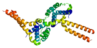
Platelet-activating factor acetylhydrolase IB subunit alpha is an enzyme that in humans is encoded by the PAFAH1B1 gene. The protein is often referred to as Lis1 and plays an important role in regulating the motor protein Dynein.

<span class="mw-page-title-main">Ganglionic eminence</span>

The ganglionic eminence (GE) is a transitory structure in the development of the nervous system that guides cell and axon migration. It is present in the embryonic and fetal stages of neural development found between the thalamus and caudate nucleus.

Gyrification is the process of forming the characteristic folds of the cerebral cortex.

<span class="mw-page-title-main">Neuronal migration disorder</span> Medical condition

Neuronal migration disorder (NMD) refers to a heterogenous group of disorders that, it is supposed, share the same etiopathological mechanism: a variable degree of disruption in the migration of neuroblasts during neurogenesis. The neuronal migration disorders are termed cerebral dysgenesis disorders, brain malformations caused by primary alterations during neurogenesis; on the other hand, brain malformations are highly diverse and refer to any insult to the brain during its formation and maturation due to intrinsic or extrinsic causes that ultimately will alter the normal brain anatomy. However, there is some controversy in the terminology because virtually any malformation will involve neuroblast migration, either primarily or secondarily.

<span class="mw-page-title-main">Ulegyria</span> Type of cortical scarring deep in the sulci

Ulegyria is a diagnosis used to describe a specific type of cortical scarring in the deep regions of the sulcus that leads to distortion of the gyri. Ulegyria is identified by its characteristic "mushroom-shaped" gyri, in which scarring causes shrinkage and atrophy in the deep sulcal regions while the surface gyri are spared. This condition is most often caused by hypoxic-ischemic brain injury in the perinatal period. The effects of ulegyria can range in severity, although it is most commonly associated with cerebral palsy, mental retardation and epilepsy. N.C. Bresler was the first to view ulegyria in 1899 and described this abnormal morphology in the brain as “mushroom-gyri." Although ulegyria was first identified in 1899, there is still limited information known or reported about the condition.

The development of the cerebral cortex, known as corticogenesis is the process during which the cerebral cortex of the brain is formed as part of the development of the nervous system of mammals including its development in humans. The cortex is the outer layer of the brain and is composed of up to six layers. Neurons formed in the ventricular zone migrate to their final locations in one of the six layers of the cortex. The process occurs from embryonic day 10 to 17 in mice and between gestational weeks seven to 18 in humans.

Cajal–Retzius cells are a heterogeneous population of morphologically and molecularly distinct reelin-producing cell types in the marginal zone/layer I of the developmental cerebral cortex and in the immature hippocampus of different species and at different times during embryogenesis and postnatal life.

<span class="mw-page-title-main">Congenital bilateral perisylvian syndrome</span> Medical condition

Congenital bilateral perisylvian syndrome (CBPS) is a rare neurological disease characterized by paralysis of certain facial muscles and epileptic seizures.

Fryns-Aftimos syndrome is a rare chromosomal condition and is associated with pachygyria, severe mental retardation, epilepsy and characteristic facial features. This syndrome is a malformation syndrome, characterized by numerous facial dysmorphias not limited to hypertelorism, iris or retinal coloboma, cleft lip, and congenital heart defects. This syndrome has been seen in 30 unrelated people. Characterized by a de novo mutation located on chromosome 7p22, there is typically no family history prior to onset. The severity of the disorder can be determined by the size of the deletion on 7p22, enveloping the ACTB gene and surrounding genes, which is consistent with a contiguous gene deletion syndrome. Confirming a diagnosis of Fryns-Aftimos syndrome typically consists of serial single-gene testing or multigene panel of genes of interest or exome sequencing.

<span class="mw-page-title-main">Microlissencephaly</span> Microcephaly combined with lissencephaly

Microlissencephaly (MLIS) is a rare congenital brain disorder that combines severe microcephaly with lissencephaly. Microlissencephaly is a heterogeneous disorder, i.e. it has many different causes and a variable clinical course. Microlissencephaly is a malformation of cortical development (MCD) that occurs due to failure of neuronal migration between the third and fifth month of gestation as well as stem cell population abnormalities. Numerous genes have been found to be associated with microlissencephaly, however, the pathophysiology is still not completely understood.

References

  1. 1 2 3 4 5 6 7 8 9 10 Guerrini R (2005). "Genetic malformations of the cerebral cortex and epilepsy". Epilepsia. 46 (Suppl 1): 32–37. doi: 10.1111/j.0013-9580.2005.461010.x . PMID   15816977.
  2. 1 2 3 4 5 6 7 8 9 10 11 12 13 14 15 16 17 Guerrini R, Marini C (2006). "Genetic malformations of cortical development". Exp Brain Res. 173 (2): 322–333. doi:10.1007/s00221-006-0501-z. PMID   16724181. S2CID   12482656.
  3. 1 2 3 4 5 Phadke, S., "et al." (2007). "Pachygyria in a Girl with Microcephalic Osteodysplastic Primordial Short Stature Type II." Brain and Development. 27:237-240.
  4. 1 2 3 4 5 6 Dobyns WB, Truwit CL (1995). "Lissencephaly and other malformations of cortical development: 1995 update". Neuropediatrics. 26 (3): 132–147. doi:10.1055/s-2007-979744. PMID   7477752.
  5. 1 2 3 4 5 6 7 8 9 Cardoso C, Leventer RJ, Matsumoto N, et al. (2000). "The location and type of mutation predict malformation severity in isolated lissencephaly caused by abnormalities within the LIS1 gene". Hum. Mol. Genet. 9 (20): 3019–3028. doi: 10.1093/hmg/9.20.3019 . PMID   11115846.
  6. 1 2 3 4 5 Kato M, Dobyns WB (2003). "Lissencephaly and the molecular basis of neuronal migration". Hum. Mol. Genet. 12 Spec No 1 (90001): R89–96. doi: 10.1093/hmg/ddg086 . PMID   12668601.
  7. 1 2 Pieh C, Goebel HH, Engle EC, Gottlob I (2003). "Congenital fibrosis syndrome associated with central nervous system abnormalities". Graefes Arch. Clin. Exp. Ophthalmol. 241 (7): 546–553. doi:10.1007/s00417-003-0703-z. PMID   12819981. S2CID   25436215.
  8. Ozawa H, Takayama C, Nishida A, Nagai T, Nishimura G, Higurashi M (2005). "Pachygyria in a girl with microcephalic osteodysplastic primordial short stature type II". Brain Dev. 27 (3): 237–240. doi:10.1016/j.braindev.2004.06.007. PMID   15737708. S2CID   26150025.