Pseudohypoaldosteronism

Last updated

Pseudohypoaldosteronism (PHA) is a condition that mimics hypoaldosteronism (presenting hyperkalemia). [1] Two major types of primary pseudohypoaldosteronism are recognized and these have major differences in etiology and presentation. [2]

Contents

Pseudohypoaldosteronism type 1 (PHA1)

Pseudohypoaldosteronism Type 1
Aldosterone-2D-skeletal.svg
In pseudohypoaldosteronism type 1, aldosterone is elevated (hyperaldosteronism), but because the body fails to respond to it, it appears similar to hypoaldosteronism.
Specialty Nephrology   OOjs UI icon edit-ltr-progressive.svg
Symptoms Failure to thrive, dehydration, hyponatremia, metabolic acidosis, hyperkalemia, and other non-specific symptoms including nausea, vomiting, extreme fatigue, and muscle weakness.
CausesMutations in the NR3C2 , SCNN1A , SCNN1B , or SCNN1G genes

Pseudohypoaldosteronism type 1 (PHA1) is characterized by the body's inability to respond adequately to aldosterone, a hormone crucial for regulating electrolyte levels. This condition often manifests with dehydration as the kidneys struggle to retain sufficient salt, leading to symptoms like increased thirst and dry mouth. Additionally, PHA1 disrupts electrolyte balance, resulting in low levels of sodium and high levels of potassium in the blood.

Mechanism

PHA1 is an heterogeneous disease, which can be caused by mutations in different genes. On one hand, mutations on the gene NR3C2 (coding the mineralocorticoid receptor) cause the synthesis of a non-functional receptor which is unable to bind aldosterone or function correctly. In the kidney, aldosterone plays an important role of regulating sodium and potassium homeostasis by its actions on distal nephron cells. [3]

On the other hand, autosomal recessive PHA1 is caused by mutations in both alleles of either SCNN1A, SCNN1B or SCNN1G. These genes code the different subunits of the epithelial sodium channel, ENaC, which is located in the collecting duct of the nephron, and is responsible for sodium reabsorption and potassium secretion (by generating the electrochemical gradient necessary for potassium efflux by ROMK channel). [3]

Onset

Symptoms

Types

Type OMIM GeneInheritanceAge of OnsetDescription
PHA1A 177735 NR3C2 (Mineralocorticoid receptor, MLR) Autosomal dominantNeonatal but improves with age. Adults are usually asymptomatic without treatment [4] .Salt wasting caused by renal unresponsiveness to mineralocorticoids. Patients often present with hyperkalaemic acidosis despite high aldosterone levels. Not all individuals with the mutation develop PHA1A suggesting that illness and volume depletion may play a role in the development of the clinically recognized PHA1A.
PHA1B 264350 SCNN1A , SCNN1B , SCNN1G (encoding epithelial sodium channel subunits)Autosomal recessiveNeonatal, persists into adulthood. [5] Renal salt wasting and high concentrations of sodium in sweat, stool, and saliva. The disorder often involves multiple organ systems and can be life threatening in the neonatal period. Patients usually present with hyponatremia, hyperkalemia, and increased plasma renin activity with high serum aldosterone concentrations. PHA1B is often mistaken for cystic fibrosis.

Treatment

Treatment of severe forms of PHA1 requires relatively large amounts of sodium chloride. [6] Potassium restriction in the diet might also contribute to decrease urinary sodium wasting. [7]

Risks

Individuals with PHA1B can have additional symptoms such as cardiac arrhythmia, shock, recurrent lung infections, or lesions on the skin due to imbalanced salts in the body especially in infancy.

A stop mutation in the SCNN1A gene has been shown to be associated with female infertility. [8]

Pseudohypoaldosteronism type 2 (PHA2)

PHA2 also known as Familial hyperkalemic hypertension or Gordon syndrome is a rare disorder characterized by abnormalities in how the body regulates sodium and potassium levels. This condition stems from mutations in specific genes involved in the regulation of sodium transport within the kidneys.

Unlike in PHA1 in which aldosterone resistance is present, in PHA2 blood volume increases occur regardless of normal or low aldosterone levels due to the enhanced activity of sodium transporters in the kidney. [9]

Mechanism

PHA2 is associated with mutations in the WNK4 , WNK1 , KLHL3 and CUL3 genes. These genes regulate the Sodium-chloride symporter (NCC) transporter, which is involved in controlling the levels of sodium and chloride in the body. Normally, the NCC transporter reabsorbs sodium and chloride in a part of the kidney called the distal convoluted tubule (DCT), however in PHA2 this process is dysregulated. Mutations in these genes lead to overactivity of NCC, causing excessive sodium and chloride reabsorption.

The hyperkalemia found in PHA2 is proposed to be a function of diminished sodium delivery to the cortical collecting tubule (potassium excretion is mediated by the renal outer medullary potassium channel (ROMK) in which sodium reabsorption plays a role). Alternatively, WNK4 mutations that result in a gain of function of the Na-Cl co-transporter may inhibit ROMK activity resulting in hyperkalemia. [10]

Onset

The age of onset is difficult to pinpoint and can range from infancy to adulthood.[ citation needed ]

Symptoms

People with PHA2 have hypertension and hyperkalemia despite having normal kidney function. Many individuals with PHA2 will develop hyperkalemia first, and will not present with hypertension until later in life. They also commonly experience both hyperchloremia and metabolic acidosis together, a condition called hyperchloremic metabolic acidosis.

People with PHA2 may experience other nonspecific symptoms including nausea, vomiting, extreme fatigue, muscle weakness, and hypercalcuria.

Some PHA2E patients present with dental abnormalities. [11] Patients with recessive KLHL3 mutations and dominant CUL3 mutations tend to have more severe phenotypes. [12]

A study in 2024 linked PHA2 to epilepsy. Epileptic seizures were seen in 3 of the 44 affected subjects. Two of the subjects had Generalized tonic–clonic seizure and one subject had migraine seizures. All three subjects had WNK4 mutations. It's speculated that the epilepsy may be caused by potassium spikes resulting in abnormal CNS neuron activity. The study also linked PHA2 to proximal renal tubular acidosis. [13] Metabolic acidosis is also known to cause epileptic seizures.

Types

Type OMIM GeneInheritanceAge Of OnsetDescription
PHA2A 145260 mapped to chromosome 1q31-q42 [14] Autosomal dominantVariesDoes not involve salt wasting.
PHA2B 614491 WNK4 Autosomal dominant10+ with a mean age of 28 [15] May involve salt wasting. [16] Patients typically do not experience hypertension until adulthood. [15] Bicarbonate is higher than other PHA2 types. Aldosterone concentrations are often normal. [17] TRPV6 may be involved. [18]
PHA2C 614492 WNK1 Autosomal dominant15+ with a mean age of 36 [15] Does not involve salt wasting. [16] Significantly less severely affected than other PHA2 types. [15] Affected patients have hypertension together with long-term hyperkalemia, hyperchloremia, normal plasma creatinine, reduced bicarbonate, and low renin levels. Aldestrone levels may be normal or elevated.
PHA2D 614495 KLHL3 Autosomal dominant or autosomal recessiveMean age at diagnosis was found to be around 24 to 26, but it varies widely. [15] May involve salt wasting. [16] Individuals with the autosomal dominant mutations typically show higher potassium levels than those with autosomal recessive mutations. Hypertension usually develops in adulthood. Patients often present with low bicarbonate (17-18). [15]
PHA2E 614496 CUL3 Autosomal dominant3-15 years old [15] Most severe manifestations of PHA2 compared to patients with other mutations. Almost all individuals present with hypertension before age 18. [15]

Treatment

PHA2 requires salt restriction and use of thiazide diuretics to block sodium chloride reabsorption and normalise blood pressure and serum potassium.[ citation needed ]

Risks

Pregnancy risks

As of 2018, at least seven reported cases of severe metabolic acidosis occurring during pregnancy have been reported in PHA2 patients. [19]

A study in 2023 also described a patient with severe preeclampsia later being diagnosed with PHA2D associated with chronic hyperkalemia and hyperchloremic metabolic acidosis. The twin babies were born healthy and discharged from the hospital. [20]

Other risks

One study noted that severe hypercalciuria from untreated PHA2 resulted in kidney stones, and osteoporosis in some patients. [21]

History

PHA1 was first described by Cheek and Perry in 1958. [22] Later pediatric endocrinologist Aaron Hanukoglu reported that there are two independent forms of PHA with different inheritance patterns: A renal form with autosomal dominant inheritance exhibiting salt loss mainly from the kidneys, and a multi-system form with autosomal recessive form exhibiting salt loss from kidney, lung, and sweat and salivary glands. [23] [24]

The hereditary lack of responsiveness to aldosterone could be due to at least two possibilities: 1. A mutation in the mineralocorticoid receptor that binds aldosterone, or 2. A mutation in a gene that is regulated by aldosterone. Linkage analysis on patients with the severe form of PHA excluded the possibility of linkage of the disease with the mineralocorticoid receptor gene region. [25] Later, the severe form of PHA was discovered to be due to mutations in the genes SCNN1A , SCNN1B , and SCNN1G that code for the epithelial sodium channel subunits, α, β, and γ, respectively. [26]

On the other hand, PHA2 was initially described by Dr. Richard Gordon [27] . Mutations in WNK1 and WNK4 as a cause for PHA2 were first described in 2001 by Richard Lifton´s laboratory [28] . Later, mutations in KLHL3 and CUL3 were also found in different PHA2 patients in 2012 [29] .

See also

Related Research Articles

<span class="mw-page-title-main">Primary aldosteronism</span> Excess production of aldosterone in the adrenal gland

Primary aldosteronism (PA), also known as primary hyperaldosteronism, refers to the excess production of the hormone aldosterone from the adrenal glands, resulting in low renin levels and high blood pressure. This abnormality is a paraneoplastic syndrome. About 35% of the cases are caused by a single aldosterone-secreting adenoma, a condition known as Conn's syndrome.

<span class="mw-page-title-main">Hyperkalemia</span> Excess potassium in the blood

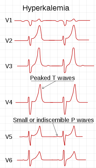
Hyperkalemia is an elevated level of potassium (K+) in the blood. Normal potassium levels are between 3.5 and 5.0 mmol/L (3.5 and 5.0 mEq/L) with levels above 5.5 mmol/L defined as hyperkalemia. Typically hyperkalemia does not cause symptoms. Occasionally when severe it can cause palpitations, muscle pain, muscle weakness, or numbness. Hyperkalemia can cause an abnormal heart rhythm which can result in cardiac arrest and death.

<span class="mw-page-title-main">Amiloride</span> Medication

Amiloride, sold under the trade name Midamor among others, is a medication typically used with other medications to treat high blood pressure or swelling due to heart failure or cirrhosis of the liver. Amiloride is classified as a potassium-sparing diuretic. Amiloride is often used together with another diuretic, such as a thiazide or loop diuretic. It is taken by mouth. Onset of action is about two hours and it lasts for about a day.

<span class="mw-page-title-main">Gitelman syndrome</span> Genetic kidney disorder

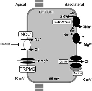
Gitelman syndrome (GS) is an autosomal recessive kidney tubule disorder characterized by low blood levels of potassium and magnesium, decreased excretion of calcium in the urine, and elevated blood pH. It is the most frequent hereditary salt-losing tubulopathy. Gitelman syndrome is caused by disease-causing variants on both alleles of the SLC12A3 gene. The SLC12A3 gene encodes the thiazide-sensitive sodium-chloride cotransporter, which can be found in the distal convoluted tubule of the kidney.

<span class="mw-page-title-main">Potassium-sparing diuretic</span> Drugs that cause diuresis without causing potassium loss in the urine and leading to hyperkalemia

Potassium-sparing diuretics or antikaliuretics refer to drugs that cause diuresis without causing potassium loss in the urine. They are typically used as an adjunct in management of hypertension, cirrhosis, and congestive heart failure. The steroidal aldosterone antagonists can also be used for treatment of primary hyperaldosteronism. Spironolactone, a steroidal aldosterone antagonist, is also used in management of female hirsutism and acne from PCOS or other causes.

<span class="mw-page-title-main">Hypoaldosteronism</span> Medical condition

Hypoaldosteronism is an endocrinological disorder characterized by decreased levels of the hormone aldosterone. Similarly, isolated hypoaldosteronism is the condition of having lowered aldosterone without corresponding changes in cortisol.

Secondary hypertension is a type of hypertension which has a specific and identifiable underlying primary cause. It is much less common than essential hypertension, affecting only 5-10% of hypertensive patients. It has many different causes including obstructive sleep apnea, kidney disease, endocrine diseases, and tumors. The cause of secondary hypertension varies significantly with age. It also can be a side effect of many medications.

<span class="mw-page-title-main">Metabolic alkalosis</span> Increase in blood pH due to imbalance in hydrogen ion and bicarbonate concentrations

Metabolic alkalosis is an acid-base disorder in which the pH of tissue is elevated beyond the normal range (7.35–7.45). This is the result of decreased hydrogen ion concentration, leading to increased bicarbonate, or alternatively a direct result of increased bicarbonate concentrations. The condition typically cannot last long if the kidneys are functioning properly.

<span class="mw-page-title-main">Renal tubular acidosis</span> Higher blood acidity due to failure of the kidneys to fully acidify urine

Renal tubular acidosis (RTA) is a medical condition that involves an accumulation of acid in the body due to a failure of the kidneys to appropriately acidify the urine. In renal physiology, when blood is filtered by the kidney, the filtrate passes through the tubules of the nephron, allowing for exchange of salts, acid equivalents, and other solutes before it drains into the bladder as urine. The metabolic acidosis that results from RTA may be caused either by insufficient secretion of hydrogen ions into the latter portions of the nephron or by failure to reabsorb sufficient bicarbonate ions from the filtrate in the early portion of the nephron. Although a metabolic acidosis also occurs in those with chronic kidney disease, the term RTA is reserved for individuals with poor urinary acidification in otherwise well-functioning kidneys. Several different types of RTA exist, which all have different syndromes and different causes. RTA is usually an incidental finding based on routine blood draws that show abnormal results. Clinically, patients may present with vague symptoms such as dehydration, mental status changes, or delayed growth in adolescents.

<span class="mw-page-title-main">Apparent mineralocorticoid excess syndrome</span> Medical condition

Apparent mineralocorticoid excess is an autosomal recessive disorder causing hypertension, hypernatremia and hypokalemia. It results from mutations in the HSD11B2 gene, which encodes the kidney isozyme of 11β-hydroxysteroid dehydrogenase type 2. In an unaffected individual, this isozyme inactivates circulating cortisol to the less active metabolite cortisone. The inactivating mutation leads to elevated local concentrations of cortisol in the aldosterone sensitive tissues like the kidney. Cortisol at high concentrations can cross-react and activate the mineralocorticoid receptor due to the non-selectivity of the receptor, leading to aldosterone-like effects in the kidney. This is what causes the hypokalemia, hypertension, and hypernatremia associated with the syndrome. Patients often present with severe hypertension and end-organ changes associated with it like left ventricular hypertrophy, retinal, renal and neurological vascular changes along with growth retardation and failure to thrive. In serum both aldosterone and renin levels are low.

<span class="mw-page-title-main">Liddle's syndrome</span> Medical condition

Liddle's syndrome, also called Liddle syndrome, is a genetic disorder inherited in an autosomal dominant manner that is characterized by early, and frequently severe, high blood pressure associated with low plasma renin activity, metabolic alkalosis, low blood potassium, and normal to low levels of aldosterone. Liddle syndrome involves abnormal kidney function, with excess reabsorption of sodium and loss of potassium from the renal tubule, and is treated with a combination of low sodium diet and potassium-sparing diuretics. It is extremely rare, with fewer than 30 pedigrees or isolated cases having been reported worldwide as of 2008.

<span class="mw-page-title-main">Oculocerebrorenal syndrome</span> Medical condition

Oculocerebrorenal syndrome is a rare X-linked recessive disorder characterized by congenital cataracts, hypotonia, intellectual disability, proximal tubular acidosis, aminoaciduria and low-molecular-weight proteinuria. Lowe syndrome can be considered a cause of Fanconi syndrome.

Pseudohyperaldosteronism is a medical condition which mimics the effects of elevated aldosterone (hyperaldosteronism) by presenting with high blood pressure, low blood potassium levels (hypokalemia), metabolic alkalosis, and low levels of plasma renin activity (PRA). However, unlike hyperaldosteronism, this conditions exhibits low or normal levels of aldosterone in the blood. Causes include genetic disorders, acquired conditions, metabolic disorders, and dietary imbalances including excessive consumption of licorice. Confirmatory diagnosis depends on the specific cause and may involve blood tests, urine tests, or genetic testing; however, all forms of this condition exhibit abnormally low concentrations of both plasma renin activity (PRA) and plasma aldosterone concentration (PAC) which differentiates this group of conditions from other forms of secondary hypertension. Treatment is tailored to the specific cause and focuses on symptom control, blood pressure management, and avoidance of triggers.

<span class="mw-page-title-main">Epithelial sodium channel</span> Group of membrane proteins

The epithelial sodium channel(ENaC), (also known as amiloride-sensitive sodium channel) is a membrane-bound ion channel that is selectively permeable to sodium ions (Na+). It is assembled as a heterotrimer composed of three homologous subunits α or δ, β, and γ, These subunits are encoded by four genes: SCNN1A, SCNN1B, SCNN1G, and SCNN1D. The ENaC is involved primarily in the reabsorption of sodium ions at the collecting ducts of the kidney's nephrons. In addition to being implicated in diseases where fluid balance across epithelial membranes is perturbed, including pulmonary edema, cystic fibrosis, COPD and COVID-19, proteolyzed forms of ENaC function as the human salt taste receptor.

<span class="mw-page-title-main">Sodium-chloride symporter</span> Protein-coding gene in the species Homo sapiens

The sodium-chloride symporter (also known as Na+-Cl cotransporter, NCC or NCCT, or as the thiazide-sensitive Na+-Cl cotransporter or TSC) is a cotransporter in the kidney which has the function of reabsorbing sodium and chloride ions from the tubular fluid into the cells of the distal convoluted tubule of the nephron. It is a member of the SLC12 cotransporter family of electroneutral cation-coupled chloride cotransporters. In humans, it is encoded by the SLC12A3 gene (solute carrier family 12 member 3) located in 16q13.

<span class="mw-page-title-main">SCNN1B</span> Protein-coding gene in the species Homo sapiens

The SCNN1B gene encodes for the β subunit of the epithelial sodium channel ENaC in vertebrates. ENaC is assembled as a heterotrimer composed of three homologous subunits α, β, and γ or δ, β, and γ. The other ENAC subunits are encoded by SCNN1A, SCNN1G, and SCNN1D.

<span class="mw-page-title-main">SCNN1A</span> Protein-coding gene in the species Homo sapiens

The SCNN1A gene encodes for the α subunit of the epithelial sodium channel ENaC in vertebrates. ENaC is assembled as a heterotrimer composed of three homologous subunits α, β, and γ or δ, β, and γ. The other ENAC subunits are encoded by SCNN1B, SCNN1G, and SCNN1D.

<span class="mw-page-title-main">SCNN1G</span> Protein-coding gene in the species Homo sapiens

The SCNN1G gene encodes for the γ subunit of the epithelial sodium channel ENaC in vertebrates. ENaC is assembled as a heterotrimer composed of three homologous subunits α, β, and γ or δ, β, and γ. The other ENAC subunits are encoded by SCNN1A, SCNN1B, and SCNN1D.

<span class="mw-page-title-main">WNK4</span> Protein-coding gene in the species Homo sapiens

Serine/threonine protein kinase WNK4 also known as With No lysine (K) protein kinase 4(WNK4), is an enzyme that in humans is encoded by the WNK4 gene. Missense mutations cause a genetic form of pseudohypoaldosteronism type 2, also called Gordon syndrome or Familial Hyperkalemic Hypertension.

<span class="mw-page-title-main">Distal renal tubular acidosis</span> Medical condition

Distal renal tubular acidosis (dRTA) is the classical form of RTA, being the first described. Distal RTA is characterized by a failure of acid secretion by the alpha intercalated cells of the distal tubule and cortical collecting duct of the distal nephron. This failure of acid secretion may be due to a number of causes. It leads to relatively alkaline urine, due to the kidney's inability to acidify the urine to a pH of less than 5.3.

References

  1. "Pseudohypoaldosteronism: Overview - eMedicine Pediatrics: General Medicine" . Retrieved 2009-03-06.
  2. Diaz-Thomas A, Pascual-Y-Baralt JF (5 August 2022). Hoffman RP (ed.). "Pseudohypoaldosteronism". MedScape. WebMD LLC. Retrieved 6 June 2024.
  3. 1 2 Riepe FG, Finkeldei J, de Sanctis L, Einaudi S, Testa A, Karges B, et al. (November 2006). "Elucidating the underlying molecular pathogenesis of NR3C2 mutants causing autosomal dominant pseudohypoaldosteronism type 1". The Journal of Clinical Endocrinology and Metabolism. 91 (11): 4552–4561. doi:10.1210/jc.2006-1161. PMID   16954160.
  4. Amin N, Alvi NS, Barth JH, Field HP, Finlay E, Tyerman K, et al. (2013-08-01). "Pseudohypoaldosteronism type 1: clinical features and management in infancy". Endocrinology, Diabetes & Metabolism Case Reports. 2013: 130010. doi:10.1530/EDM-13-0010. PMC   3922296 . PMID   24616761.
  5. Bandhakavi M, Kirk J, Hogler W, Barrett T, Shaw N (November 2008). "Long-term outcome of autosomal recessive pseudohypoaldosteronism". Endocrine Abstracts. 17. ISSN   1470-3947.
  6. Hanukoglu A, Hanukoglu I (October 2010). "Clinical improvement in patients with autosomal recessive pseudohypoaldosteronism and the necessity for salt supplementation". Clinical and Experimental Nephrology. 14 (5): 518–519. doi:10.1007/s10157-010-0326-8. PMID   20661616. S2CID   9764720.
  7. Adachi M, Tajima T, Muroya K (2020). "Dietary potassium restriction attenuates urinary sodium wasting in the generalized form of pseudohypoaldosteronism type 1". CEN Case Reports. 9 (2): 133–137. doi:10.1007/s13730-019-00441-0. ISSN   2192-4449. PMC   7148393 . PMID   31900739.
  8. Boggula VR, Hanukoglu I, Sagiv R, Enuka Y, Hanukoglu A (October 2018). "Expression of the epithelial sodium channel (ENaC) in the endometrium - Implications for fertility in a patient with pseudohypoaldosteronism". The Journal of Steroid Biochemistry and Molecular Biology. 183: 137–141. doi:10.1016/j.jsbmb.2018.06.007. PMID   29885352. S2CID   47010706.
  9. O'Shaughnessy KM (November 2015). "Gordon Syndrome: a continuing story". Pediatric Nephrology. 30 (11): 1903–1908. doi:10.1007/s00467-014-2956-7. PMID   25503323. S2CID   195676310.
  10. Garovic VD, Hilliard AA, Turner ST (November 2006). "Monogenic forms of low-renin hypertension". Nature Clinical Practice. Nephrology. 2 (11). Nature Clinical Practice Nephrology: 624–630. doi:10.1038/ncpneph0309. PMID   17066054. S2CID   27864633.
  11. Perrier R, Boscardin E, Malsure S, Sergi C, Maillard MP, Loffing J, et al. (August 2016). "Severe Salt-Losing Syndrome and Hyperkalemia Induced by Adult Nephron-Specific Knockout of the Epithelial Sodium Channel α-Subunit". Journal of the American Society of Nephrology. 27 (8): 2309–2318. doi:10.1681/ASN.2015020154. PMC   4978035 . PMID   26701978.
  12. Fernandez CJ (2023). Monogenic hypertension with hyperkalemic acidosis, low renin, and variable aldosterone. pp. 1–117. doi:10.1016/C2021-0-01825-3. ISBN   978-0-323-96120-2.
  13. Shirin N, Rabinowitz G, Blatt I, Karlish SJ, Farfel Z, Mayan H (2024). "Association of Familial Hyperkalemia and Hypertension with Proximal Renal Tubular Acidosis and Epileptic Seizures". Nephron. 148 (3): 179–184. doi: 10.1159/000531868 . PMID   37666233.
  14. Mansfield TA, Simon DB, Farfel Z, Bia M, Tucci JR, Lebel M, et al. (June 1997). "Multilocus linkage of familial hyperkalaemia and hypertension, pseudohypoaldosteronism type II, to chromosomes 1q31-42 and 17p11-q21". Nature Genetics. 16 (2): 202–205. doi:10.1038/ng0697-202. PMID   9171836.
  15. 1 2 3 4 5 6 7 8 Boyden LM, Choi M, Choate KA, Nelson-Williams CJ, Farhi A, Toka HR, et al. (January 2012). "Mutations in kelch-like 3 and cullin 3 cause hypertension and electrolyte abnormalities". Nature. 482 (7383): 98–102. Bibcode:2012Natur.482...98B. doi:10.1038/nature10814. PMC   3278668 . PMID   22266938.
  16. 1 2 3 Susa K, Sohara E, Rai T, Zeniya M, Mori Y, Mori T, et al. (October 2014). "Impaired degradation of WNK1 and WNK4 kinases causes PHAII in mutant KLHL3 knock-in mice". Human Molecular Genetics. 23 (19): 5052–5060. doi: 10.1093/hmg/ddu217 . PMID   24821705.
  17. Farfel Z, Iaina A, Levi J, Gafni J (December 1978). "Proximal renal tubular acidosis: association with familial normaldosteronemic hyperpotassemia and hypertension". Archives of Internal Medicine. 138 (12): 1837–1840. doi:10.1001/archinte.1978.03630370047021. PMID   718349.
  18. Yang SS, Hsu YJ, Chiga M, Rai T, Sasaki S, Uchida S, et al. (April 2010). "Mechanisms for hypercalciuria in pseudohypoaldosteronism type II-causing WNK4 knock-in mice". Endocrinology. 151 (4): 1829–1836. doi: 10.1210/en.2009-0951 . PMID   20181799.
  19. Awad S, Keely E, Abujrad H (2018). "Resolution of Metabolic Abnormalities During Pregnancy in a Patient with Gordon Syndrome and KLHL3 Mutation". AACE Clinical Case Reports. 4 (3): 235–239. doi: 10.4158/AACR-2017-0006 .
  20. Cater TL, Espinosa LB (2023). "THU598 Pseudohypoaldosteronism Type 2: A New Variant Of A Rare Disease". Journal of the Endocrine Society. 7 (S1): A322–A323. doi: 10.1210/jendso/bvad114.595 . PMC   10555376 .
  21. D'ambrosio V, Mcknight O, Wan E, Speller R, Moss R, Siew K, et al. (June 2023). "#6875 Complications and Treatment of Hypercalciuria in Familial Hyperkalaemic Hypertension (FHHT)". Nephrology Dialysis Transplantation. 38 (S1): i241–i242. doi: 10.1093/ndt/gfad063c_6875 . S2CID   259396958.
  22. Cheek DB, Perry JW (June 1958). "A salt wasting syndrome in infancy". Archives of Disease in Childhood. 33 (169): 252–256. doi:10.1136/adc.33.169.252. PMC   2012226 . PMID   13545877.
  23. Hanukoglu A (November 1991). "Type I pseudohypoaldosteronism includes two clinically and genetically distinct entities with either renal or multiple target organ defects". The Journal of Clinical Endocrinology and Metabolism. 73 (5): 936–944. doi:10.1210/jcem-73-5-936. PMID   1939532.
  24. Hanukoglu I, Hanukoglu A (April 2016). "Epithelial sodium channel (ENaC) family: Phylogeny, structure-function, tissue distribution, and associated inherited diseases". Gene. 579 (2): 95–132. doi:10.1016/j.gene.2015.12.061. PMC   4756657 . PMID   26772908.
  25. Chung E, Hanukoglu A, Rees M, Thompson R, Dillon M, Hanukoglu I, et al. (November 1995). "Exclusion of the locus for autosomal recessive pseudohypoaldosteronism type 1 from the mineralocorticoid receptor gene region on human chromosome 4q by linkage analysis". The Journal of Clinical Endocrinology and Metabolism. 80 (11): 3341–3345. doi:10.1210/jcem.80.11.7593448. PMID   7593448.
  26. Chang SS, Grunder S, Hanukoglu A, Rösler A, Mathew PM, Hanukoglu I, et al. (March 1996). "Mutations in subunits of the epithelial sodium channel cause salt wasting with hyperkalaemic acidosis, pseudohypoaldosteronism type 1". Nature Genetics. 12 (3): 248–253. doi:10.1038/ng0396-248. PMID   8589714. S2CID   8185511.
  27. Gordon RD, Geddes RA, Pawsey CG, O'Halloran MW (November 1970). "Hypertension and severe hyperkalaemia associated with suppression of renin and aldosterone and completely reversed by dietary sodium restriction". Australasian Annals of Medicine. 19 (4): 287–294. doi:10.1111/imj.1970.19.4.287. PMID   5490655.
  28. Wilson FH, Disse-Nicodème S, Choate KA, Ishikawa K, Nelson-Williams C, Desitter I, et al. (August 2001). "Human hypertension caused by mutations in WNK kinases". Science. 293 (5532): 1107–1112. doi:10.1126/science.1062844. PMID   11498583.
  29. Boyden LM, Choi M, Choate KA, Nelson-Williams CJ, Farhi A, Toka HR, et al. (January 2012). "Mutations in kelch-like 3 and cullin 3 cause hypertension and electrolyte abnormalities". Nature. 482 (7383): 98–102. Bibcode:2012Natur.482...98B. doi:10.1038/nature10814. PMC   3278668 . PMID   22266938.