KCNE1

Last updated
KCNE1
Available structures
PDB Human UniProt search: PDBe RCSB
Identifiers
Aliases KCNE1 , ISK, JLNS, JLNS2, LQT2/5, LQT5, MinK, potassium voltage-gated channel subfamily E regulatory subunit 1
External IDs OMIM: 176261 GeneCards: KCNE1
Orthologs
SpeciesHumanMouse
Entrez
Ensembl
UniProt
RefSeq (mRNA)

n/a

RefSeq (protein)

n/a

Location (UCSC) Chr 21: 34.45 – 34.51 Mb n/a
PubMed search [2] n/a
Wikidata
View/Edit Human

Potassium voltage-gated channel subfamily E member 1 is a protein that in humans is encoded by the KCNE1 gene. [3] [4]

Contents

Voltage-gated potassium channels (Kv) represent the most complex class of voltage-gated ion channels from both functional and structural standpoints. Their diverse functions include regulating neurotransmitter release, heart rate, insulin secretion, neuronal excitability, epithelial electrolyte transport, smooth muscle contraction, and cell volume. [5]

KCNE1 is one of five members of the KCNE family of Kv channel ancillary or β subunits. It is also known as minK (minimal potassium channel subunit).

Function

KCNE1 is primarily known for modulating the cardiac and epithelial Kv channel alfa subunit, KCNQ1. KCNQ1 and KCNE1 form a complex in human ventricular cardiomyocytes that generates the slowly activating K+ current, IKs. Together with the rapidly activating K+ current (IKr), IKs is important for human ventricular repolarization. [6] [7] KCNQ1 is also essential for the normal function of many different epithelial tissues, but in these non-excitable cells it is thought to be predominantly regulated by KCNE2 or KCNE3. [8]

KCNE1 slows the activation of KCNQ1 5-10 fold, increases its unitary conductance 4-fold, eliminates its inactivation, and alters the manner in which KCNQ1 is regulated by other proteins, lipids and small molecules. The association of KCNE1 with KCNQ1 was discovered 8 years after Takumi and colleagues reported the isolation of a fraction of RNA from rat kidney that, when injected into Xenopus oocytes, produced an unusually slow-activating, voltage-dependent, potassium-selective current. Takumi et al discovered the KCNE1 gene [9] and it was correctly predicted to encode a single-transmembrane domain protein with an extracellular N-terminal domain and a cytosolic C-terminal domain. The ability of KCNE1 to generate this current was confusing because of its simple primary structure and topology, contrasting with the 6-transmembrane domain topology of other known Kv α subunits such as Shaker from Drosophila, cloned 2 years earlier. The mystery was solved when KCNQ1 was cloned and found to co-assemble with KCNE1, and it was shown that Xenopus laevis oocytes endogenously express KCNQ1, which is upregulated by exogenous expression of KCNE1 to generate the characteristic slowly activating current., [6] [7] KCNQ1 is also essential for the normal function of many different epithelial tissues, but in these non-excitable cells it is thought to be predominantly regulated by KCNE2 or KCNE3. [8]

KCNE1 is also reported to regulate two other KCNQ family α subunits, KCNQ4 and KCNQ5. KCNE1 increased both their peak currents in oocyte expression studies, and slowed the activation of the latter., [10] [11]

KCNE1 also regulates hERG, which is the Kv α subunit that generates ventricular IKr. KCNE1 doubled hERG current when the two were expressed in mammalian cells, although the mechanism for this remains unknown. [12]

Although KCNE1 had no effect when co-expressed with the Kv1.1 α subunit in Chinese Hamster ovary (CHO) cells, KCNE1 traps the N-type (rapidly inactivating) Kv1.4 α subunit in the ER/Golgi when co-expressed with it. KCNE1 (and KCNE2) also has this effect on the two other canonical N-type Kv α subunits, Kv3.3 and Kv3.4. This appears to be a mechanism for ensuring that homomeric N-type channels do not reach the cell surface, as this mode of suppression by KCNE1 or KCNE2 is relieved by co-expression of same-subfamily delayed rectifier (slowly inactivating) α subunits. Thus, Kv1.1 rescued Kv1.4, Kv3.1 rescued Kv3.4; in each of these cases the resultant channels at the membrane were heteromers (e.g., Kv3.1-Kv3.4) and displayed intermediate inactivation kinetics to those of either α subunit alone., [13] [14]

KCNE1 also regulates the gating kinetics of Kv2.1, Kv3.1 and Kv3.2, in each case slowing their activation and deactivation, and accelerating inactivation of the latter two., [15] [16] No effects were observed upon oocyte co-expression of KCNE1 and Kv4.2, [17] but KCNE1 was found to slow the gating and increase macroscopic current of Kv4.3 in HEK cells. [18] In contrast, channels formed by Kv4.3 and the cytosolic ancillary subunit KChIP2 exhibited faster activation and altered inactivation when co-expressed with KCNE1 in CHO cells. [19] Finally, KCNE1 inhibited Kv12.2 in Xenopus oocytes. [20]


Structure

The large majority of studies into the structural basis for KCNE1 modulation of Kv channels focus on its interaction with KCNQ1 (previously named KvLQT1). Residues in the transmembrane domain of KCNE1 lies close to the selectivity filter of KCNQ1 within heteromeric KCNQ1-KCNE1 channel complexes., [21] [22] The C-terminal domain of KCNE1, specifically from amino acids 73 to 79 is necessary for stimulation of slow delayed potassium rectifier current by SGK1. [23] The interaction of KCNE1 with an alpha helix in the S6 KvLQT1 domain contributes to the higher affinity this channel has for benzodiazepine L7 and chromanol 293B by repositioning amino acid residues to allow for this. KCNE1 destabilizes the S4-S5 alpha-helix linkage in the KCNQ1 channel protein in addition to destabilizing the S6 alpha helix, leading to slower activation of this channel when associated with KCNE1. [24] Variable stohiometries have been discussed but there are probably 2 KCNE1 subunits and 4 KCNQ1 subunits in a plasma membrane IKs complex. [25]

The transmembrane segment of KCNE1 is α-helical when in a membrane environment., [26] [27] The transmembrane segment of KCNE1 has been suggested to interact with the KCNQ1 pore domain (S5/S6) and with the S4 domain of the KCNQ1 (KvLQT1) channel. [21] KCNE1 may bind to the outer part of the KCNQ1 pore domain, and slide from this position into the “activation cleft” which leads to greater current amplitudes [23]

KCNE1 slows KCNQ1 activation several-fold, and there are ongoing discussions about the precise mechanisms underlying this. In a study in which KCNQ1 voltage sensor movement was monitored by site-directed fluorimetry and also by measuring the charge displacement associated with movement of charges within the S4 segment of the voltage sensor (gating current), KCNE1 was found to slow S4 movement so much that the gating current was no longer measurable. Fluorimetry measurements indicated that KCNQ1-KCNE1 channel S4 movement was 30-fold slower than that of the well-studied DrosophilaShaker Kv channel. [28] Nakajo and Kubo found that KCNE1 either slowed KCNQ1 S4 movement upon membrane depolarization, or altered S4 equilibrium at a given membrane potential. [29] The Kass lab deduced that while homomeric KCNQ1 channels can open after the movement of a single S4 segment, KCNQ1-KCNE1 channels can only open after all four S4 segments have been activated. [30] The intracellular C-terminal domain of KCNE1 is thought to sit on the KCNQ1 S4-S5 linker, a segment of KCNQ1 crucial for communicating S4 status to the pore and thus control activation. [31]

Tissue distribution

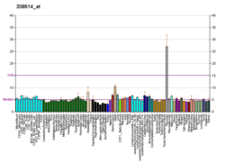
KCNE1 is expressed in human heart (atria and ventricles), whereas in adult mouse heart its expression appears limited to the atria and/or conduction system. [32] KCNE1 is also expressed in human and musine inner ear [33] and kidneys. [34] KCNE1 has been detected in mouse brain [35] but this finding is a subject of ongoing debate.

Clinical significance

Inherited or sporadic KCNE gene mutations can cause Romano–Ward syndrome (heterozygotes) and Jervell Lange-Nielsens syndrome (homozygotes). Both these syndromes are characterized by Long QT syndrome, a delay in ventricular repolarization. In addition, Jervell and Lange-Nielsen syndrome also involves bilateral sensorineural deafness. Mutation D76N in the KCNE1 protein can lead to long QT syndrome due to structural changes in the KvLQT1/KCNE1 complex, and people with these mutations are advised to avoid triggers of cardiac arrhythmia and prolonged QT intervals, such as stress or strenuous exercise. [23]

While loss-of-function mutations in KCNE1 cause Long QT syndrome, gain-of-function KCNE1 mutations are associated with early-onset atrial fibrillation. [36] A common KCNE1 polymorphism, S38G, is associated with altered predisposition to lone atrial fibrillation [37] and postoperative atrial fibrillation. [38] Atrial KCNE1 expression was downregulated in a porcine model of post-operative atrial fibrillation following lung lobectomy. [39]

Recently an analysis of 32 KCNE1 variants shows that putative/confirmed loss-of-function KCNE1 variants predispose to QT-prolongation, however the low ECG penetrance observed suggests they do not manifest clinically in the majority of individuals, aligning with the mild phenotype observed for JLNS2 patients. [40]

See also

Notes

Related Research Articles

<span class="mw-page-title-main">Long QT syndrome</span> Medical condition

Long QT syndrome (LQTS) is a condition affecting repolarization (relaxing) of the heart after a heartbeat, giving rise to an abnormally lengthy QT interval. It results in an increased risk of an irregular heartbeat which can result in fainting, drowning, seizures, or sudden death. These episodes can be triggered by exercise or stress. Some rare forms of LQTS are associated with other symptoms and signs including deafness and periods of muscle weakness.

<span class="mw-page-title-main">BK channel</span> Family of transport proteins

BK channels (big potassium), are large conductance calcium-activated potassium channels, also known as Maxi-K, slo1, or Kca1.1. BK channels are voltage-gated potassium channels that conduct large amounts of potassium ions (K+) across the cell membrane, hence their name, big potassium. These channels can be activated (opened) by either electrical means, or by increasing Ca2+ concentrations in the cell. BK channels help regulate physiological processes, such as circadian behavioral rhythms and neuronal excitability. BK channels are also involved in many processes in the body, as it is a ubiquitous channel. They have a tetrameric structure that is composed of a transmembrane domain, voltage sensing domain, potassium channel domain, and a cytoplasmic C-terminal domain, with many X-ray structures for reference. Their function is to repolarize the membrane potential by allowing for potassium to flow outward, in response to a depolarization or increase in calcium levels.

<span class="mw-page-title-main">Repolarization</span> Change in membrane potential

In neuroscience, repolarization refers to the change in membrane potential that returns it to a negative value just after the depolarization phase of an action potential which has changed the membrane potential to a positive value. The repolarization phase usually returns the membrane potential back to the resting membrane potential. The efflux of potassium (K+) ions results in the falling phase of an action potential. The ions pass through the selectivity filter of the K+ channel pore.

<span class="mw-page-title-main">Jervell and Lange-Nielsen syndrome</span> Medical condition

Jervell and Lange-Nielsen syndrome (JLNS) is a rare type of long QT syndrome associated with severe, bilateral sensorineural hearing loss. Those with JLNS are at risk of abnormal heart rhythms called arrhythmias, which can lead to fainting, seizures, or sudden death. JLNS, like other forms of long QT syndrome, causes the cardiac muscle to take longer than usual to recharge between beats. It is caused by genetic variants responsible for producing ion channels that carry transport potassium out of cells. The condition is usually diagnosed using an electrocardiogram, but genetic testing can also be used. Treatment includes lifestyle measures, beta blockers, and implantation of a defibrillator in some cases. It was first described by Anton Jervell and Fred Lange-Nielsen in 1957.

<span class="mw-page-title-main">Romano–Ward syndrome</span> Medical condition

Romano–Ward syndrome is the most common form of congenital Long QT syndrome (LQTS), a genetic heart condition that affects the electrical properties of heart muscle cells. Those affected are at risk of abnormal heart rhythms which can lead to fainting, seizures, or sudden death. Romano–Ward syndrome can be distinguished clinically from other forms of inherited LQTS as it affects only the electrical properties of the heart, while other forms of LQTS can also affect other parts of the body.

<span class="mw-page-title-main">KvLQT1</span> Protein-coding gene in the species Homo sapiens

Kv7.1 (KvLQT1) is a potassium channel protein whose primary subunit in humans is encoded by the KCNQ1 gene. It's mutation causes Long QT syndrome, Kv7.1 is a voltage and lipid-gated potassium channel present in the cell membranes of cardiac tissue and in inner ear neurons among other tissues. In the cardiac cells, Kv7.1 mediates the IKs (or slow delayed rectifying K+) current that contributes to the repolarization of the cell, terminating the cardiac action potential and thereby the heart's contraction. It is a member of the KCNQ family of potassium channels.

Voltage-gated calcium channels (VGCCs), also known as voltage-dependent calcium channels (VDCCs), are a group of voltage-gated ion channels found in the membrane of excitable cells (e.g., muscle, glial cells, neurons, etc.) with a permeability to the calcium ion Ca2+. These channels are slightly permeable to sodium ions, so they are also called Ca2+–Na+ channels, but their permeability to calcium is about 1000-fold greater than to sodium under normal physiological conditions.

hERG Mammalian protein found in humans

hERG is a gene that codes for a protein known as Kv11.1, the alpha subunit of a potassium ion channel. This ion channel is best known for its contribution to the electrical activity of the heart: the hERG channel mediates the repolarizing IKr current in the cardiac action potential, which helps coordinate the heart's beating.

<span class="mw-page-title-main">Azimilide</span> Chemical compound

Azimilide is a class ΙΙΙ antiarrhythmic drug. The agents from this heterogeneous group have an effect on the repolarization, they prolong the duration of the action potential and the refractory period. Also they slow down the spontaneous discharge frequency of automatic pacemakers by depressing the slope of diastolic depolarization. They shift the threshold towards zero or hyperpolarize the membrane potential. Although each agent has its own properties and will have thus a different function.

<span class="mw-page-title-main">Voltage-gated potassium channel</span> Class of transport proteins

Voltage-gated potassium channels (VGKCs) are transmembrane channels specific for potassium and sensitive to voltage changes in the cell's membrane potential. During action potentials, they play a crucial role in returning the depolarized cell to a resting state.

<span class="mw-page-title-main">KCNE2</span> Protein-coding gene in the species Homo sapiens

Potassium voltage-gated channel subfamily E member 2 (KCNE2), also known as MinK-related peptide 1 (MiRP1), is a protein that in humans is encoded by the KCNE2 gene on chromosome 21. MiRP1 is a voltage-gated potassium channel accessory subunit associated with Long QT syndrome. It is ubiquitously expressed in many tissues and cell types. Because of this and its ability to regulate multiple different ion channels, KCNE2 exerts considerable influence on a number of cell types and tissues. Human KCNE2 is a member of the five-strong family of human KCNE genes. KCNE proteins contain a single membrane-spanning region, extracellular N-terminal and intracellular C-terminal. KCNE proteins have been widely studied for their roles in the heart and in genetic predisposition to inherited cardiac arrhythmias. The KCNE2 gene also contains one of 27 SNPs associated with increased risk of coronary artery disease. More recently, roles for KCNE proteins in a variety of non-cardiac tissues have also been explored.

<span class="mw-page-title-main">KvLQT2</span> Protein-coding gene in the species Homo sapiens

Kv7.2 (KvLQT2) is a voltage- and lipid-gated potassium channel protein coded for by the gene KCNQ2.

<span class="mw-page-title-main">KvLQT3</span> Protein-coding gene in the species Homo sapiens

Kv7.3 (KvLQT3) is a potassium channel protein coded for by the gene KCNQ3.

<span class="mw-page-title-main">KCNJ5</span> Protein-coding gene in the species Homo sapiens

G protein-activated inward rectifier potassium channel 4(GIRK-4) is a protein that in humans is encoded by the KCNJ5 gene and is a type of G protein-gated ion channel.

<span class="mw-page-title-main">KCNE3</span> Protein-coding gene in the species Homo sapiens

Potassium voltage-gated channel, Isk-related family, member 3 (KCNE3), also known as MinK-related peptide 2(MiRP2) is a protein that in humans is encoded by the KCNE3 gene.

<span class="mw-page-title-main">KCND3</span> Protein-coding gene in the species Homo sapiens

Potassium voltage-gated channel subfamily D member 3 also known as Kv4.3 is a protein that in humans is encoded by the KCND3 gene. It contributes to the cardiac transient outward potassium current (Ito1), the main contributing current to the repolarizing phase 1 of the cardiac action potential.

<span class="mw-page-title-main">KCNH1</span> Protein-coding gene in the species Homo sapiens

Potassium voltage-gated channel subfamily H member 1 is a protein that in humans is encoded by the KCNH1 gene.

<span class="mw-page-title-main">KCNMB3</span> Protein-coding gene in the species Homo sapiens

Calcium-activated potassium channel subunit beta-3 is a protein that in humans is encoded by the KCNMB3 gene.

<span class="mw-page-title-main">KCNE4</span> Protein-coding gene in the species Homo sapiens

Potassium voltage-gated channel subfamily E member 4, originally named MinK-related peptide 3 or MiRP3 when it was discovered, is a protein that in humans is encoded by the KCNE4 gene.

<span class="mw-page-title-main">KCNE5</span> Protein-coding gene in the species Homo sapiens

KCNE1-like also known as KCNE1L is a protein that in humans is encoded by the KCNE1L gene.

References

  1. 1 2 3 GRCh38: Ensembl release 89: ENSG00000180509 - Ensembl, May 2017
  2. "Human PubMed Reference:". National Center for Biotechnology Information, U.S. National Library of Medicine.
  3. Chevillard C, Attali B, Lesage F, Fontes M, Barhanin J, Lazdunski M, Mattei MG (Jan 1993). "Localization of a potassium channel gene (KCNE1) to 21q22.1-q22.2 by in situ hybridization and somatic cell hybridization" (PDF). Genomics. 15 (1): 243–5. doi:10.1006/geno.1993.1051. PMID   8432548.
  4. "Entrez Gene: KCNE1 potassium voltage-gated channel, Isk-related family, member 1".
  5. Millar, I. D.; Hartley, J. A.; Haigh, C.; Grace, A. A.; White, S. J.; Kibble, J. D.; Robson, L. (2004). "Volume regulation is defective in renal proximal tubule cells isolated from KCNE1 knockout mice". Experimental Physiology. 89 (2): 173–180. doi:10.1113/expphysiol.2003.026674. ISSN   0958-0670.
  6. 1 2 Sanguinetti MC, Curran ME, Zou A, Shen J, Spector PS, Atkinson DL, Keating MT (Nov 1996). "Coassembly of K(V)LQT1 and minK (IsK) proteins to form cardiac I(Ks) potassium channel". Nature. 384 (6604): 80–3. doi:10.1038/384080a0. PMID   8900283. S2CID   4277239.
  7. 1 2 Barhanin J, Lesage F, Guillemare E, Fink M, Lazdunski M, Romey G (Nov 1996). "K(V)LQT1 and lsK (minK) proteins associate to form the I(Ks) cardiac potassium current". Nature. 384 (6604): 78–80. doi:10.1038/384078a0. PMID   8900282. S2CID   4366973.
  8. 1 2 Abbott GW (Jun 2015). "The KCNE2 K(+) channel regulatory subunit: Ubiquitous influence, complex pathobiology". Gene. 569 (2): 162–72. doi:10.1016/j.gene.2015.06.061. PMC   4917011 . PMID   26123744.
  9. Takumi T, Ohkubo H, Nakanishi S (Nov 1988). "Cloning of a membrane protein that induces a slow voltage-gated potassium current". Science. 242 (4881): 1042–5. Bibcode:1988Sci...242.1042T. doi:10.1126/science.3194754. PMID   3194754.
  10. Strutz-Seebohm N, Seebohm G, Fedorenko O, Baltaev R, Engel J, Knirsch M, Lang F (2006). "Functional coassembly of KCNQ4 with KCNE-beta- subunits in Xenopus oocytes". Cellular Physiology and Biochemistry. 18 (1–3): 57–66. doi: 10.1159/000095158 . PMID   16914890.
  11. Roura-Ferrer M, Etxebarria A, Solé L, Oliveras A, Comes N, Villarroel A, Felipe A (2009). "Functional implications of KCNE subunit expression for the Kv7.5 (KCNQ5) channel". Cellular Physiology and Biochemistry. 24 (5–6): 325–34. doi: 10.1159/000257425 . PMID   19910673.
  12. McDonald TV, Yu Z, Ming Z, Palma E, Meyers MB, Wang KW, Goldstein SA, Fishman GI (Jul 1997). "A minK-HERG complex regulates the cardiac potassium current I(Kr)". Nature. 388 (6639): 289–92. Bibcode:1997Natur.388..289M. doi: 10.1038/40882 . PMID   9230439. S2CID   4395891.
  13. Kanda VA, Lewis A, Xu X, Abbott GW (Sep 2011). "KCNE1 and KCNE2 inhibit forward trafficking of homomeric N-type voltage-gated potassium channels". Biophysical Journal. 101 (6): 1354–63. Bibcode:2011BpJ...101.1354K. doi:10.1016/j.bpj.2011.08.015. PMC   3177047 . PMID   21943416.
  14. Kanda VA, Lewis A, Xu X, Abbott GW (Sep 2011). "KCNE1 and KCNE2 provide a checkpoint governing voltage-gated potassium channel α-subunit composition". Biophysical Journal. 101 (6): 1364–75. Bibcode:2011BpJ...101.1364K. doi:10.1016/j.bpj.2011.08.014. PMC   3177048 . PMID   21943417.
  15. McCrossan ZA, Lewis A, Panaghie G, Jordan PN, Christini DJ, Lerner DJ, Abbott GW (Sep 2003). "MinK-related peptide 2 modulates Kv2.1 and Kv3.1 potassium channels in mammalian brain". The Journal of Neuroscience. 23 (22): 8077–91. doi:10.1523/JNEUROSCI.23-22-08077.2003. PMC   6740484 . PMID   12954870.
  16. Lewis A, McCrossan ZA, Abbott GW (Feb 2004). "MinK, MiRP1, and MiRP2 diversify Kv3.1 and Kv3.2 potassium channel gating". The Journal of Biological Chemistry. 279 (9): 7884–92. doi: 10.1074/jbc.M310501200 . PMID   14679187.
  17. Zhang M, Jiang M, Tseng GN (May 2001). "minK-related peptide 1 associates with Kv4.2 and modulates its gating function: potential role as beta subunit of cardiac transient outward channel?". Circulation Research. 88 (10): 1012–9. doi: 10.1161/hh1001.090839 . PMID   11375270.
  18. Deschênes I, Tomaselli GF (Sep 2002). "Modulation of Kv4.3 current by accessory subunits". FEBS Letters. 528 (1–3): 183–8. doi: 10.1016/s0014-5793(02)03296-9 . PMID   12297301. S2CID   41910930.
  19. Radicke S, Cotella D, Graf EM, Banse U, Jost N, Varró A, Tseng GN, Ravens U, Wettwer E (Sep 2006). "Functional modulation of the transient outward current Ito by KCNE beta-subunits and regional distribution in human non-failing and failing hearts". Cardiovascular Research. 71 (4): 695–703. doi: 10.1016/j.cardiores.2006.06.017 . PMID   16876774.
  20. Clancy SM, Chen B, Bertaso F, Mamet J, Jegla T (22 July 2009). "KCNE1 and KCNE3 beta-subunits regulate membrane surface expression of Kv12.2 K(+) channels in vitro and form a tripartite complex in vivo". PLOS ONE. 4 (7): e6330. Bibcode:2009PLoSO...4.6330C. doi: 10.1371/journal.pone.0006330 . PMC   2710002 . PMID   19623261.
  21. 1 2 Tristani-Firouzi M, Sanguinetti MC (Jul 1998). "Voltage-dependent inactivation of the human K+ channel KvLQT1 is eliminated by association with minimal K+ channel (minK) subunits". The Journal of Physiology. 510 (Pt 1): 37–45. doi:10.1111/j.1469-7793.1998.037bz.x. PMC   2231024 . PMID   9625865.
  22. Tai KK, Goldstein SA (Feb 1998). "The conduction pore of a cardiac potassium channel". Nature. 391 (6667): 605–8. Bibcode:1998Natur.391..605T. doi:10.1038/35416. PMID   9468141. S2CID   4415584.
  23. 1 2 3 Seebohm G, Strutz-Seebohm N, Ureche ON, Henrion U, Baltaev R, Mack AF, Korniychuk G, Steinke K, Tapken D, Pfeufer A, Kääb S, Bucci C, Attali B, Merot J, Tavare JM, Hoppe UC, Sanguinetti MC, Lang F (Dec 2008). "Long QT syndrome-associated mutations in KCNQ1 and KCNE1 subunits disrupt normal endosomal recycling of IKs channels". Circulation Research. 103 (12): 1451–7. doi: 10.1161/CIRCRESAHA.108.177360 . PMID   19008479.
  24. Strutz-Seebohm N, Pusch M, Wolf S, Stoll R, Tapken D, Gerwert K, Attali B, Seebohm G (2011). "Structural basis of slow activation gating in the cardiac I Ks channel complex". Cellular Physiology and Biochemistry. 27 (5): 443–52. doi: 10.1159/000329965 . PMID   21691061.
  25. Plant LD, Xiong D, Dai H, Goldstein SA (Apr 2014). "Individual IKs channels at the surface of mammalian cells contain two KCNE1 accessory subunits". Proceedings of the National Academy of Sciences of the United States of America. 111 (14): E1438–46. Bibcode:2014PNAS..111E1438P. doi: 10.1073/pnas.1323548111 . PMC   3986162 . PMID   24591645.
  26. Mercer EA, Abbott GW, Brazier SP, Ramesh B, Haris PI, Srai SK (Jul 1997). "Synthetic putative transmembrane region of minimal potassium channel protein (minK) adopts an alpha-helical conformation in phospholipid membranes". The Biochemical Journal. 325 (2): 475–9. doi:10.1042/bj3250475. PMC   1218584 . PMID   9230130.
  27. Tian C, Vanoye CG, Kang C, Welch RC, Kim HJ, George AL, Sanders CR (Oct 2007). "Preparation, functional characterization, and NMR studies of human KCNE1, a voltage-gated potassium channel accessory subunit associated with deafness and long QT syndrome". Biochemistry. 46 (41): 11459–72. doi:10.1021/bi700705j. PMC   2565491 . PMID   17892302.
  28. Ruscic KJ, Miceli F, Villalba-Galea CA, Dai H, Mishina Y, Bezanilla F, Goldstein SA (Feb 2013). "IKs channels open slowly because KCNE1 accessory subunits slow the movement of S4 voltage sensors in KCNQ1 pore-forming subunits". Proceedings of the National Academy of Sciences of the United States of America. 110 (7): E559–66. Bibcode:2013PNAS..110E.559R. doi: 10.1073/pnas.1222616110 . PMC   3574954 . PMID   23359697.
  29. Nakajo K, Kubo Y (Sep 2007). "KCNE1 and KCNE3 stabilize and/or slow voltage sensing S4 segment of KCNQ1 channel". The Journal of General Physiology. 130 (3): 269–81. doi:10.1085/jgp.200709805. PMC   2151641 . PMID   17698596.
  30. Osteen JD, Gonzalez C, Sampson KJ, Iyer V, Rebolledo S, Larsson HP, Kass RS (Dec 2010). "KCNE1 alters the voltage sensor movements necessary to open the KCNQ1 channel gate". Proceedings of the National Academy of Sciences of the United States of America. 107 (52): 22710–5. Bibcode:2010PNAS..10722710O. doi: 10.1073/pnas.1016300108 . PMC   3012494 . PMID   21149716.
  31. Kang C, Tian C, Sönnichsen FD, Smith JA, Meiler J, George AL, Vanoye CG, Kim HJ, Sanders CR (Aug 2008). "Structure of KCNE1 and implications for how it modulates the KCNQ1 potassium channel". Biochemistry. 47 (31): 7999–8006. doi:10.1021/bi800875q. PMC   2580054 . PMID   18611041.
  32. Temple J, Frias P, Rottman J, Yang T, Wu Y, Verheijck EE, Zhang W, Siprachanh C, Kanki H, Atkinson JB, King P, Anderson ME, Kupershmidt S, Roden DM (Jul 2005). "Atrial fibrillation in KCNE1-null mice". Circulation Research. 97 (1): 62–9. doi: 10.1161/01.RES.0000173047.42236.88 . PMID   15947250.
  33. Nicolas M, Demêmes D, Martin A, Kupershmidt S, Barhanin J (Mar 2001). "KCNQ1/KCNE1 potassium channels in mammalian vestibular dark cells". Hearing Research. 153 (1–2): 132–45. doi:10.1016/s0378-5955(00)00268-9. PMID   11223304. S2CID   34273800.
  34. Sugimoto T, Tanabe Y, Shigemoto R, Iwai M, Takumi T, Ohkubo H, Nakanishi S (Jan 1990). "Immunohistochemical study of a rat membrane protein which induces a selective potassium permeation: its localization in the apical membrane portion of epithelial cells". The Journal of Membrane Biology. 113 (1): 39–47. doi:10.1007/bf01869604. PMID   2154581. S2CID   25369134.
  35. Goldman AM, Glasscock E, Yoo J, Chen TT, Klassen TL, Noebels JL (Oct 2009). "Arrhythmia in heart and brain: KCNQ1 mutations link epilepsy and sudden unexplained death". Science Translational Medicine. 1 (2): 2ra6. doi:10.1126/scitranslmed.3000289. PMC   2951754 . PMID   20368164.
  36. Olesen MS, Bentzen BH, Nielsen JB, Steffensen AB, David JP, Jabbari J, Jensen HK, Haunsø S, Svendsen JH, Schmitt N (3 April 2012). "Mutations in the potassium channel subunit KCNE1 are associated with early-onset familial atrial fibrillation". BMC Medical Genetics. 13: 24. doi: 10.1186/1471-2350-13-24 . PMC   3359244 . PMID   22471742.
  37. Han HG, Wang HS, Yin Z, Jiang H, Fang M, Han J (20 October 2014). "KCNE1 112G>a polymorphism and atrial fibrillation risk: a meta-analysis". Genetics and Molecular Research. 13 (4): 8367–77. doi: 10.4238/2014.October.20.12 . PMID   25366730.
  38. Voudris KV, Apostolakis S, Karyofillis P, Doukas K, Zaravinos A, Androutsopoulos VP, Michalis A, Voudris V, Spandidos DA (Feb 2014). "Genetic diversity of the KCNE1 gene and susceptibility to postoperative atrial fibrillation". American Heart Journal. 167 (2): 274–280.e1. doi:10.1016/j.ahj.2013.09.020. PMID   24439990.
  39. Heerdt PM, Kant R, Hu Z, Kanda VA, Christini DJ, Malhotra JK, Abbott GW (Sep 2012). "Transcriptomic analysis reveals atrial KCNE1 down-regulation following lung lobectomy". Journal of Molecular and Cellular Cardiology. 53 (3): 350–3. doi:10.1016/j.yjmcc.2012.05.010. PMC   3418454 . PMID   22641150.
  40. Roberts JD, Asaki SY, Mazzanti A, Bos JM, Tuleta I, Muir AR, Crotti L, Krahn AD, Kutyifa V, Shoemaker MB, Johnsrude CL, Aiba T, Marcondes L, Baban A, Udupa S, Dechert B, Fischbach P, Knight LM, Vittinghoff E, Kukavica D, Stallmeyer B, Giudicessi JR, Spazzolini C, Shimamoto K, Tadros R, Cadrin-Tourigny J, Duff HJ, Simpson CS, Roston TM, Wijeyeratne YD, El Hajjaji I, Yousif MD, Gula LJ, Leong-Sit P, Chavali N, Landstrom AP, Marcus GM, Dittmann S, Wilde AA, Behr ER, Tfelt-Hansen J, Scheinman MM, Perez MV, Kaski JP, Gow RM, Drago F, Aziz PF, Abrams DJ, Gollob MH, Skinner JR, Shimizu W, Kaufman ES, Roden DM, Zareba W, Schwartz PJ, Schulze-Bahr E, Etheridge SP, Priori SG, Ackerman MJ (16 January 2020). "An International Multi-Center Evaluation of Type 5 Long QT Syndrome: A Low Penetrant Primary Arrhythmic Condition". Circulation. 141 (6): 429–439. doi:10.1161/CIRCULATIONAHA.119.043114. PMC   7035205 . PMID   31941373.

Further reading