Two-pore channel

Last updated
two pore segment channel 1
Identifiers
SymbolTPCN1
IUPHAR 392
NCBI gene 53373
HGNC 18182
OMIM 609666
RefSeq NM_017901
UniProt Q9ULQ1
Other data
Locus Chr. 12 q24.21
Search for
Structures Swiss-model
Domains InterPro
two pore segment channel 2
Identifiers
SymbolTPCN2
IUPHAR 393
NCBI gene 219931
HGNC 20820
RefSeq NM_139075
UniProt Q8NHX9
Other data
Locus Chr. 11 q13.1
Search for
Structures Swiss-model
Domains InterPro

Two-pore channels (TPCs) are eukaryotic intracellular voltage-gated and ligand gated cation selective ion channels. [1] There are two known paralogs in the human genome, TPC1s and TPC2s. [2] In humans, TPC1s are sodium selective and TPC2s conduct sodium ions, calcium ions and possibly hydrogen ions. Plant TPC1s are non-selective channels. Expression of TPCs are found in both plant vacuoles and animal acidic organelles. [3] These organelles consist of endosomes and lysosomes. [3] TPCs are formed from two transmembrane non-equivalent tandem Shaker-like, pore-forming subunits, dimerized to form quasi-tetramers. Quasi-tetramers appear very similar to tetramers, but are not quite the same. [1] [3] Some key roles of TPCs include calcium dependent responses in muscle contraction(s), hormone secretion, fertilization, and differentiation.[ citation needed ] Disorders linked to TPCs include membrane trafficking, Parkinson's disease, Ebola, and fatty liver. [4] [5] [6] [7]

Contents

As implied by their name, TPC channels possess two pores and were named for their two Shaker-like repeats, which each have a pore domain. [1] [8] [9] [10] [11] This contrasts with two-pore-domain potassium channels, which confusingly have only one pore and were named for the fact that each subunit has two P (pore) domains in its primary sequence. [12] [13]

History and discovery

Although much is left to be discovered about TPC function, they have been extensively studied thus far. Many questions have been raised about the specific function of TPC channels, as well as the ions and molecules that appear to be most closely affiliated with these channels. Some of these ions are sodium, calcium, and NAADP. Present knowledge of TPCs has come from experiments done on mice and plants, especially Arabidopsis thaliana . [14] Additionally, because of the localization of these channels in mammals, it is difficult to use electrophysiological recordings on them. Therefore, these TPC channels have to be expressed in alternative compartments or organelles of the cell, such as plant vacuoles to be studied using the electrophysiological methods – especially the patch clamp technique. In order to clearly visualize the plant vacuoles, scientists have relied on fluorescent microscopy in their experiments. Using these techniques, scientists have been able to collect significant qualitative data in order to make conclusions about mammalian TPC functions. Specifically, scientists were able to conclude that human TPC are predominantly voltage-dependent sodium channels, and that PI(3,5)P2, an endolysosome-specific phosphoinositide (PIP), is a direct activator of TPC channels while NAADP is actually not an activator as it was once previously assumed to be. [15]

Structure and domains

At the mouth of the TPC pore, there are four amino acid residues with negative charges that can interact with ions that pass through. This site is too wide to select ions. Below the group of negative charges is the selectivity filter which is largely hydrophobic. There are two non-identical Shaker-like pore forming subunits. Subunit 1 consists of voltage sensing domain 1 (VSD1) and subunit 2 consists of the voltage sensing domain 2 (VSD2). The two subunit domains are separated by an EF-hand domain that has a calcium ion binding motif. This binding motif can facilitate channel activation by cytosolic calcium ions. Each of the two subunits are built from 12 transmembrane helices. The two central pore domains are combined from the voltage sensing domains, VSD1 and VSD2. Both the N-terminal domain (NTD) and C-terminal domain (CTD) extend out on the cytosolic side, along with the EF-hand domain in the center that extends into the cytoplasm. The EF-hand domain extends into the cytosol, positioned between VSD1 and VSD2, where it can be activated by cytosolic calcium. The VSD2 domain is voltage sensitive active and can be inhibited by calcium in the lumen. This is a conformation change from the activation state to the inactive state. Two rings of hydrophobic residues seal the pore cavity from the cytoplasm; this results in forming the pore gate. Voltage sensors, selectivity filter, and the gate work together in a coordinated manner to open and close TPCs for regulation of ion conductance. [1]

A depiction of Two-Pore Channel 2 (TPC2). There are two domains, labelled I and II. A pore exists in each domain, as labeled by P. Adapted from image in Grimm, C. et al. "Role Of TRPML And Two-Pore Channels In Endolysosomal Cation Homeostasis". Journal of Pharmacology and Experimental Therapeutics 342.2 (2012): 236-244. Web. Two-Pore Channel TPC2.jpg
A depiction of Two-Pore Channel 2 (TPC2). There are two domains, labelled I and II. A pore exists in each domain, as labeled by P. Adapted from image in Grimm, C. et al. "Role Of TRPML And Two-Pore Channels In Endolysosomal Cation Homeostasis". Journal of Pharmacology and Experimental Therapeutics 342.2 (2012): 236–244. Web.

The VSD2 domain contains a normal voltage sensing motif, arginine residues R1, R2 and R3 and alpha helix S10, [16] in respect to other voltage-gated ion channels structures, but this domain adopts a distinct conformation in the resting state of a voltage sensor. Luminal calcium acts as a TPC1 inhibitor, preventing ion conductance. There are two calcium binding sites for VSD2 on the luminal side. The first site does not affect the channel. Site 2, composed of residues in VSD2 and the pore domain, inhibits the channel by shifting the voltage dependence to more positive voltages. [1]

Activation of TPCs is induced by a decrease in transmembrane potential, or by an increase in calcium concentrations in the cytosol. Low pH of the lumen and low calcium concentration could cause inhibition of these channels. TPCs are also phosphorylation-gated channels in both animals as well as plants. Sites of phosphorylation are at the N-terminal and C-terminal domains. These terminals are positioned to provide allosteric change in order to be activated by calcium from the cytosol. [1]

Human and plant TPCs are multi-modal for conductance. The mechanism for channel opening is likely contributed to a combination of calcium concentrations, voltage, and phosphoregulation integration, in order to govern the conduction of ions through TPCs. [1]

Biological roles (function/dysfunction)

Two-pore channels were analyzed by using cell biological methods, endolysosomal patch clamp techniques, and a variety of other methods to study their functions. From these, it was suggested that TPCs have some power in controlling the luminal pH in endolysosomal vesicles. When TPC2 expression is decreased or knocked out, there is a resultant elevation in production of melanin and thus melanosomal pH, and when TPC2 expression is increased, there is less production of melanin. [17]

TPCs also are involved in nutrient detection as they become active constitutively on identifying the status of the nutrients. This is done by direct communication between the TPCs and mammalian/mechanistic targets of rapamycin (mTORs), which are associated with detecting levels of oxygen, nutrients, and energy in the cells and thus help with regulation of metabolism. This is how the TPCs play a role in this physiological regulation through this interaction. [17]

TPCs regulate sodium and calcium ion conductance, intravasicular pH, and trafficking excitability. The second messenger nicotinic acid adenine dinucleotide phosphate (NAADP) has been shown to mediate calcium release from these acidic organelles through TPCs. [3] [18] TPC2s are NAADP-gated calcium release channels where these TPC currents can be blocked by NAADP antagonists. [18] TCP2 plays a critical role in the endocytosis allowing SARS-CoV-2 virus to enter cells. [19]

Various ailments can occur from the knockdown of these channels, from metabolic and general infectious diseases to even cancer. The pathological conditions due to this lacking of TPCs are covered in the following sections. [17]

Membrane trafficking

TPCs play an integral role in membrane trafficking pathways. They are sectioned in endosomes and lysosomes, especially functioning in endo-lysosomal fusions. TPC trafficking activity has been noted to be conserved; but modifying TPCs affects transportation in the endocytotic pathway. The exact roles of TPCs are specific to cell type and context. These channels are permeable to calcium, making them function as Ca2+ ion channels. When stimulated by NAADP – a second messenger for TPCs –, calcium is released into the cytosol. The influx of calcium is what regulates the fusion between the endosome and lysosomes and what mediates trafficking events. When the function of TPCs are lost, substrates accumulate creating congestion. When the function of TPCs are increased, the lysosome becomes enlarged – which logically relates to increased fusion events with the endosome to lysosome. [4]

Parkinson's disease

One implication of membrane trafficking dysfunction leads to Parkinson's disease. Mutations to LRRK2 enzyme alter autophagy dependent upon NAADP and TPC2. The mutation increases the amount of Ca2+ flow through TPC2 by NAADP evoked signals. This increase in signaling leads to an increase in size of the lysosomes due to the increased rate and amount of fusion. The lysosome, therefore, is not able to break down components the way it should. This inability is associated with the onset of the disease. As TPC2 plays a vital role in this specific mechanism of Parkinson's disease development, it may potentially be a therapeutic target. [4]

Ebola

The Ebolavirus takes advantage of host cell endocytotic membrane trafficking, leaving TPCs as a potential drug target. Ebolavirus enter cells through micropinocytosis with endosomal vesicles. After entrance into the endosomal vesicle, Ebolavirus membrane fuses with the endosomal membrane to release the viral contents into the cytosol before the endosome can fuse with the lysosome. For the movement of the virus in endosomes, Ca2+ is necessary. As NAADP regulates maturation of endosomes by the calcium release through TPCs, normal functioning of TPCs allows the Ebolavirus to escape. Therefore, when TPCs are not functioning, the Ebolavirus cannot escape before the fusion of the endosome with the lysosome. In fact, when mice are treated with tetradine the infection is inhibited. This is because tetradine blocks TPC functioning of calcium release and thus, the Ebolaviruses is contained within the endosomal network destined to be degraded by the lysosome. [5] [6]

Fatty liver

TPCs have been implicated in fatty liver diseases, such as NAFLD and NASH. As TPC2 is a cation channel for endocytotic membrane trafficking, TPCs contribute in trafficking LDL molecules for their breakdown and recycling. This primarily occurs within the liver. The degradation pathway causes LDL to end up in endosomes and lysosomes – where TPCs are located. The TPC mechanism once again allows the efflux of calcium for the fusion of the endosomes and lysosomes (where LDL is degraded). When TPCs are not present, or are not functioning properly, the degradation pathway results in defected trafficking. Without the fusion event LDL accumulates in liver cells. The loss of TPCs have been found to be a cause of the yellow coloration of liver, an expression of fatty liver which indicates liver damage. [7]

Related Research Articles

<span class="mw-page-title-main">Ion channel</span> Pore-forming membrane protein

Ion channels are pore-forming membrane proteins that allow ions to pass through the channel pore. Their functions include establishing a resting membrane potential, shaping action potentials and other electrical signals by gating the flow of ions across the cell membrane, controlling the flow of ions across secretory and epithelial cells, and regulating cell volume. Ion channels are present in the membranes of all cells. Ion channels are one of the two classes of ionophoric proteins, the other being ion transporters.

<span class="mw-page-title-main">BK channel</span> Family of transport proteins

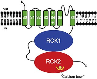
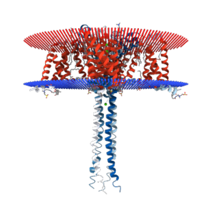
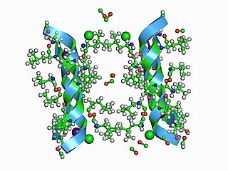
BK channels (big potassium), are large conductance calcium-activated potassium channels, also known as Maxi-K, slo1, or Kca1.1. BK channels are voltage-gated potassium channels that conduct large amounts of potassium ions (K+) across the cell membrane, hence their name, big potassium. These channels can be activated (opened) by either electrical means, or by increasing Ca2+ concentrations in the cell. BK channels help regulate physiological processes, such as circadian behavioral rhythms and neuronal excitability. BK channels are also involved in many processes in the body, as it is a ubiquitous channel. They have a tetrameric structure that is composed of a transmembrane domain, voltage sensing domain, potassium channel domain, and a cytoplasmic C-terminal domain, with many X-ray structures for reference. Their function is to repolarize the membrane potential by allowing for potassium to flow outward, in response to a depolarization or increase in calcium levels.

<span class="mw-page-title-main">Cardiac action potential</span> Biological process in the heart

The cardiac action potential is a brief change in voltage across the cell membrane of heart cells. This is caused by the movement of charged atoms between the inside and outside of the cell, through proteins called ion channels. The cardiac action potential differs from action potentials found in other types of electrically excitable cells, such as nerves. Action potentials also vary within the heart; this is due to the presence of different ion channels in different cells.

Calcium release-activated channels (CRAC) are specialized plasma membrane Ca2+ ion channels. When calcium ions (Ca2+) are depleted from the endoplasmic reticulum (a major store of Ca2+) of mammalian cells, the CRAC channel is activated to slowly replenish the level of calcium in the endoplasmic reticulum. The Ca2+ Release-activated Ca2+ (CRAC) Channel (CRAC-C) Family (TC# 1.A.52) is a member of the Cation Diffusion Facilitator (CDF) Superfamily. These proteins typically have between 4 and 6 transmembrane α-helical spanners (TMSs). The 4 TMS CRAC channels arose by loss of 2TMSs from 6TMS CDF carriers, an example of 'reverse' evolution'.

<span class="mw-page-title-main">Voltage-gated ion channel</span> Type of ion channel transmembrane protein

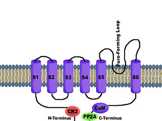
Voltage-gated ion channels are a class of transmembrane proteins that form ion channels that are activated by changes in the electrical membrane potential near the channel. The membrane potential alters the conformation of the channel proteins, regulating their opening and closing. Cell membranes are generally impermeable to ions, thus they must diffuse through the membrane through transmembrane protein channels. They have a crucial role in excitable cells such as neuronal and muscle tissues, allowing a rapid and co-ordinated depolarization in response to triggering voltage change. Found along the axon and at the synapse, voltage-gated ion channels directionally propagate electrical signals. Voltage-gated ion-channels are usually ion-specific, and channels specific to sodium (Na+), potassium (K+), calcium (Ca2+), and chloride (Cl) ions have been identified. The opening and closing of the channels are triggered by changing ion concentration, and hence charge gradient, between the sides of the cell membrane.

<span class="mw-page-title-main">Chloride channel</span> Class of transport proteins

Chloride channels are a superfamily of poorly understood ion channels specific for chloride. These channels may conduct many different ions, but are named for chloride because its concentration in vivo is much higher than other anions. Several families of voltage-gated channels and ligand-gated channels have been characterized in humans.

Voltage-gated calcium channels (VGCCs), also known as voltage-dependent calcium channels (VDCCs), are a group of voltage-gated ion channels found in the membrane of excitable cells (e.g., muscle, glial cells, neurons, etc.) with a permeability to the calcium ion Ca2+. These channels are slightly permeable to sodium ions, so they are also called Ca2+–Na+ channels, but their permeability to calcium is about 1000-fold greater than to sodium under normal physiological conditions.

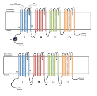
Sodium channels are integral membrane proteins that form ion channels, conducting sodium ions (Na+) through a cell's membrane. They belong to the superfamily of cation channels.

<span class="mw-page-title-main">P2X purinoreceptor</span>

The P2X receptors, also ATP-gated P2X receptor cation channel family, is a protein family that consists of cation-permeable ligand-gated ion channels that open in response to the binding of extracellular adenosine 5'-triphosphate (ATP). They belong to a larger family of receptors known as the ENaC/P2X superfamily. ENaC and P2X receptors have similar 3-D structures and are homologous. P2X receptors are present in a diverse array of organisms including humans, mouse, rat, rabbit, chicken, zebrafish, bullfrog, fluke, and amoeba.

<span class="mw-page-title-main">SK channel</span> Protein subfamily of calcium-activated potassium channels

SK channels are a subfamily of calcium-activated potassium channels. They are so called because of their small single channel conductance in the order of 10 pS. SK channels are a type of ion channel allowing potassium cations to cross the cell membrane and are activated (opened) by an increase in the concentration of intracellular calcium through N-type calcium channels. Their activation limits the firing frequency of action potentials and is important for regulating afterhyperpolarization in the neurons of the central nervous system as well as many other types of electrically excitable cells. This is accomplished through the hyperpolarizing leak of positively charged potassium ions along their concentration gradient into the extracellular space. This hyperpolarization causes the membrane potential to become more negative. SK channels are thought to be involved in synaptic plasticity and therefore play important roles in learning and memory.

<span class="mw-page-title-main">Nicotinic acid adenine dinucleotide phosphate</span> Chemical compound

Nicotinic acid adenine dinucleotide phosphate, (NAADP), is a Ca2+-mobilizing second messenger synthesised in response to extracellular stimuli. Like its mechanistic cousins, IP3 and cyclic adenosine diphosphoribose (Cyclic ADP-ribose), NAADP binds to and opens Ca2+ channels on intracellular organelles, thereby increasing the intracellular Ca2+ concentration which, in turn, modulates sundry cellular processes (see Calcium signalling). Structurally, it is a dinucleotide that only differs from the house-keeping enzyme cofactor, NADP by a hydroxyl group (replacing the nicotinamide amino group) and yet this minor modification converts it into the most potent Ca2+-mobilizing second messenger yet described. NAADP acts across phyla from plants to humans.

<span class="mw-page-title-main">N-type calcium channel</span> Protein family

N-type calcium channels also called Cav2.2 channels are voltage gated calcium channels that are localized primarily on the nerve terminals and dendrites as well as neuroendocrine cells. The calcium N-channel consists of several subunits: the primary subunit α1B and the auxiliary subunits α2δ and β. The α1B subunit forms the pore through which the calcium enters and helps to determine most of the channel's properties. These channels play an important role in the neurotransmission during development. In the adult nervous system, N-type calcium channels are critically involved in the release of neurotransmitters, and in pain pathways. N-type calcium channels are the target of ziconotide, the drug prescribed to relieve intractable cancer pain. There are many known N-type calcium channel blockers that function to inhibit channel activity, although the most notable blockers are ω-conotoxins.

<span class="mw-page-title-main">L-type calcium channel</span> Family of transport proteins

The L-type calcium channel is part of the high-voltage activated family of voltage-dependent calcium channel. "L" stands for long-lasting referring to the length of activation. This channel has four isoforms: Cav1.1, Cav1.2, Cav1.3, and Cav1.4.

Ca<sub>v</sub>1.3 Protein-coding gene in the species Homo sapiens

Calcium channel, voltage-dependent, L type, alpha 1D subunit is a protein that in humans is encoded by the CACNA1D gene. Cav1.3 channels belong to the Cav1 family, which form L-type calcium currents and are sensitive to selective inhibition by dihydropyridines (DHP).

<span class="mw-page-title-main">KCNMB2</span> Protein-coding gene in the species Homo sapiens

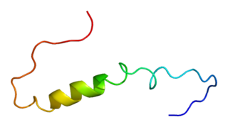
Calcium-activated potassium channel subunit beta-2 is a protein that in humans is encoded by the KCNMB2 gene.

<span class="mw-page-title-main">TPCN2</span> Protein-coding gene in the species Homo sapiens

Two pore segment channel 2 (TPC2) is a protein which in humans is encoded by the TPCN2 gene. TPC2 is an ion channel, however, in contrast to other calcium and sodium channels which have four homologous domains, each containing 6 transmembrane segments, TPCN1 only contains two domain.

Vesicle fusion is the merging of a vesicle with other vesicles or a part of a cell membrane. In the latter case, it is the end stage of secretion from secretory vesicles, where their contents are expelled from the cell through exocytosis. Vesicles can also fuse with other target cell compartments, such as a lysosome. Exocytosis occurs when secretory vesicles transiently dock and fuse at the base of cup-shaped structures at the cell plasma membrane called porosome, the universal secretory machinery in cells. Vesicle fusion may depend on SNARE proteins in the presence of increased intracellular calcium (Ca2+) concentration.

<span class="mw-page-title-main">Gating (electrophysiology)</span>

In electrophysiology, the term gating refers to the opening (activation) or closing of ion channels. This change in conformation is a response to changes in transmembrane voltage.

A calcium channel opener is a type of drug which facilitates ion transmission through calcium channels.

<span class="mw-page-title-main">Gap junction modulation</span>

Gap junction modulation describes the functional manipulation of gap junctions, specialized channels that allow direct electrical and chemical communication between cells without exporting material from the cytoplasm. Gap junctions play an important regulatory role in various physiological processes including signal propagation in cardiac muscles and tissue homeostasis of the liver. Modulation is required, since gap junctions must respond to their environment, whether through an increased expression or permeability. Impaired or altered modulation can have significant health implications and are associated with the pathogenesis of the liver, heart and intestines.

References

  1. 1 2 3 4 5 6 7 Kintzer AF, Stroud RM (March 2016). "Structure, inhibition and regulation of two-pore channel TPC1 from Arabidopsis thaliana". Nature. 531 (7593): 258–62. Bibcode:2016Natur.531..258K. doi:10.1038/nature17194. PMC   4863712 . PMID   26961658. Other than Ca2+ and Na+ channels that are formed by four intramolecular repeats, together forming the tetrameric channel's pore, the new channel had just two Shaker-like repeats, each of which was equipped with one pore domain. Because of this unusual topology, this channel, present in animals as well as plants, was named Two Pore Channel1 (TPC1).
  2. Yu FH, Catterall WA (October 2004). "The VGL-chanome: a protein superfamily specialized for electrical signaling and ionic homeostasis". Science's STKE. 2004 (253): re15. doi:10.1126/stke.2532004re15. PMID   15467096. S2CID   19506706.
  3. 1 2 3 4 Patel S (July 2015). "Function and dysfunction of two-pore channels". Science Signaling. 8 (384): re7. doi:10.1126/scisignal.aab3314. PMID   26152696. S2CID   27822899.
  4. 1 2 3 Marchant JS, Patel S (June 2015). "Two-pore channels at the intersection of endolysosomal membrane traffic". Biochemical Society Transactions. 43 (3): 434–41. doi:10.1042/BST20140303. PMC   4730950 . PMID   26009187.
  5. 1 2 Falasca L, Agrati C, Petrosillo N, Di Caro A, Capobianchi MR, Ippolito G, Piacentini M (August 2015). "Molecular mechanisms of Ebola virus pathogenesis: focus on cell death". Cell Death and Differentiation. 22 (8): 1250–9. doi:10.1038/cdd.2015.67. PMC   4495366 . PMID   26024394.
  6. 1 2 Sakurai Y, Kolokoltsov AA, Chen CC, Tidwell MW, Bauta WE, Klugbauer N, Grimm C, Wahl-Schott C, Biel M, Davey RA (February 2015). "Ebola virus. Two-pore channels control Ebola virus host cell entry and are drug targets for disease treatment". Science. 347 (6225): 995–8. doi:10.1126/science.1258758. PMC   4550587 . PMID   25722412.
  7. 1 2 Grimm C, Holdt LM, Chen CC, Hassan S, Müller C, Jörs S, Cuny H, Kissing S, Schröder B, Butz E, Northoff B, Castonguay J, Luber CA, Moser M, Spahn S, Lüllmann-Rauch R, Fendel C, Klugbauer N, Griesbeck O, Haas A, Mann M, Bracher F, Teupser D, Saftig P, Biel M, Wahl-Schott C (August 2014). "High susceptibility to fatty liver disease in two-pore channel 2-deficient mice". Nature Communications. 5 (2): 4699. Bibcode:2014NatCo...5.4699G. CiteSeerX   10.1.1.659.8695 . doi:10.1038/ncomms5699. PMID   25144390. S2CID   9153781.
  8. Spalding EP, Harper JF (December 2011). "The ins and outs of cellular Ca(2+) transport". Current Opinion in Plant Biology. 14 (6): 715–20. doi:10.1016/j.pbi.2011.08.001. PMC   3230696 . PMID   21865080. The best candidate for a vacuolar Ca2+ release channel is TPC1, a homolog of a mammalian voltage-gated Ca2+ channel that possesses two pores and twelve membrane spans.
  9. Brown BM, Nguyen HM, Wulff H (2019-01-30). "Recent advances in our understanding of the structure and function of more unusual cation channels". F1000Research. 8: 123. doi: 10.12688/f1000research.17163.1 . PMC   6354322 . PMID   30755796. Organellar two-pore channels (TPCs) are an interesting type of channel that, as the name suggests, has two pores.
  10. Jammes F, Hu HC, Villiers F, Bouten R, Kwak JM (November 2011). "Calcium-permeable channels in plant cells". The FEBS Journal. 278 (22): 4262–76. doi: 10.1111/j.1742-4658.2011.08369.x . PMID   21955583. S2CID   205884593. The Arabidopsis two‐pore channel (AtTPC1) has been predicted to have 12 transmembrane helices and two pores (red lines).
  11. Robert Hooper (September 2011). Molecular characterisation of NAADP-gated two-pore channels (PDF) (Thesis). It is believed that TPCs, with their two pores, dimerise to form a functional channel.
  12. "Two P domain potassium channels". Guide to Pharmacology . Retrieved 2019-05-28.
  13. Rang, HP (2003). Pharmacology (8 ed.). Edinburgh: Churchill Livingstone. p. 59. ISBN   978-0-443-07145-4.
  14. Lagostena L, Festa M, Pusch M, Carpaneto A (March 2017). "The human two-pore channel 1 is modulated by cytosolic and luminal calcium". Scientific Reports. 7: 43900. Bibcode:2017NatSR...743900L. doi:10.1038/srep43900. PMC   5333365 . PMID   28252105.
  15. Wang X, Zhang X, Dong XP, Samie M, Li X, Cheng X, Goschka A, Shen D, Zhou Y, Harlow J, Zhu MX, Clapham DE, Ren D, Xu H (October 2012). "TPC proteins are phosphoinositide- activated sodium-selective ion channels in endosomes and lysosomes". Cell. 151 (2): 372–83. doi:10.1016/j.cell.2012.08.036. PMC   3475186 . PMID   23063126.
  16. Vargas E, Bezanilla F, Roux B (December 2011). "In search of a consensus model of the resting state of a voltage-sensing domain". Neuron. 72 (5): 713–20. doi:10.1016/j.neuron.2011.09.024. PMC   3268064 . PMID   22153369.
  17. 1 2 3 Grimm C, Chen CC, Wahl-Schott C, Biel M (2017-01-01). "Two-Pore Channels: Catalyzers of Endolysosomal Transport and Function". Frontiers in Pharmacology. 8: 45. doi: 10.3389/fphar.2017.00045 . PMC   5293812 . PMID   28223936.
  18. 1 2 Galione A (January 2011). "NAADP receptors". Cold Spring Harbor Perspectives in Biology. 3 (1): a004036. doi:10.1101/cshperspect.a004036. PMC   3003455 . PMID   21047915.
  19. Jin X, Zhang Y, Alharbi A, Parrington J (2020). "Targeting Two-Pore Channels: Current Progress and Future Challenges". Trends in Pharmacological Sciences . 41 (8): 582–594. doi:10.1016/j.tips.2020.06.002. PMC   7365084 . PMID   32679067.