CACNA1G

Last updated
CACNA1G
Identifiers
Aliases CACNA1G , Ca(V)T.1, Cav3.1, NBR13, SCA42, calcium voltage-gated channel subunit alpha1 G, SCA42ND
External IDs OMIM: 604065 MGI: 1201678 HomoloGene: 22544 GeneCards: CACNA1G
Orthologs
SpeciesHumanMouse
Entrez
Ensembl
UniProt
RefSeq (mRNA)

NM_001112813
NM_001177888
NM_001177890
NM_009783

RefSeq (protein)

n/a

Location (UCSC) Chr 17: 50.56 – 50.63 Mb Chr 11: 94.3 – 94.37 Mb
PubMed search [3] [4]
Wikidata
View/Edit Human View/Edit Mouse

Calcium channel, voltage-dependent, T type, alpha 1G subunit, also known as CACNA1G or Cav3.1 is a protein which in humans is encoded by the CACNA1G gene. [5] [6] [7] It is one of the primary targets in the pharmacology of absence seizure.

Function

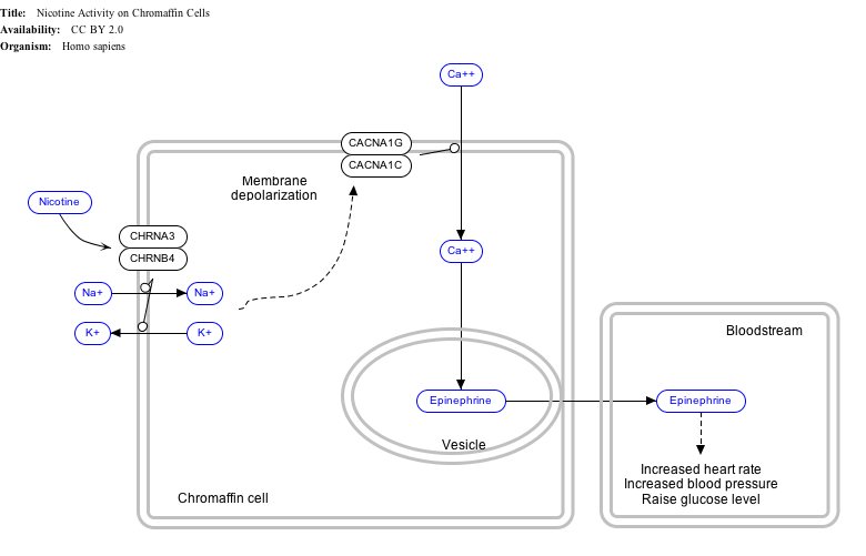
Cav3.1 is a type of low-voltage-activated calcium channel, also known as "T-type" for its transient on and off. [5] It is expressed in thalamocortical relay nucleus, and is responsible for the slow-wave sleep and absence seizure. [8] During a slow-wave sleep, Cav3.1 is put into burst mode, and a self-sustaining synchronous cycle between cortex and thalamus is formed, sensory inputs are isolated from cortex; while awake the thalamus should instead relay sensory inputs from outside the central nervous system. The mechanism of absence seizure has a lot in common with slow-wave sleep. Therefore, a blocker that inhibits the burst mode activation of Cav3.1 is effective in treating absence seizures. Common drugs including ethosuximide, as well as trimethadione. [8]


Interactive pathway map

Click on genes, proteins and metabolites below to link to respective Wikipedia articles. [§ 1]

[[File:
NicotineChromaffinCells WP1603.png Go to articlego to articleGo to articleGo to articleGo to articleGo to articlego to articlego to articleGo to articleGo to articlego to articleGo to articleGo to article
[[
]]
[[
]]
[[
]]
[[
]]
[[
]]
[[
]]
[[
]]
[[
]]
[[
]]
[[
]]
[[
]]
[[
]]
[[
]]
[[
]]
NicotineChromaffinCells WP1603.png Go to articlego to articleGo to articleGo to articleGo to articleGo to articlego to articlego to articleGo to articleGo to articlego to articleGo to articleGo to article
|alt=Nicotine Activity on Chromaffin Cells edit]]
Nicotine Activity on Chromaffin Cells edit
  1. The interactive pathway map can be edited at WikiPathways: "NicotineActivityonChromaffinCells_WP1603".

See also

Related Research Articles

Voltage-gated calcium channels (VGCCs), also known as voltage-dependent calcium channels (VDCCs), are a group of voltage-gated ion channels found in the membrane of excitable cells (e.g., muscle, glial cells, neurons, etc.) with a permeability to the calcium ion Ca2+. These channels are slightly permeable to sodium ions, so they are also called Ca2+-Na+ channels, but their permeability to calcium is about 1000-fold greater than to sodium under normal physiological conditions.

Ca<sub>v</sub>1.2

Calcium channel, voltage-dependent, L type, alpha 1C subunit is a protein that in humans is encoded by the CACNA1C gene. Cav1.2 is a subunit of L-type voltage-dependent calcium channel.

<span class="mw-page-title-main">Childhood absence epilepsy</span> Neurological disorder causing absence seizures

Childhood absence epilepsy (CAE), is one of the most frequent pediatric epilepsy syndromes. CAE is an idiopathic generalized epilepsy that occurs in otherwise normal children. The only seizure type at the time of diagnosis is the typical absence seizure. CAE is a well-known pediatric epilepsy syndrome affecting 10–17% of all children with epilepsy. It was previously known as pyknolepsy. The word pyknolepsy originates from the Greek piknoz (picnós), which means recurrent or grouped. The usual age of onset of CAE is between 4 and 10 years, with peak between 5 and 7 years.

T-type calcium channels are low voltage activated calcium channels that become inactivated during cell membrane hyperpolarization but then open to depolarization. The entry of calcium into various cells has many different physiological responses associated with it. Within cardiac muscle cell and smooth muscle cells voltage-gated calcium channel activation initiates contraction directly by allowing the cytosolic concentration to increase. Not only are T-type calcium channels known to be present within cardiac and smooth muscle, but they also are present in many neuronal cells within the central nervous system. Different experimental studies within the 1970s allowed for the distinction of T-type calcium channels from the already well-known L-type calcium channels. The new T-type channels were much different from the L-type calcium channels due to their ability to be activated by more negative membrane potentials, had small single channel conductance, and also were unresponsive to calcium antagonist drugs that were present. These distinct calcium channels are generally located within the brain, peripheral nervous system, heart, smooth muscle, bone, and endocrine system.

The R-type calcium channel is a type of voltage-dependent calcium channel. Like the others of this class, the α1 subunit forms the pore through which calcium enters the cell and determines most of the channel's properties. This α1 subunit is also known as the calcium channel, voltage-dependent, R type, alpha 1E subunit (CACNA1E) or Cav2.3 which in humans is encoded by the CACNA1E gene. They are strongly expressed in cortex, hippocampus, striatum, amygdala and interpeduncular nucleus.

The P-type calcium channel is a type of voltage-dependent calcium channel. Similar to many other high-voltage-gated calcium channels, the α1 subunit determines most of the channel's properties. The 'P' signifies cerebellar Purkinje cells, referring to the channel's initial site of discovery. P-type calcium channels play a similar role to the N-type calcium channel in neurotransmitter release at the presynaptic terminal and in neuronal integration in many neuronal types.

<span class="mw-page-title-main">L-type calcium channel</span> Family of transport proteins

The L-type calcium channel is part of the high-voltage activated family of voltage-dependent calcium channel. "L" stands for long-lasting referring to the length of activation. This channel has four isoforms: Cav1.1, Cav1.2, Cav1.3, and Cav1.4.

Ca<sub>v</sub>2.1 Protein-coding gene in the species Homo sapiens

Cav2.1, also called the P/Q voltage-dependent calcium channel, is a calcium channel found mainly in the brain. Specifically, it is found on the presynaptic terminals of neurons in the brain and cerebellum. Cav2.1 plays an important role in controlling the release of neurotransmitters between neurons. It is composed of multiple subunits, including alpha-1, beta, alpha-2/delta, and gamma subunits. The alpha-1 subunit is the pore-forming subunit, meaning that the calcium ions flow through it. Different kinds of calcium channels have different isoforms (versions) of the alpha-1 subunit. Cav2.1 has the alpha-1A subunit, which is encoded by the CACNA1A gene. Mutations in CACNA1A have been associated with various neurologic disorders, including familial hemiplegic migraine, episodic ataxia type 2, and spinocerebellar ataxia type 6.

Ca<sub>v</sub>1.4 Protein-coding gene in the species Homo sapiens

Cav1.4 also known as the calcium channel, voltage-dependent, L type, alpha 1F subunit (CACNA1F), is a human gene.

Ca<sub>v</sub>1.1 Mammalian protein found in Homo sapiens

Cav1.1 also known as the calcium channel, voltage-dependent, L type, alpha 1S subunit, (CACNA1S), is a protein which in humans is encoded by the CACNA1S gene. It is also known as CACNL1A3 and the dihydropyridine receptor.

<span class="mw-page-title-main">CACNB4</span>

Voltage-dependent L-type calcium channel subunit beta-4 is a protein that in humans is encoded by the CACNB4 gene.

<span class="mw-page-title-main">CACNG2</span>

Calcium channel, voltage-dependent, gamma subunit 2, also known as CACNG2 or stargazin is a protein that in humans is encoded by the CACNG2 gene.

Ca<sub>v</sub>1.3

Calcium channel, voltage-dependent, L type, alpha 1D subunit is a protein that in humans is encoded by the CACNA1D gene. Cav1.3 channels belong to the Cav1 family, which form L-type calcium currents and are sensitive to selective inhibition by dihydropyridines (DHP).

<span class="mw-page-title-main">KCNMB2</span>

Calcium-activated potassium channel subunit beta-2 is a protein that in humans is encoded by the KCNMB2 gene.

<span class="mw-page-title-main">CACNA2D1</span>

Voltage-dependent calcium channel subunit alpha-2/delta-1 is a protein that in humans is encoded by the CACNA2D1 gene.

<span class="mw-page-title-main">CACNG3</span>

Voltage-dependent calcium channel gamma-3 subunit is a protein that in humans is encoded by the CACNG3 gene.

<span class="mw-page-title-main">Calcium channel, voltage-dependent, T type, alpha 1H subunit</span>

Calcium channel, voltage-dependent, T type, alpha 1H subunit, also known as CACNA1H, is a protein which in humans is encoded by the CACNA1H gene.

<span class="mw-page-title-main">CACNA2D2</span>

Voltage-dependent calcium channel subunit alpha2delta-2 is a protein that in humans is encoded by the CACNA2D2 gene.

<span class="mw-page-title-main">CACNA1I</span>

Calcium channel, voltage-dependent, T type, alpha 1I subunit, also known as CACNA1I or Cav3.3 is a protein which in humans is encoded by the CACNA1I gene.

<span class="mw-page-title-main">CACNA1B</span> Protein-coding gene in the species Homo sapiens

The voltage-dependent N-type calcium channel subunit alpha-1B is a protein that in humans is encoded by the CACNA1B gene. The α1B protein, together with β and α2δ subunits forms N-type calcium channel PMID 26386135. It is a R-type calcium channel.

References

  1. 1 2 3 GRCh38: Ensembl release 89: ENSG00000006283 - Ensembl, May 2017
  2. 1 2 3 GRCm38: Ensembl release 89: ENSMUSG00000020866 - Ensembl, May 2017
  3. "Human PubMed Reference:". National Center for Biotechnology Information, U.S. National Library of Medicine.
  4. "Mouse PubMed Reference:". National Center for Biotechnology Information, U.S. National Library of Medicine.
  5. 1 2 "Entrez Gene: CACNA1H calcium channel, voltage-dependent, T type, alpha 1H subunit".
  6. Perez-Reyes E, Cribbs LL, Daud A, Lacerda AE, Barclay J, Williamson MP, Fox M, Rees M, Lee JH (February 1998). "Molecular characterization of a neuronal low-voltage-activated T-type calcium channel". Nature. 391 (6670): 896–900. Bibcode:1998Natur.391..896P. doi:10.1038/36110. PMID   9495342. S2CID   4373283.
  7. Catterall WA, Perez-Reyes E, Snutch TP, Striessnig J (December 2005). "International Union of Pharmacology. XLVIII. Nomenclature and structure-function relationships of voltage-gated calcium channels". Pharmacol. Rev. 57 (4): 411–25. doi:10.1124/pr.57.4.5. PMID   16382099. S2CID   10386627.
  8. 1 2 Kopecky, Benjamin J.; Liang, Ruqiang; Bao, Jianxin (2014). "T-type Calcium Channel Blockers as Neuroprotective Agents". Pflügers Archiv. 466 (4): 757–765. doi:10.1007/s00424-014-1454-x. ISSN   0031-6768. PMC   4005039 . PMID   24563219.

This article incorporates text from the United States National Library of Medicine, which is in the public domain.