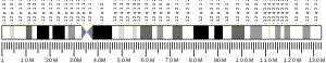
The gene is located on the Watson (plus) strand of the short arm of chromosome 12 (12p13.32). The gene itself is 8,348 bases in length and encodes a protein of 495 amino acids (predicted molecular weight 56.466 kilodaltons).
Alternative names
The recommended name for this protein is potassium voltage-gated channel subfamily A member 1 but a number of alternatives have been used in the literature including HuK1 (human K+ channel I), RBK1 (rubidium potassium channel 1), MBK (mouse brain K+ channel), voltage gated potassium channel HBK1, voltage gated potassium channel subunit Kv1.1, voltage-gated K+ channel HuKI and AEMK (associated with myokymia with periodic ataxia).
Structure
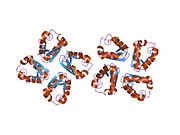
The protein is believed to have six domains (S1-S6) with the loop between S5 and S6 forming the channel pore. This region also has a conserved selectivity filter motif. The functional channel is a homotetramer. The N-terminus of the protein associates with β subunits. These subunits regulate channel inactivation as well as its expression. The C-terminus is associated with a PDZ domain protein involved in channel targeting.[9][10]
Function
The protein functions as a potassium selective channel through which the potassium ion may pass in consensus with the electrochemical gradient. They play a role in repolarisation of membranes.[9]
A to I RNA editing is catalyzed by a family of adenosine deaminases acting on RNA (ADARs) that specifically recognize adenosines within double-stranded regions of pre-mRNAs (e.g. Potassium channel RNA editing signal) and deaminate them to inosine. Inosines are recognised as guanosine by the cells translational machinery. There are three members of the ADAR family ADARs 1-3 with ADAR1 and ADAR2 being the only enzymatically active members. ADAR3 is thought to have a regulatory role in the brain. ADAR1 and ADAR2 are widely expressed in tissues while ADAR3 is restricted to the brain. The double stranded regions of RNA are formed by base-pairing between residues in the region close to the editing site with residues usually in a neighboring intron but can sometimes be an exonic sequence too. The region that base pairs with the editing region is known as an Editing Complementary Sequence (ECS).
Location
The modified residue is found at amino acid 400 of the final protein. This is located in the sixth transmembrane region found, which corresponds to the inner vestibule of the pore. A stem loop hairpin structure mediates the RNA editing. ADAR2 is likely to be the preferred editing enzyme at the I/V site. Editing results in a codon alteration from ATT to GTT, resulting in an amino acid change from isoleucine to valine. ADAR2 enzyme is the major editing enzyme. The MFOLD programme predicted that the minimum region required for editing would form an imperfect inverted repeat hairpin. This region is composed of a 114 base pairs. Similar regions have been identified in mouse and rat. The edited adenosine is found in a 6-base pair duplex region. Mutation experiment in the region near the 6-base pair duplex have shown that the specific bases in this region were also essential for editing to occur. The region required for editing is unusual in that the hairpin structure is formed by exonic sequences only. In the majority of A to I editing the ECS is found within an intronic sequence.[11]
Conservation
The editing is highly conserved having been observed in squid, fruit fly, mouse, and rat.[11]
Editing results in a codon (I/V) change from (ATT) to (GTT) resulting in translation of a valine instead of an isoleucine at the position of the editing site. Valine has a larger side-chain. RNA editing at this position occurs at a highly conserved ion conducting pore of the channel. This may affect the channels role in the process of fast inactivation.[13]
Function
Voltage-dependent potassium channels modulate excitability by opening and closing a potassium selective pore in response to voltage. The flow of potassium ions is interrupted by interaction of an inactivating particle, an auxiliary protein in humans but an intrinsic part of the channel in other species. The I to V amino acid change is thought to disrupt the hydrophobic interaction between the inactivating particle and the pore lining. This interrupts the process of fast inactivation. Activation kinetics are unaffected by RNA editing.[11] Changes in inactivation kinetics affect the duration and frequency of the action potential. An edited channel passes more current and has a shorter action potential than the non-edited type due to the inability of the inactivating particle to interact with the residue in the ion-conducting pore of the channel. This was determined by electrophysiology analysis.[14] The length of time the membrane is depolarised is decreased, which also reduces the efficiency of transmitter release.[12] Since editing can cause amino acid changes in 1- 4 in potassium channel tetramers, it can have a wide variety of effects on channel inactivation.
Dysregulation
Changes in the process of fast inactivation are known to have behavioral and neurological consequences in vivo.[11]
↑ "Human PubMed Reference:". National Center for Biotechnology Information, U.S. National Library of Medicine.
↑ "Mouse PubMed Reference:". National Center for Biotechnology Information, U.S. National Library of Medicine.
↑ Curran ME, Landes GM, Keating MT (April 1992). "Molecular cloning, characterization, and genomic localization of a human potassium channel gene". Genomics. 12 (4): 729–737. doi:10.1016/0888-7543(92)90302-9. PMID1349297.
↑ Albrecht B, Weber K, Pongs O (1995). "Characterization of a voltage-activated K-channel gene cluster on human chromosome 12p13". Receptors & Channels. 3 (3): 213–220. PMID8821794.
↑ Gutman GA, Chandy KG, Grissmer S, Lazdunski M, McKinnon D, Pardo LA, etal. (December 2005). "International Union of Pharmacology. LIII. Nomenclature and molecular relationships of voltage-gated potassium channels". Pharmacological Reviews. 57 (4): 473–508. doi:10.1124/pr.57.4.10. PMID16382104. S2CID219195192.
1 2 3 4 5 Bhalla T, Rosenthal JJ, Holmgren M, Reenan R (October 2004). "Control of human potassium channel inactivation by editing of a small mRNA hairpin". Nature Structural & Molecular Biology. 11 (10): 950–956. doi:10.1038/nsmb825. PMID15361858. S2CID34081059.
↑ Bhalla T, Rosenthal JJ, Holmgren M, Reenan R (October 2004). "Control of human potassium channel inactivation by editing of a small mRNA hairpin". Nature Structural & Molecular Biology. 11 (10): 950–956. doi:10.1038/nsmb825. PMID15361858. S2CID34081059.
Imbrici P, Cusimano A, D'Adamo MC, De Curtis A, Pessia M (June 2003). "Functional characterization of an episodic ataxia type-1 mutation occurring in the S1 segment of hKv1.1 channels". Pflugers Archiv. 446 (3): 373–379. doi:10.1007/s00424-002-0962-2. PMID12799903. S2CID21478393.
Kinali M, Jungbluth H, Eunson LH, Sewry CA, Manzur AY, Mercuri E, etal. (October 2004). "Expanding the phenotype of potassium channelopathy: severe neuromyotonia and skeletal deformities without prominent Episodic Ataxia". Neuromuscular Disorders. 14 (10): 689–693. doi:10.1016/j.nmd.2004.06.007. PMID15351427. S2CID44972020.
Demos MK, Macri V, Farrell K, Nelson TN, Chapman K, Accili E, etal. (April 2009). "A novel KCNA1 mutation associated with global delay and persistent cerebellar dysfunction". Movement Disorders. 24 (5): 778–782. doi:10.1002/mds.22467. PMID19205071. S2CID25655998.
Imbrici P, Gualandi F, D'Adamo MC, Masieri MT, Cudia P, De Grandis D, etal. (December 2008). "A novel KCNA1 mutation identified in an Italian family affected by episodic ataxia type 1". Neuroscience. 157 (3): 577–587. doi:10.1016/j.neuroscience.2008.09.022. PMID18926884. S2CID15772885.
This page is based on this Wikipedia article Text is available under the CC BY-SA 4.0 license; additional terms may apply. Images, videos and audio are available under their respective licenses.