Calcium-activated potassium channel subunit alpha-1

Last updated
KCNMA1
BK-cartoon wp.jpg
Available structures
PDB Ortholog search: PDBe RCSB
Identifiers
Aliases KCNMA1 , BKTM, KCa1.1, MaxiK, SAKCA, SLO, SLO-ALPHA, SLO1, bA205K10.1, mSLO1, hSlo, potassium calcium-activated channel subfamily M alpha 1, CADEDS, PNKD3, IEG16, LIWAS
External IDs OMIM: 600150 MGI: 99923 HomoloGene: 1693 GeneCards: KCNMA1
Orthologs
SpeciesHumanMouse
Entrez
Ensembl
UniProt
RefSeq (mRNA)
RefSeq (protein)
Location (UCSC) Chr 10: 76.87 – 77.64 Mb Chr 14: 23.34 – 24.06 Mb
PubMed search [3] [4]
Wikidata
View/Edit Human View/Edit Mouse

Calcium-activated potassium channel subunit alpha-1 also known as large conductance calcium-activated potassium channel, subfamily M, alpha member 1 (KCa1.1), or BK channel alpha subunit, [5] is a voltage gated potassium channel encoded by the KCNMA1 gene and characterized by their large conductance of potassium ions (K+) through cell membranes. [6]

Function

BK channels are activated (opened) by changes in membrane electrical potential and/or by increases in concentration of intracellular calcium ion (Ca2+). [7] [8] Opening of BK channels allows K+ to passively flow through the channel, down the electrochemical gradient. Under typical physiological conditions, this results in an efflux of K+ from the cell, which leads to cell membrane hyperpolarization (a decrease in the electrical potential across the cell membrane) and a decrease in cell excitability (a decrease in the probability that the cell will transmit an action potential).

BK channels are essential for the regulation of several key physiological processes including smooth muscle tone and neuronal excitability. [6] They control the contraction of smooth muscle and are involved with the electrical tuning of hair cells in the cochlea. BK channels also contribute to the behavioral effects of ethanol in the worm C. elegans under high concentrations (> 100 mM, or approximately 0.50% BAC). [9] It remains to be determined if BK channels contribute to intoxication in humans.

Structure

BK channels have a tetrameric structure. Each monomer of the channel-forming alpha subunit is the product of the KCNMA1 gene. Modulatory beta subunits (encoded by KCNMB1, KCNMB2, KCNMB3, or KCNMB4) can associate with the tetrameric channel. Alternatively spliced transcript variants encoding different isoforms have been identified. [6]

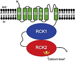
Each BK channel alpha subunit consists of (from N- to C-terminal):

  1. A unique transmembrane domain (S0) [10] that precedes the 6 transmembrane domains (S1-S6) conserved in all voltage-dependent K+ channels.
  2. A voltage sensing domain (S1-S4).
  3. A K+ channel pore domain (S5, selectivity filter, and S6).
  4. A cytoplasmic C-terminal domain (CTD) consisting of a pair of RCK domains that assemble into an octameric gating ring on the intracellular side of the tetrameric channel. [8] [11] [12] [13] [14] [15] [16] The CTD contains four primary binding sites for Ca2+, called "calcium bowls", encoded within the second RCK domain of each monomer. [8] [11] [15] [16]
Calcium-activated BK potassium channel alpha subunit
Identifiers
SymbolBK_channel_a
Pfam PF03493
InterPro IPR003929
Available protein structures:
Pfam   structures / ECOD  
PDB RCSB PDB; PDBe; PDBj
PDBsum structure summary

Available X-ray structures include:

Pharmacology

BK channels are pharmacological targets for the treatment of stroke. Various pharmaceutical companies developed synthetic molecules activating these channels [17] in order to prevent excessive neurotoxic calcium entry in neurons. [18] But BMS-204352 (MaxiPost) a molecule developed by Bristol-Myers Squibb failed to improve clinical outcome in stroke patients compared to placebo. [19] BK channels have also been found to be activated by exogenous pollutants and endogenous gasotransmitters carbon monoxide [20] [21] and hydrogen sulphide. [22]

BK channels are blocked by tetraethylammonium (TEA), paxilline [23] and iberiotoxin. [24]

Researchers have identified a rare disease in humans caused by mutations in the gene.  KCNMA1-linked channelopathy can cause neurological conditions like seizures and movement disorders. [25] An episode of the Diagnosis TV show, based on a column in the New York Times, was about a young girl with a KCNMA1 disorder that caused transient episodes of muscle weakness. [26]

See also

Related Research Articles

<span class="mw-page-title-main">Ion channel</span> Pore-forming membrane protein

Ion channels are pore-forming membrane proteins that allow ions to pass through the channel pore. Their functions include establishing a resting membrane potential, shaping action potentials and other electrical signals by gating the flow of ions across the cell membrane, controlling the flow of ions across secretory and epithelial cells, and regulating cell volume. Ion channels are present in the membranes of all cells. Ion channels are one of the two classes of ionophoric proteins, the other being ion transporters.

<span class="mw-page-title-main">BK channel</span> Family of transport proteins

BK channels (big potassium), are large conductance calcium-activated potassium channels, also known as Maxi-K, slo1, or Kca1.1. BK channels are voltage-gated potassium channels that conduct large amounts of potassium ions (K+) across the cell membrane, hence their name, big potassium. These channels can be activated (opened) by either electrical means, or by increasing Ca2+ concentrations in the cell. BK channels help regulate physiological processes, such as circadian behavioral rhythms and neuronal excitability. BK channels are also involved in many processes in the body, as it is a ubiquitous channel. They have a tetrameric structure that is composed of a transmembrane domain, voltage sensing domain, potassium channel domain, and a cytoplasmic C-terminal domain, with many X-ray structures for reference. Their function is to repolarize the membrane potential by allowing for potassium to flow outward, in response to a depolarization or increase in calcium levels.

<span class="mw-page-title-main">Voltage-gated ion channel</span> Type of ion channel transmembrane protein

Voltage-gated ion channels are a class of transmembrane proteins that form ion channels that are activated by changes in the electrical membrane potential near the channel. The membrane potential alters the conformation of the channel proteins, regulating their opening and closing. Cell membranes are generally impermeable to ions, thus they must diffuse through the membrane through transmembrane protein channels. They have a crucial role in excitable cells such as neuronal and muscle tissues, allowing a rapid and co-ordinated depolarization in response to triggering voltage change. Found along the axon and at the synapse, voltage-gated ion channels directionally propagate electrical signals. Voltage-gated ion-channels are usually ion-specific, and channels specific to sodium (Na+), potassium (K+), calcium (Ca2+), and chloride (Cl) ions have been identified. The opening and closing of the channels are triggered by changing ion concentration, and hence charge gradient, between the sides of the cell membrane.

Voltage-gated calcium channels (VGCCs), also known as voltage-dependent calcium channels (VDCCs), are a group of voltage-gated ion channels found in the membrane of excitable cells (e.g., muscle, glial cells, neurons, etc.) with a permeability to the calcium ion Ca2+. These channels are slightly permeable to sodium ions, so they are also called Ca2+–Na+ channels, but their permeability to calcium is about 1000-fold greater than to sodium under normal physiological conditions.

Calcium-activated potassium channels are potassium channels gated by calcium, or that are structurally or phylogenetically related to calcium gated channels. They were first discovered in 1958 by Gardos who saw that calcium levels inside of a cell could affect the permeability of potassium through that cell membrane. Then in 1970, Meech was the first to observe that intracellular calcium could trigger potassium currents. In humans they are divided into three subtypes: large conductance or BK channels, which have very high conductance which range from 100 to 300 pS, intermediate conductance or IK channels, with intermediate conductance ranging from 25 to 100 pS, and small conductance or SK channels with small conductances from 2-25 pS.

Ca<sub>v</sub>1.2 Protein-coding gene in humans

Calcium channel, voltage-dependent, L type, alpha 1C subunit is a protein that in humans is encoded by the CACNA1C gene. Cav1.2 is a subunit of L-type voltage-dependent calcium channel.

<span class="mw-page-title-main">SK channel</span> Protein subfamily of calcium-activated potassium channels

SK channels are a subfamily of calcium-activated potassium channels. They are so called because of their small single channel conductance in the order of 10 pS. SK channels are a type of ion channel allowing potassium cations to cross the cell membrane and are activated (opened) by an increase in the concentration of intracellular calcium through N-type calcium channels. Their activation limits the firing frequency of action potentials and is important for regulating afterhyperpolarization in the neurons of the central nervous system as well as many other types of electrically excitable cells. This is accomplished through the hyperpolarizing leak of positively charged potassium ions along their concentration gradient into the extracellular space. This hyperpolarization causes the membrane potential to become more negative. SK channels are thought to be involved in synaptic plasticity and therefore play important roles in learning and memory.

Ca<sub>v</sub>2.1 Protein-coding gene in the species Homo sapiens

Cav2.1, also called the P/Q voltage-dependent calcium channel, is a calcium channel found mainly in the brain. Specifically, it is found on the presynaptic terminals of neurons in the brain and cerebellum. Cav2.1 plays an important role in controlling the release of neurotransmitters between neurons. It is composed of multiple subunits, including alpha-1, beta, alpha-2/delta, and gamma subunits. The alpha-1 subunit is the pore-forming subunit, meaning that the calcium ions flow through it. Different kinds of calcium channels have different isoforms (versions) of the alpha-1 subunit. Cav2.1 has the alpha-1A subunit, which is encoded by the CACNA1A gene. Mutations in CACNA1A have been associated with various neurologic disorders, including familial hemiplegic migraine, episodic ataxia type 2, and spinocerebellar ataxia type 6.

<span class="mw-page-title-main">Potassium channel tetramerisation domain</span>

K+ channel tetramerisation domain is the N-terminal, cytoplasmic tetramerisation domain (T1) of voltage-gated K+ channels. It defines molecular determinants for subfamily-specific assembly of alpha-subunits into functional tetrameric channels. It is distantly related to the BTB/POZ domain Pfam PF00651.

<span class="mw-page-title-main">KCNMB1</span> Protein-coding gene in the species Homo sapiens

Calcium-activated potassium channel subunit beta-1 is a protein that in humans is encoded by the KCNMB1 gene.

<span class="mw-page-title-main">KCNB1</span> Protein-coding gene in the species Homo sapiens

Potassium voltage-gated channel, Shab-related subfamily, member 1, also known as KCNB1 or Kv2.1, is a protein that, in humans, is encoded by the KCNB1 gene.

<span class="mw-page-title-main">CACNB4</span> Protein-coding gene in the species Homo sapiens

Voltage-dependent L-type calcium channel subunit beta-4 is a protein that in humans is encoded by the CACNB4 gene.

Ca<sub>v</sub>1.3 Protein-coding gene in the species Homo sapiens

Calcium channel, voltage-dependent, L type, alpha 1D subunit is a protein that in humans is encoded by the CACNA1D gene. Cav1.3 channels belong to the Cav1 family, which form L-type calcium currents and are sensitive to selective inhibition by dihydropyridines (DHP).

<span class="mw-page-title-main">KCNMB2</span> Protein-coding gene in the species Homo sapiens

Calcium-activated potassium channel subunit beta-2 is a protein that in humans is encoded by the KCNMB2 gene.

<span class="mw-page-title-main">KCNMB3</span> Protein-coding gene in the species Homo sapiens

Calcium-activated potassium channel subunit beta-3 is a protein that in humans is encoded by the KCNMB3 gene.

<span class="mw-page-title-main">KCNN2</span> Protein-coding gene in the species Homo sapiens

Potassium intermediate/small conductance calcium-activated channel, subfamily N, member 2, also known as KCNN2, is a protein which in humans is encoded by the KCNN2 gene. KCNN2 is an ion channel protein also known as KCa2.2.

<span class="mw-page-title-main">KCNN1</span> Protein-coding gene in the species Homo sapiens

Potassium intermediate/small conductance calcium-activated channel, subfamily N, member 1 , also known as KCNN1 is a human gene encoding the KCa2.1 protein.

<span class="mw-page-title-main">KCNMB4</span> Protein-coding gene in humans

Calcium-activated potassium channel subunit beta-4 is a protein that in humans is encoded by the KCNMB4 gene.

<span class="mw-page-title-main">Isopimaric acid</span> Chemical compound

Isopimaric acid (IPA) is a toxin which acts as a large conductance Ca2+-activated K+ channel (BK channel) opener.

<span class="mw-page-title-main">Calcium-activated potassium channel beta subunit</span>

In molecular biology, the calcium-activated potassium channel beta subunit is a family of proteins comprising the beta subunits of calcium-activated potassium channels.

References

  1. 1 2 3 GRCh38: Ensembl release 89: ENSG00000156113 - Ensembl, May 2017
  2. 1 2 3 GRCm38: Ensembl release 89: ENSMUSG00000063142 - Ensembl, May 2017
  3. "Human PubMed Reference:". National Center for Biotechnology Information, U.S. National Library of Medicine.
  4. "Mouse PubMed Reference:". National Center for Biotechnology Information, U.S. National Library of Medicine.
  5. "HomoloGene - NCBI". www.ncbi.nlm.nih.gov.
  6. 1 2 3 "Entrez Gene: KCNMA1 potassium large conductance calcium-activated channel, subfamily M, alpha member 1".
  7. Miller C (2000). "An overview of the potassium channel family". Genome Biology. 1 (4): REVIEWS0004. doi: 10.1186/gb-2000-1-4-reviews0004 . PMC   138870 . PMID   11178249.
  8. 1 2 3 4 Yuan P, Leonetti MD, Pico AR, Hsiung Y, MacKinnon R (July 2010). "Structure of the human BK channel Ca2+-activation apparatus at 3.0 A resolution". Science. 329 (5988): 182–6. Bibcode:2010Sci...329..182Y. doi:10.1126/science.1190414. PMC   3022345 . PMID   20508092.
  9. Davies AG, Pierce-Shimomura JT, Kim H, VanHoven MK, Thiele TR, Bonci A, et al. (December 2003). "A central role of the BK potassium channel in behavioral responses to ethanol in C. elegans". Cell. 115 (6): 655–66. doi: 10.1016/S0092-8674(03)00979-6 . PMID   14675531. S2CID   8120562.
  10. Wallner M, Meera P, Toro L (December 1996). "Determinant for beta-subunit regulation in high-conductance voltage-activated and Ca(2+)-sensitive K+ channels: an additional transmembrane region at the N terminus". Proceedings of the National Academy of Sciences of the United States of America. 93 (25): 14922–7. Bibcode:1996PNAS...9314922W. doi: 10.1073/pnas.93.25.14922 . PMC   26238 . PMID   8962157.
  11. 1 2 3 Wu Y, Yang Y, Ye S, Jiang Y (July 2010). "Structure of the gating ring from the human large-conductance Ca(2+)-gated K(+) channel". Nature. 466 (7304): 393–7. Bibcode:2010Natur.466..393W. doi:10.1038/nature09252. PMC   2910425 . PMID   20574420.
  12. Jiang Y, Pico A, Cadene M, Chait BT, MacKinnon R (March 2001). "Structure of the RCK domain from the E. coli K+ channel and demonstration of its presence in the human BK channel". Neuron. 29 (3): 593–601. doi: 10.1016/S0896-6273(01)00236-7 . PMID   11301020. S2CID   17880955.
  13. Pico A. 2003. RCK domain model of calcium activation in BK channels. PhD thesis. The Rockfeller University, New York.
  14. Yusifov T, Savalli N, Gandhi CS, Ottolia M, Olcese R (January 2008). "The RCK2 domain of the human BKCa channel is a calcium sensor". Proceedings of the National Academy of Sciences of the United States of America. 105 (1): 376–81. Bibcode:2008PNAS..105..376Y. doi: 10.1073/pnas.0705261105 . PMC   2224220 . PMID   18162557.
  15. 1 2 Schreiber M, Salkoff L (September 1997). "A novel calcium-sensing domain in the BK channel". Biophysical Journal. 73 (3): 1355–63. Bibcode:1997BpJ....73.1355S. doi:10.1016/S0006-3495(97)78168-2. PMC   1181035 . PMID   9284303.
  16. 1 2 3 Yuan P, Leonetti MD, Hsiung Y, MacKinnon R (December 2011). "Open structure of the Ca2+ gating ring in the high-conductance Ca2+-activated K+ channel". Nature. 481 (7379): 94–7. Bibcode:2012Natur.481...94Y. doi:10.1038/nature10670. PMC   3319005 . PMID   22139424.
  17. Gribkoff VK, Winquist RJ (May 2005). "Voltage-gated cation channel modulators for the treatment of stroke". Expert Opinion on Investigational Drugs. 14 (5): 579–92. doi:10.1517/13543784.14.5.579. PMID   15926865. S2CID   10236998.
  18. Gribkoff VK, Starrett JE, Dworetzky SI (April 2001). "Maxi-K potassium channels: form, function, and modulation of a class of endogenous regulators of intracellular calcium". The Neuroscientist. 7 (2): 166–77. doi:10.1177/107385840100700211. PMID   11496927. S2CID   8791803.
  19. Jensen BS (2002). "BMS-204352: a potassium channel opener developed for the treatment of stroke". CNS Drug Reviews. 8 (4): 353–60. doi:10.1111/j.1527-3458.2002.tb00233.x. PMC   6741660 . PMID   12481191.
  20. Dubuis E, Potier M, Wang R, Vandier C (February 2005). "Continuous inhalation of carbon monoxide attenuates hypoxic pulmonary hypertension development presumably through activation of BKCa channels". Cardiovascular Research. 65 (3): 751–61. doi: 10.1016/j.cardiores.2004.11.007 . PMID   15664403.
  21. Hou S, Xu R, Heinemann SH, Hoshi T (March 2008). "The RCK1 high-affinity Ca2+ sensor confers carbon monoxide sensitivity to Slo1 BK channels". Proceedings of the National Academy of Sciences of the United States of America. 105 (10): 4039–43. Bibcode:2008PNAS..105.4039H. doi: 10.1073/pnas.0800304105 . PMC   2268785 . PMID   18316727.
  22. Sitdikova GF, Weiger TM, Hermann A (February 2010). "Hydrogen sulfide increases calcium-activated potassium (BK) channel activity of rat pituitary tumor cells". Pflügers Archiv. 459 (3): 389–97. doi:10.1007/s00424-009-0737-0. PMID   19802723. S2CID   23073556.
  23. "Paxilline, from Fermentek".
  24. Candia S, Garcia ML, Latorre R (August 1992). "Mode of action of iberiotoxin, a potent blocker of the large conductance Ca(2+)-activated K+ channel". Biophysical Journal. 63 (2): 583–90. Bibcode:1992BpJ....63..583C. doi:10.1016/S0006-3495(92)81630-2. PMC   1262182 . PMID   1384740.
  25. Bailey CS, Moldenhauer HJ, Park SM, Keros S, Meredith AL (October 2019). "KCNMA1-linked channelopathy". The Journal of General Physiology. 151 (10): 1173–1189. doi:10.1085/jgp.201912457. PMC   6785733 . PMID   31427379.
  26. Sanders L (2018-09-11). "A Diagnosis Update: New Information on a Young Girl's Rare Genetic Condition". The New York Times. Retrieved 2019-11-02.

Further reading