Familial hemiplegic migraine

Last updated
Familial hemiplegic migraine
Specialty Neurology   OOjs UI icon edit-ltr-progressive.svg

Familial hemiplegic migraine (FHM) is an autosomal dominant type of hemiplegic migraine that typically includes weakness of half the body which can last for hours, days, or weeks. It can be accompanied by other symptoms, such as ataxia, coma, and paralysis. Migraine attacks may be provoked by minor head trauma. Some cases of minor head trauma in patients with hemiplegic migraine can develop into delayed cerebral edema, a life-threatening medical emergency. [1] Clinical overlap occurs in some FHM patients with episodic ataxia type 2 and spinocerebellar ataxia type 6, benign familial infantile epilepsy, and alternating hemiplegia of childhood.

Contents

Three genetic loci for FHM are known. FHM1, which accounts for about 50% of FHM patients, is caused by mutations in a gene coding for the P/Q-type calcium channel α subunit, CACNA1A . FHM1 is also associated with cerebellar degeneration. FHM2, which accounts for less than 25% of cases, is caused by mutations in the Na+
/K+
-ATPase
gene ATP1A2. FHM3 is a rare subtype of FHM and is caused by mutations in a sodium channel α-subunit coding gene, SCN1A. These three subtypes do not account for all cases of FHM, suggesting the existence of at least one other locus (FHM4).

Also, nonfamilial cases of hemiplegic migraine are seen, termed sporadic hemiplegic migraine. These cases seem to have the same causes as the familial cases and represent de novo mutations. Sporadic cases are also clinically identical to familial cases with the exception of a lack of known family history of attacks.

Signs and symptoms

FHM signs overlap significantly with those of migraine with aura. In short, FHM is typified by migraine with aura associated with hemiparesis, and in FHM1, cerebellar degeneration, which can result in episodic or progressive ataxia. FHM can also present with the same signs as benign familial infantile convulsions and alternating hemiplegia of childhood. Other symptoms are altered consciousness (in fact, some cases seem related to head trauma), gaze-evoked nystagmus, and coma. Aura symptoms, such as numbness and blurring of vision, typically persist for 30–60 minutes, but can last for weeks to months. An attack resembles a stroke, but unlike a stroke, it resolves in time. These signs typically first manifest themselves in the first or second decade of life.[ citation needed ]

Causes

See the equivalent section in the main migraine article.

FHM mutations are believed to lead to migraine susceptibility by lowering the threshold for cortical-spreading-depression generation. The FHM1 and FHM3 mutations occur in ion channels expressed in neurons. These mutations may lead to both the hyper- and hypoexcitable neurons that might underlie cortical-spreading-depression. How the mutations seen in FHM2 patients might lead to FHM symptoms is even less clear, as the gene mutated in FHM2 is expressed primarily in astrocytes. One proposal states that the depolarization of astrocytes caused by haploinsufficiency of the ATP1A2 Na+
/K+
-ATPase
causes increased release of compounds such as adenosine from astrocytes. These compounds then interact with neighboring neurons, altering their excitability and leading to cortical-spreading-depression and migraine.[ citation needed ]

Pathophysiology

FHM1 (CACNA1A)

The first discovered FHM locus was the CACNA1A gene (originally named CACNL1A4), which encodes the P/Q-type calcium channel CaV2.1. Currently, 17 mutations in this channel are known (table 1), and these mutations are distributed throughout the channel. Some of these mutations result in patients with notable cerebellar degeneration or other dysfunction, including one mutation (S218L), which may be related to severe responses to mild concussion, up to and including delayed cerebral edema, coma, and death. [2] Fifteen of these mutants have received at least some further analysis at the electrophysiological level to attempt to determine how they might lead to the FHM1 phenotype. Contradiction in the literature is increasing as to the end result of these mutations on channel kinetics and neuronal excitability.[ citation needed ]

A good example of this contradiction can be seen in the literature regarding the R192Q mutation. [3] The first investigation of this mutation, using the rabbit isoform of the channel expressed in oocytes, found that it did not alter any measured channel properties. [4] A subsequent report, using human channels expressed in HEK293 cells, found a small, hyperpolarizing shift in the midpoint for activation, a result common among FHM1 mutants. [5] This shift results in channels that open at more negative potentials, thus have a higher open probability than wild-type channels at most potentials. This report also found that the R192Q mutant produced almost twice as much whole-cell current compared to wild-type channels. This is not due to a change in single channel conductance, but to an equivalent increase in channel density. A subsequent group noticed that this mutation is in a region important for modulation by G protein-coupled receptors (GPCRs). [6] GPCR activation leads to inhibition of wild-type CaV2.1 currents. R192Q mutant channel currents are also decreased by GPCR activation, but by a smaller amount. A more recent group has confirmed some of these results by creating a R192Q knock-in mouse. [7] They confirmed that the R192Q mutant activates at more negative potentials and that neurons producing these channels have much larger whole-cell current. This resulted in a much larger quantal content (the number of neurotransmitter packets released per action potential) and generally enhanced neurotransmitter release in R192Q-expressing neurons versus wild-type. Consequently, these mutant mice were more susceptible to cortical-spreading-depression than their wild-type counterparts. The most recent experiments on this mutant, however, have contradicted some of these results. [8] In CaV2.1 knockout neurons transfected with human channels, P/Q-type currents from mutant channels are actually smaller than their wild-type counterpart. They also found a significant decrease in calcium influx during depolarization, leading to decreased quantal content, in mutant versus wild-type expressing neurons. Neurons expressing mutant channels were also less able to mediate inhibitory input and have smaller inhibitory postsynaptic currents through P/Q-type channels. Further testing with this and other mutants is required to determine their end effect on human physiology.[ citation needed ]

Table 1. Summary of mutations in CACNA1A found in patients diagnosed with FHM type 1
MutationPositionEffectCerebellar signsReference
Nucleotide Amino acid
c.G575AR192QD1S4Decreases G-protein mediated inhibition, activates at more negative potentials, increased expression, faster recovery from inactivation. In mice: greater current, activates at more negative potentials, enhances transmitter release ? [3] [4] [5] [6] [7] [8]
c.G584AR195KD1S4No [9]
c.C653TS218LD1S4-5Increases sojourns to subconductances, activates at more negative potentials, decreased slow inactivation, increased fast inactivationYes [2] [10]
c.G1748AR583Q* D2S4Activates at more negative potentials, faster current decay, faster inactivation, slower recovery from inactivationYes [9] [11] [12] [13] [14]
c.C1997TT666M D2-pore Activates at more negative potentials, faster current decay, slowed recovery from inactivation, smaller single channel conductance, higher i*Po, slower recovery from inactivation, Increased G-protein mediated inhibition, decreased gating charge (fewer channels available to open) Yes [3] [4] [5] [8] [9] [13] [15] [16] [17]
c.T2141CV714A D2S6 Activates at more negative potentials, faster current decay, faster recovery from inactivation, decreases expression, faster recovery from inactivation, increases G-protein-mediated inhibition No [3] [4] [5] [8] [13]
c.C2145GD715E D2S6 Activates at more negative potentials, faster current decay, faster inactivation Yes [9] [11] [15]
c.A4003GK1335E D3S3-4 Activates at more negative potentials, inactivates at more negative potentials, slowed recovery from inactivation, increased frequency dependent rundown No [9] [18]
c.G4037AR1346Q D3S4 Yes [19]
c.A4151GY1384C D3S5 Yes [9] [20]
c.G4366TV1456L D3-pore Activates at more negative potentials, slower current decay, slower recovery from inactivation No [12] [21]
c.C4636TR1546X** D4S1 Decreased current Yes [22] [23] [24]
c.C4999TR1667W D4S4 Yes [9]
c.T5047CW1683R D4S4-5 Activates at more negative potentials, inactivates at more negative potentials, slowed recovery from inactivation, increased frequency dependent rundown Yes [9] [18]
c.G5083AV1695I D4S5 Slowed recovery from inactivation, increased frequency dependent rundown No [9] [18]
c.T5126CI1709T D4S5 Yes [25] [26]
c.A5428CI1810L D4S6 Activates at more negative potentials, faster recovery from inactivation, decreased expression, faster recovery from inactivation, Increased G-protein mediated inhibition Yes [3] [4] [5] [8] [13]
*
**
Also diagnosed as spinocerebellar ataxia type-6
Also diagnosed as episodic ataxia type-2
Sequence numbering according to NCBI reference sequence NM_000068.2. Cerebellar signs refers to findings of cerebellar degeneration or ataxia upon clinical examination.

FHM2 (ATP1A2)

The crystal structure of the Na
/K
-ATPase with FHM2 mutations noted in purple: The N-terminus is colored blue and the C-terminus red. The approximate location of the cell membrane is noted. The original pdb file is available here. ATP1A2 structure with FHM2 mutations.png
The crystal structure of the Na
/K
-ATPase
with FHM2 mutations noted in purple: The N-terminus is colored blue and the C-terminus red. The approximate location of the cell membrane is noted. The original pdb file is available here.

The second subtype of familial hemiplegic migraine, FHM2, is caused by mutations in the gene ATP1A2 that encodes a Na+
/K+
-ATPase
. This Na+
/K+
-ATPase is heavily expressed in astrocytes and helps to set and maintain their reversal potential. Twenty-nine known mutations in this gene are associated with FHM2 (table 2), many clustering in the large intracellular loop between membrane-spanning segments 4 and 5 (figure 1). Twelve of these mutations have been studied by expression in model cells. All but one have shown either complete loss of function or more complex decreases in ATPase activity or potassium sensitivity. Astrocytes expressing these mutant ion pumps will have much higher resting potentials and are believed to lead to disease through a poorly understood mechanism.[ citation needed ]

Table 2. Summary of mutations in ATP1A2 found in patients diagnosed with FHM type 2
MutationLocationPhysiological resultReference(s)
E174KM2-3No change [27]
T263MM2-3 [28]
G301RM3 [29]
T345AM4-5Decreased K influx [30] [31]
T376MM4-5 [28]
R383HM4-5 [32]
T378NM4-5 [33]
C515YM4-5Loss of function (haploinsufficiency) [27]
R548HM4-5 [34]
R593WM4-5Loss of function (haploinsufficiency) [35]
A606TM4-5 [28]
G615RM4-5Loss of function (haploinsufficiency) [36]
V628MM4-5Loss of function (haploinsufficiency) [35]
R689QM4-5Decreased catalytic turnover [31] [37] [38]
E700KM4-5 [39]
D718NM4-5Loss of function (haploinsufficiency) [32]
M731TM4-5Decreased catalytic turnover [31] [37] [38]
R763HM4-5Loss of function (haploinsufficiency) [32]
L764PM4-5Loss of function (haploinsufficiency) [31] [40] [41]
P796RM5-6 [32]
M829RM6-7 [28]
R834QM6-7 [28]
W887RM7-8Loss of function (haploinsufficiency) [27] [31] [40] [41]
E902KM7-8 [32]
935K_940SdelinsIM8-9 [28]
R937PM8-9 [28]
S966LfsX998M9 [28]
P979LM9-10 [32]
X1021RextX28C-Terminus [32]
Numbering according to the NCBI reference sequence NM_000702.2.

FHM3 (SCN1A)

The final known locus FHM3 is the SCN1A gene, which encodes a sodium channel α subunit. The only study so far that has found mutations in this gene discovered the same Q1489K mutation in three of 20 families (15%) with 11 other kindreds (55%) already having mutations in CACNA1A or ATP1A2. This mutation is located in a highly conserved region of an intracellular loop connecting domains three and four. This mutation results in a greatly hastened (two- to four-fold) recovery from inactivation compared to wild-type. [42] As this channel is important for action potential generation in neurons, the Q1489K mutant is expected to result in hyperexcitable neurons.[ citation needed ]

FHM4 (1q31)

The final locus for FHM maps to the q-arm of chromosome 1. A number of attractive candidate genes occur in this area, though no mutations in them have yet been linked to FHM4. [43]

Other genetic associations

Other genes associated with this condition are proline-rich transmembrane protein 2 ( PRRT2 ) and SLC4A4 , which encodes the electrogenic NaHCO3 cotransporter NBCe1. [44] [45]

Diagnosis

Diagnosis of FHM is made according to these criteria:[ citation needed ]

  • Aura with motor weakness accompanied by either reversible visual symptoms (flickering lights, spots, lines, etc.), reversible sensory symptoms (pins and needles, numbness, etc.) or speech symptoms
  • At least two occurrences of:
  • One or more aura symptoms that develop over at least 5 minutes
  • These symptoms lasting more than 5 minutes and less than 24 hours
  • Headache beginning within 60 minutes of aura onset: These headaches can last 4–72 hours, occur on only one side of the head, pulsate, be of moderate to severe intensity, and may be aggravated by common physical activities such as walking. These headaches must also be accompanied by nausea/vomiting, phonophobia (avoidance of sound due to hypersensitivity), and/or photophobia (avoidance of light due to hypersensitivity).

Sporadic forms follow the same diagnostic criteria, with the exception of family history.

In all cases, family and patient histories are used for diagnosis. Brain-imaging techniques, such as MRI, CAT scan, and SPECT, [46] are used to look for signs of other familial conditions such as CADASIL or mitochondrial disease, and for evidence of cerebellar degeneration. With the discovery of causative genes, genetic sequencing can also be used to verify diagnosis (though not all genetic loci are known).

Screening

Prenatal screening is not typically done for FHM, but it may be performed if requested. As penetrance is high, individuals found to carry mutations should be expected to develop signs of FHM at some point in life.[ citation needed ]

Management

See the equivalent section in the main migraine article.

People with FHM are encouraged to avoid activities that may trigger their attacks. Minor head trauma is a common attack precipitant, so FHM sufferers should avoid contact sports. Acetazolamide or standard drugs are often used to treat attacks, though those leading to vasoconstriction should be avoided due to the risk of stroke.[ citation needed ]

Epidemiology

Migraine itself is a very common disorder, occurring in 15–20% of the population. Hemiplegic migraine, be it familial or spontaneous, is less prevalent, at 0.01% prevalence according to one report. [47] Women are three times more likely to be affected than males.[ citation needed ]

See also

Related Research Articles

In emergency medicine, a lucid interval is a temporary improvement in a patient's condition after a traumatic brain injury, after which the condition deteriorates. A lucid interval is especially indicative of an epidural hematoma. An estimated 20 to 50% of patients with epidural hematoma experience such a lucid interval.

<span class="mw-page-title-main">Hyperekplexia</span> Genetic disorder causing an exaggerated startle response

Hyperekplexia is a very rare neurologic disorder, classically characterised by a pronounced startle responses to tactile or acoustic stimuli and an ensuing period of hypertonia. The hypertonia may be predominantly truncal, attenuated during sleep, or less prominent after one year of age.

Generalized epilepsy with febrile seizures plus (GEFS+) is a syndromic autosomal dominant disorder where affected individuals can exhibit numerous epilepsy phenotypes. GEFS+ can persist beyond early childhood. GEFS+ is also now believed to encompass three other epilepsy disorders: severe myoclonic epilepsy of infancy (SMEI), which is also known as Dravet's syndrome, borderline SMEI (SMEB), and intractable epilepsy of childhood (IEC). There are at least six types of GEFS+, delineated by their causative gene. Known causative gene mutations are in the sodium channel α subunit genes SCN1A, an associated β subunit SCN1B, and in a GABAA receptor γ subunit gene, in GABRG2 and there is another gene related with calcium channel the PCDH19 which is also known as Epilepsy Female with Mental Retardation. Penetrance for this disorder is estimated at 60%.

Benign familial neonatal seizures (BFNS), also referred to as benign familial neonatal epilepsy (BFNE), is a rare autosomal dominant inherited form of seizures. This condition manifests in newborns as brief and frequent episodes of tonic-clonic seizures with asymptomatic periods in between. Characteristically, seizure activity spontaneously ends during infancy and does not affect childhood development. However, some studies have reported that a minority of children with BFNS consequently develop intellectual disability. Additionally, BFNS increases lifetime susceptibility to seizures as approximately 14% of those afflicted go on to develop epilepsy later in life. There are three known genetic causes of BFNE, two being the voltage-gated potassium channels KCNQ2 (BFNC1) and KCNQ3 (BFNC2) and the third being a chromosomal inversion (BFNC3). There is no obvious correlation between most of the known mutations and clinical variability seen in BFNE.

<span class="mw-page-title-main">Hemiplegic migraine</span> Medical condition

Hemiplegic migraine is a type of migraine headache characterized by motor weakness affecting only one side of the body, accompanied by aura. There is often an impairment in vision, speech, or sensation. It can run in the family, called familial hemiplegic migraine, or in a single individual, called sporadic hemiplegic migraine. The symptoms can be similar to a stroke, and may be precipitated by minor head trauma. People with FHM are advised to avoid activities that may trigger their attacks.

<span class="mw-page-title-main">Sporadic hemiplegic migraine</span> Medical condition

Sporadic hemiplegic migraine (SHM) is a form of hemiplegic migraine headache isolated cases of which are observed. It is a rare disease. It is considered to be a separate type of migraine.

<span class="mw-page-title-main">Spinocerebellar ataxia type 6</span> Medical condition

Spinocerebellar ataxia type 6 (SCA6) is a rare, late-onset, autosomal dominant disorder, which, like other types of SCA, is characterized by dysarthria, oculomotor disorders, peripheral neuropathy, and ataxia of the gait, stance, and limbs due to cerebellar dysfunction. Unlike other types, SCA 6 is not fatal. This cerebellar function is permanent and progressive, differentiating it from episodic ataxia type 2 (EA2) where said dysfunction is episodic. In some SCA6 families, some members show these classic signs of SCA6 while others show signs more similar to EA2, suggesting that there is some phenotypic overlap between the two disorders. SCA6 is caused by mutations in CACNA1A, a gene encoding a calcium channel α subunit. These mutations tend to be trinucleotide repeats of CAG, leading to the production of mutant proteins containing stretches of 20 or more consecutive glutamine residues; these proteins have an increased tendency to form intracellular agglomerations. Unlike many other polyglutamine expansion disorders expansion length is not a determining factor for the age that symptoms present.

Episodic ataxia (EA) is an autosomal dominant disorder characterized by sporadic bouts of ataxia with or without myokymia. There are seven types recognized but the majority are due to two recognized entities. Ataxia can be provoked by psychological stress or startle, or heavy exertion, including exercise. Symptoms can first appear in infancy. There are at least six loci for EA, of which 4 are known genes. Some patients with EA also have migraine or progressive cerebellar degenerative disorders, symptomatic of either familial hemiplegic migraine or spinocerebellar ataxia. Some patients respond to acetazolamide though others do not.

<span class="mw-page-title-main">Spinocerebellar ataxia type-13</span> Medical condition

Spinocerebellar ataxia type 13 (SCA13) is a rare autosomal dominant disorder, which, like other types of SCA, is characterized by dysarthria, nystagmus, and ataxia of gait, stance and the limbs due to cerebellar dysfunction. Patients with SCA13 also tend to present with epilepsy, an inability to run, and increased reflexes. This cerebellar dysfunction is permanent and progressive. SCA13 is caused by mutations in KCNC3, a gene encoding a voltage-gated potassium channel KV3.3. There are two known mutations in this gene causative for SCA13. Unlike many other types of SCA, these are not polyglutamine expansions but, rather, point mutations resulting in channels with no current or altered kinetics.

The R-type calcium channel is a type of voltage-dependent calcium channel. Like the others of this class, the α1 subunit forms the pore through which calcium enters the cell and determines most of the channel's properties. This α1 subunit is also known as the calcium channel, voltage-dependent, R type, alpha 1E subunit (CACNA1E) or Cav2.3 which in humans is encoded by the CACNA1E gene. They are strongly expressed in cortex, hippocampus, striatum, amygdala and interpeduncular nucleus.

The P-type calcium channel is a type of voltage-dependent calcium channel. Similar to many other high-voltage-gated calcium channels, the α1 subunit determines most of the channel's properties. The 'P' signifies cerebellar Purkinje cells, referring to the channel's initial site of discovery. P-type calcium channels play a similar role to the N-type calcium channel in neurotransmitter release at the presynaptic terminal and in neuronal integration in many neuronal types.

Ca<sub>v</sub>2.1 Protein-coding gene in the species Homo sapiens

Cav2.1, also called the P/Q voltage-dependent calcium channel, is a calcium channel found mainly in the brain. Specifically, it is found on the presynaptic terminals of neurons in the brain and cerebellum. Cav2.1 plays an important role in controlling the release of neurotransmitters between neurons. It is composed of multiple subunits, including alpha-1, beta, alpha-2/delta, and gamma subunits. The alpha-1 subunit is the pore-forming subunit, meaning that the calcium ions flow through it. Different kinds of calcium channels have different isoforms (versions) of the alpha-1 subunit. Cav2.1 has the alpha-1A subunit, which is encoded by the CACNA1A gene. Mutations in CACNA1A have been associated with various neurologic disorders, including familial hemiplegic migraine, episodic ataxia type 2, and spinocerebellar ataxia type 6.

SCN1A Protein-coding gene in the species Homo sapiens

Sodium channel protein type 1 subunit alpha (SCN1A), is a protein which in humans is encoded by the SCN1A gene.
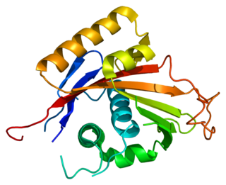
<span class="mw-page-title-main">ATP1A2</span> Protein-coding gene in the species Homo sapiens

Sodium/potassium-transporting ATPase subunit alpha-2 is a protein which in humans is encoded by the ATP1A2 gene.
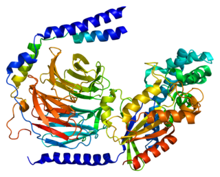
<span class="mw-page-title-main">GNG2</span> Protein-coding gene in the species Homo sapiens

Guanine nucleotide-binding protein G(I)/G(S)/G(O) subunit gamma-2 is a protein that in humans is encoded by the GNG2 gene.

<span class="mw-page-title-main">ATP1A3</span> Protein-coding gene in the species Homo sapiens

Sodium/potassium-transporting ATPase subunit alpha-3 is an enzyme that in humans is encoded by the ATP1A3 gene.

<span class="mw-page-title-main">GRID2</span> Protein-coding gene in the species Homo sapiens

Glutamate receptor, ionotropic, delta 2, also known as GluD2, GluRδ2, or δ2, is a protein that in humans is encoded by the GRID2 gene. This protein together with GluD1 belongs to the delta receptor subtype of ionotropic glutamate receptors. They possess 14–24% sequence homology with AMPA, kainate, and NMDA subunits, but, despite their name, do not actually bind glutamate or various other glutamate agonists.

<span class="mw-page-title-main">PRRT2</span> Protein-coding gene in the species Homo sapiens

Proline-rich transmembrane protein 2 is a protein that in humans is encoded by the PRRT2 gene.

Migraine is often hereditary. It is estimated that 60% of migraine cases are caused by genetics. The role of natural selection in the development of migraines is not known. Fitness-impairing disorders, including migraines, tend to disappear as a result of natural selection, and their frequency decreases to near the rate of spontaneous mutation. However, it is estimated that migraines affect 15-20% of the population and is increasing. This could suggest that a central nervous system (CNS) susceptible to severe, intermittent headache has been linked to an important survival or reproductive advantage. Five possible evolutionary explanations exist: i) migraine as a defence mechanism, ii) migraine as a result of conflicts with other organisms, iii) migraine as a result of novel environmental factors, iv) migraine as a compromise between genetic harms and benefits, and v) headache as a design constraint. These considerations allow the treatment and prevention of migraine to be approached from an evolutionary medicine perspective.

A heme transporter is a protein that delivers heme to the various parts of a biological cell that require it.

References

  1. Stam AH, Luijckx GJ, Poll-Thé BT, Ginjaar IB, Frants RR, Haan J, Ferrari MD, Terwindt GM, van den Maagdenberg AM (2009). "Early seizures and cerebral oedema after trivial head trauma associated with the CACNA1A S218L mutation". J Neurol Neurosurg Psychiatry. 80 (10): 1125–1129. doi:10.1136/jnnp.2009.177279. PMID   19520699. S2CID   1000464.
  2. 1 2 Kors E, Terwindt G, Vermeulen F, Fitzsimons R, Jardine P, Heywood P, Love S, van den Maagdenberg A, Haan J, Frants R, Ferrari M (2001). "Delayed cerebral edema and fatal coma after minor head trauma: role of the CACNA1A calcium channel subunit gene and relationship with familial hemiplegic migraine". Ann Neurol. 49 (6): 753–60. doi:10.1002/ana.1031. PMID   11409427. S2CID   23340019.
  3. 1 2 3 4 5 Ophoff R, Terwindt G, Vergouwe M, van Eijk R, Oefner P, Hoffman S, Lamerdin J, Mohrenweiser H, Bulman D, Ferrari M, Haan J, Lindhout D, van Ommen G, Hofker M, Ferrari M, Frants R (1996). "Familial hemiplegic migraine and episodic ataxia type-2 are caused by mutations in the Ca2+
    channel gene CACNL1A4"
    . Cell. 87 (3): 543–52. doi:10.1016/S0092-8674(00)81373-2. hdl: 1765/57576 . PMID   8898206. S2CID   16840573.
  4. 1 2 3 4 5 Kraus R, Sinnegger M, Glossmann H, Hering S, Striessnig J (1998). "Familial hemiplegic migraine mutations change alpha1A Ca2+
    channel kinetics"
    . J Biol Chem. 273 (10): 5586–90. doi: 10.1074/jbc.273.10.5586 . PMID   9488686.
  5. 1 2 3 4 5 Hans M, Luvisetto S, Williams M, Spagnolo M, Urrutia A, Tottene A, Brust P, Johnson E, Harpold M, Stauderman K, Pietrobon D (1999). "Functional consequences of mutations in the human alpha1A calcium channel subunit linked to familial hemiplegic migraine". J Neurosci. 19 (5): 1610–9. doi:10.1523/JNEUROSCI.19-05-01610.1999. PMC   6782159 . PMID   10024348.
  6. 1 2 Melliti K, Grabner M, Seabrook G (2003). "The familial hemiplegic migraine mutation R192q reduces G-protein-mediated inhibition of p/q-type (Cav2.1) calcium channels expressed in human embryonic kidney cells". J Physiol. 546 (Pt 2): 337–47. doi:10.1113/jphysiol.2002.026716. PMC   2342512 . PMID   12527722.
  7. 1 2 van den Maagdenberg A, Pietrobon D, Pizzorusso T, Kaja S, Broos L, Cesetti T, van de Ven R, Tottene A, van der Kaa J, Plomp J, Frants R, Ferrari M (2004). "A Cacna1a knockin migraine mouse model with increased susceptibility to cortical spreading depression". Neuron. 41 (5): 701–10. doi: 10.1016/S0896-6273(04)00085-6 . hdl: 11577/1481527 . PMID   15003170. S2CID   1456789.
  8. 1 2 3 4 5 Cao Y, Tsien R (2005). "Effects of familial hemiplegic migraine type 1 mutations on neuronal P/Q-type Ca2+
    channel activity and inhibitory synaptic transmission"
    . Proc Natl Acad Sci USA. 102 (7): 2590–5. doi: 10.1073/pnas.0409896102 . PMC   548328 . PMID   15699344.
  9. 1 2 3 4 5 6 7 8 9 Ducros A, Denier C, Joutel A, Cecillon M, Lescoat C, Vahedi K, Darcel F, Vicaut E, Bousser M, Tournier-Lasserve E (2001). "The clinical spectrum of familial hemiplegic migraine associated with mutations in a neuronal calcium channel". N Engl J Med. 345 (1): 17–24. doi: 10.1056/NEJM200107053450103 . PMID   11439943.
  10. Tottene A, Pivotto F, Fellin T, Cesetti T, van den Maagdenberg A, Pietrobon D (2005). "Specific kinetic alterations of human CaV2.1 calcium channels produced by mutation S218L causing familial hemiplegic migraine and delayed cerebral edema and coma after minor head trauma". J Biol Chem. 280 (18): 17678–86. doi: 10.1074/jbc.M501110200 . hdl: 11577/1481524 . PMID   15743764.
  11. 1 2 Battistini S, Stenirri S, Piatti M, Gelfi C, Righetti P, Rocchi R, Giannini F, Battistini N, Guazzi G, Ferrari M, Carrera P (1999). "A new CACNA1A gene mutation in acetazolamide-responsive familial hemiplegic migraine and ataxia". Neurology. 53 (1): 38–43. doi:10.1212/WNL.53.1.38. PMID   10408534. S2CID   32390280.
  12. 1 2 Kraus R, Sinnegger M, Koschak A, Glossmann H, Stenirri S, Carrera P, Striessnig J (2000). "Three new familial hemiplegic migraine mutants affect P/Q-type Ca(2+) channel kinetics". J Biol Chem. 275 (13): 9239–43. doi: 10.1074/jbc.275.13.9239 . PMID   10734061.
  13. 1 2 3 4 Tottene A, Fellin T, Pagnutti S, Luvisetto S, Striessnig J, Fletcher C, Pietrobon D (2002). "Familial hemiplegic migraine mutations increase Ca2+
    influx through single human CaV2.1 channels and decrease maximal CaV2.1 current density in neurons"
    . Proc Natl Acad Sci USA. 99 (20): 13284–9. doi: 10.1073/pnas.192242399 . PMC   130625 . PMID   12235360.
  14. Alonso I, Barros J, Tuna A, Coelho J, Sequeiros J, Silveira I, Coutinho P (2003). "Phenotypes of spinocerebellar ataxia type 6 and familial hemiplegic migraine caused by a unique CACNA1A missense mutation in patients from a large family". Arch Neurol. 60 (4): 610–4. doi: 10.1001/archneur.60.4.610 . hdl: 10400.16/349 . PMID   12707077.
  15. 1 2 Ducros A, Denier C, Joutel A, Vahedi K, Michel A, Darcel F, Madigand M, Guerouaou D, Tison F, Julien J, Hirsch E, Chedru F, Bisgård C, Lucotte G, Després P, Billard C, Barthez M, Ponsot G, Bousser M, Tournier-Lasserve E (1999). "Recurrence of the T666M calcium channel CACNA1A gene mutation in familial hemiplegic migraine with progressive cerebellar ataxia". Am J Hum Genet. 64 (1): 89–98. doi:10.1086/302192. PMC   1377706 . PMID   9915947.
  16. Friend K, Crimmins D, Phan T, Sue C, Colley A, Fung V, Morris J, Sutherland G, Richards R (1999). "Detection of a novel missense mutation and second recurrent mutation in the CACNA1A gene in individuals with EA-2 and FHM". Hum Genet. 105 (3): 261–5. doi:10.1007/s004390051099 (inactive 2024-04-26). PMID   10987655.{{cite journal}}: CS1 maint: DOI inactive as of April 2024 (link)
  17. Barrett C, Cao Y, Tsien R (2005). "Gating deficiency in a familial hemiplegic migraine type 1 mutant P/Q-type calcium channel". J Biol Chem. 280 (25): 24064–71. doi: 10.1074/jbc.M502223200 . PMID   15795222.
  18. 1 2 3 Müllner C, Broos L, van den Maagdenberg A, Striessnig J (2004). "Familial hemiplegic migraine type 1 mutations K1336E, W1684R, and V1696I alter Cav2.1 Ca2+
    channel gating: evidence for beta-subunit isoform-specific effects"
    . J Biol Chem. 279 (50): 51844–50. doi: 10.1074/jbc.M408756200 . PMID   15448138.
  19. Alonso I, Barros J, Tuna A, Seixas A, Coutinho P, Sequeiros J, Silveira I (2004). "A novel R1347Q mutation in the predicted voltage sensor segment of the P/Q-type calcium-channel alpha-subunit in a family with progressive cerebellar ataxia and hemiplegic migraine". Clin Genet. 65 (1): 70–2. doi:10.1111/j..2004.00187.x. PMID   15032980. S2CID   45747611.
  20. Vahedi K, Denier C, Ducros A, Bousson V, Levy C, Chabriat H, Haguenau M, Tournier-Lasserve E, Bousser M (2000). "CACNA1A gene de novo mutation causing hemiplegic migraine, coma, and cerebellar atrophy". Neurology. 55 (7): 1040–2. doi:10.1212/WNL.55.7.1040. PMID   11061267. S2CID   26855561.
  21. Carrera P, Piatti M, Stenirri S, Grimaldi L, Marchioni E, Curcio M, Righetti P, Ferrari M, Gelfi C (1999). "Genetic heterogeneity in Italian families with familial hemiplegic migraine". Neurology. 53 (1): 26–33. doi:10.1212/WNL.53.1.26. PMID   10408532. S2CID   25604212.
  22. Denier C, Ducros A, Vahedi K, Joutel A, Thierry P, Ritz A, Castelnovo G, Deonna T, Gérard P, Devoize J, Gayou A, Perrouty B, Soisson T, Autret A, Warter J, Vighetto A, Van Bogaert P, Alamowitch S, Roullet E, Tournier-Lasserve E (1999). "High prevalence of CACNA1A truncations and broader clinical spectrum in episodic ataxia type 2". Neurology. 52 (9): 1816–21. doi:10.1212/WNL.52.9.1816. PMID   10371528. S2CID   39421883.
  23. Jen J, Yue Q, Nelson S, Yu H, Litt M, Nutt J, Baloh R (1999). "A novel nonsense mutation in CACNA1A causes episodic ataxia and hemiplegia". Neurology. 53 (1): 34–7. doi:10.1212/WNL.53.1.34. PMID   10408533. S2CID   22046224.
  24. Jen J, Wan J, Graves M, Yu H, Mock A, Coulin C, Kim G, Yue Q, Papazian D, Baloh R (2001). "Loss-of-function EA2 mutations are associated with impaired neuromuscular transmission". Neurology. 57 (10): 1843–8. doi:10.1212/WNL.57.10.1843. PMID   11723274. S2CID   23258478.
  25. Beauvais K, Cavé-Riant F, De Barace C, Tardieu M, Tournier-Lasserve E, Furby A (2004). "New CACNA1A gene mutation in a case of familial hemiplegic migraine with status epilepticus". Eur Neurol. 52 (1): 58–61. doi:10.1159/000079546. PMID   15240985. S2CID   35019951.
  26. Kors E, Vanmolkot K, Haan J, Kheradmand Kia S, Stroink H, Laan L, Gill D, Pascual J, van den Maagdenberg A, Frants R, Ferrari M (2004). "Alternating hemiplegia of childhood: no mutations in the second familial hemiplegic migraine gene ATP1A2". Neuropediatrics. 35 (5): 293–6. doi:10.1055/s-2004-821082. PMID   15534763. S2CID   260241252.
  27. 1 2 3 Todt U, Dichgans M, Jurkat-Rott K, Heinze A, Zifarelli G, Koenderink J, Goebel I, Zumbroich V, Stiller A, Ramirez A, Friedrich T, Göbel H, Kubisch C (2005). "Rare missense variants in ATP1A2 in families with clustering of common forms of migraine". Hum Mutat. 26 (4): 315–21. doi: 10.1002/humu.20229 . PMID   16110494. S2CID   15851518.
  28. 1 2 3 4 5 6 7 8 Riant F, De Fusco M, Aridon P, Ducros A, Ploton C, Marchelli F, Maciazek J, Bousser M, Casari G, Tournier-Lasserve E (2005). "ATP1A2 mutations in 11 families with familial hemiplegic migraine". Hum Mutat. 26 (3): 281. doi: 10.1002/humu.9361 . PMID   16088919.
  29. Spadaro M, Ursu S, Lehmann-Horn F, Veneziano L, Liana V, Antonini G, Giovanni A, Giunti P, Paola G, Frontali M, Jurkat-Rott K (2004). "A G301R Na+
    /K+
    -ATPase mutation causes familial hemiplegic migraine type 2 with cerebellar signs". Neurogenetics. 5 (3): 177–85. doi:10.1007/s10048-004-0183-2. PMID   15459825. S2CID   1480291.
  30. Kaunisto M, Harno H, Vanmolkot K, Gargus J, Sun G, Hämäläinen E, Liukkonen E, Kallela M, van den Maagdenberg A, Frants R, Färkkilä M, Palotie A, Wessman M (2004). "A novel missense ATP1A2 mutation in a Finnish family with familial hemiplegic migraine type 2". Neurogenetics. 5 (2): 141–6. doi:10.1007/s10048-004-0178-z. PMID   15133718. S2CID   27379120.
  31. 1 2 3 4 5 Segall L, Scanzano R, Kaunisto M, Wessman M, Palotie A, Gargus J, Blostein R (2004). "Kinetic alterations due to a missense mutation in the Na,K-ATPase alpha2 subunit cause familial hemiplegic migraine type 2". J Biol Chem. 279 (42): 43692–6. doi: 10.1074/jbc.M407471200 . PMID   15308625.
  32. 1 2 3 4 5 6 7 Jurkat-Rott K, Freilinger T, Dreier J, Herzog J, Göbel H, Petzold G, Montagna P, Gasser T, Lehmann-Horn F, Dichgans M (2004). "Variability of familial hemiplegic migraine with novel A1A2 Na+
    /K+
    -ATPase variants". Neurology. 62 (10): 1857–61. doi:10.1212/01.WNL.0000127310.11526.FD. PMID   15159495. S2CID   43023377.
  33. Swoboda K, Kanavakis E, Xaidara A, Johnson J, Leppert M, Schlesinger-Massart M, Ptacek L, Silver K, Youroukos S (2004). "Alternating hemiplegia of childhood or familial hemiplegic migraine? A novel ATP1A2 mutation". Ann Neurol. 55 (6): 884–7. doi:10.1002/ana.20134. PMID   15174025. S2CID   13430399.
  34. Ambrosini A, D'Onofrio M, Grieco G, Di Mambro A, Montagna G, Fortini D, Nicoletti F, Nappi G, Sances G, Schoenen J, Buzzi M, Santorelli F, Pierelli F (2005). "Familial basilar migraine associated with a new mutation in the ATP1A2 gene". Neurology. 65 (11): 1826–8. doi:10.1212/01.wnl.0000187072.71931.c0. PMID   16344534. S2CID   12870819.
  35. 1 2 Vanmolkot K, Kors E, Turk U, Turkdogan D, Keyser A, Broos L, Kia S, van den Heuvel J, Black D, Haan J, Frants R, Barone V, Ferrari M, Casari G, Koenderink J, van den Maagdenberg A (2006). "Two de novo mutations in the Na,K-ATPase gene ATP1A2 associated with pure familial hemiplegic migraine". Eur J Hum Genet. 14 (5): 555–60. doi: 10.1038/sj.ejhg.5201607 . PMID   16538223.
  36. Vanmolkot K, Stroink H, Koenderink J, Kors E, van den Heuvel J, van den Boogerd E, Stam A, Haan J, De Vries B, Terwindt G, Frants R, Ferrari M, van den Maagdenberg A (2006). "Severe episodic neurological deficits and permanent mental retardation in a child with a novel FHM2 ATP1A2 mutation". Ann Neurol. 59 (2): 310–4. doi:10.1002/ana.20760. PMID   16437583. S2CID   8626672.
  37. 1 2 Vanmolkot K, Kors E, Hottenga J, Terwindt G, Haan J, Hoefnagels W, Black D, Sandkuijl L, Frants R, Ferrari M, van den Maagdenberg A (2003). "Novel mutations in the Na+
    , K+
    -ATPase pump gene ATP1A2 associated with familial hemiplegic migraine and benign familial infantile convulsions". Ann Neurol. 54 (3): 360–6. doi:10.1002/ana.10674. PMID   12953268. S2CID   43526424.
  38. 1 2 Segall L, Mezzetti A, Scanzano R, Gargus J, Purisima E, Blostein R (2005). "Alterations in the α2 isoform of Na,K-ATPase associated with familial hemiplegic migraine type 2". Proc Natl Acad Sci USA. 102 (31): 11106–11. Bibcode:2005PNAS..10211106S. doi: 10.1073/pnas.0504323102 . PMC   1178013 . PMID   16037212.
  39. Pierelli F, Grieco G, Pauri F, Pirro C, Fiermonte G, Ambrosini A, Costa A, Buzzi M, Valoppi M, Caltagirone C, Nappi G, Santorelli F (2006). "A novel ATP1A2 mutation in a family with FHM type II". Cephalalgia. 26 (3): 324–8. doi:10.1111/j.1468-2982.2006.01002.x. PMID   16472340. S2CID   33708885.
  40. 1 2 De Fusco M, Marconi R, Silvestri L, Atorino L, Rampoldi L, Morgante L, Ballabio A, Aridon P, Casari G (2003). "Haploinsufficiency of ATP1A2 encoding the Na+
    /K+
    pump alpha2 subunit associated with familial hemiplegic migraine type 2". Nat Genet. 33 (2): 192–6. doi:10.1038/ng1081. PMID   12539047. S2CID   1296597.
  41. 1 2 Koenderink J, Zifarelli G, Qiu L, Schwarz W, De Pont J, Bamberg E, Friedrich T (2005). "Na,K-ATPase mutations in familial hemiplegic migraine lead to functional inactivation". Biochim Biophys Acta. 1669 (1): 61–8. doi: 10.1016/j.bbamem.2005.01.003 . PMID   15843000.
  42. Dichgans M, Freilinger T, Eckstein G, Babini E, Lorenz-Depiereux B, Biskup S, Ferrari M, Herzog J, van den Maagdenberg A, Pusch M, Strom T (2005). "Mutation in the neuronal voltage-gated sodium channel SCN1A in familial hemiplegic migraine". Lancet. 366 (9483): 371–7. doi:10.1016/S0140-6736(05)66786-4. PMID   16054936. S2CID   30365311.
  43. Gardner K, Barmada M, Ptacek L, Hoffman E (1997). "A new locus for hemiplegic migraine maps to chromosome 1q31". Neurology. 49 (5): 1231–8. doi:10.1212/WNL.49.5.1231. PMID   9371899. S2CID   32611926.
  44. Ducros, A (Apr 22, 2013). "[Genetics of migraine.]". Revue Neurologique. 169 (5): 360–71. doi:10.1016/j.neurol.2012.11.010. PMID   23618705.
  45. Suzuki, M.; Van Paesschen, W.; Stalmans, I.; Horita, S.; Yamada, H.; Bergmans, B. A.; Legius, E.; Riant, F.; De Jonghe, P.; Li, Y.; Sekine, T.; Igarashi, T.; Fujimoto, I.; Mikoshiba, K.; Shimadzu, M.; Shiohara, M.; Braverman, N.; Al-Gazali, L.; Fujita, T.; Seki, G. (23 August 2010). "Defective membrane expression of the Na+-HCO3- cotransporter NBCe1 is associated with familial migraine". Proceedings of the National Academy of Sciences. 107 (36): 15963–15968. Bibcode:2010PNAS..10715963S. doi: 10.1073/pnas.1008705107 . PMC   2936614 . PMID   20798035.
  46. Arias-Rivas S, Rodríguez-Yañez M, Cortés J, Pardo-Parrado M, Aguiar P, Leira R, Castillo J, Blanco M (2012). "Familial hemiplegic migraine with prolonged global aura: follow-up findings of subtraction ictal SPECT co-registered to MRI (SISCOM)". Cephalalgia. 32 (13): 1013–1014. doi:10.1177/0333102412457093. PMID   22933508. S2CID   18838384.
  47. Lykke Thomsen L, Kirchmann Eriksen M, Faerch Romer S, Andersen I, Ostergaard E, Keiding N, Olesen J, Russell M (2002). "An epidemiological survey of hemiplegic migraine". Cephalalgia. 22 (5): 361–75. doi:10.1046/j.1468-2982.2002.00371.x. PMID   12110112. S2CID   22040734.