Aura (symptom)

Last updated
Aura
Migraine aura.jpg
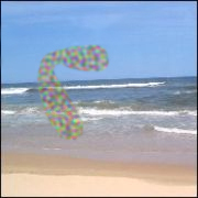
Artist's depiction of zig-zag lines experienced as part of a migraine aura phenomenon
Specialty Neurology, neuro-ophthalmology
Types Scintillating scotoma
Differential diagnosis Persistent aura without infarction, retinal migraine, visual snow

An aura is a perceptual disturbance experienced by some with epilepsy or migraine. An epileptic aura is a seizure. [1]

Contents

Epileptic and migraine auras are due to the involvement of specific areas of the brain, which are those that determine the symptoms of the aura. Therefore, if the visual area is affected, the aura will consist of visual symptoms, while if a sensory one, then sensory symptoms will occur.

Epileptic auras are subjective sensory or psychic phenomena due to a focal seizure, i.e. a seizure that originates from that area of the brain responsible for the function which then expresses itself with the symptoms of the aura. It is important because it makes it clear where the alteration causing the seizure is located. An epileptic aura is in most cases followed by other manifestations of a seizure, for example a convulsion, since the epileptic discharge spreads to other parts of the brain. Rarely it remains isolated. Auras, when they occur, allow some people who have epilepsy time to prevent injury to themselves and/or others when they lose consciousness.

Migraine

The aura of migraine is visual in the vast majority of cases, because dysfunction starts from the visual cortex. The aura is usually followed, after a time varying from minutes to an hour, by the migraine headache. However, the migraine aura can manifest itself in isolation, that is, without being followed by headache. The aura can stay for the duration of the migraine; depending on the type of aura, it can leave the person disoriented and confused. It is common for people with migraines to experience more than one type of aura during the migraine. Most people who have auras have the same type of aura every time.[ citation needed ]

Auras can also be confused with sudden onset of panic, panic attacks or anxiety attacks, which creates difficulties in diagnosis. The differential diagnosis of patients who experience symptoms of paresthesias, derealization, dizziness, chest pain, tremors, and palpitations can be quite challenging. [2]

Seizures

An epileptic aura is the consequence of the activation of functional cortex by abnormal neuronal discharge. [3] In addition to being a warning sign for an impending seizure, the nature of an aura can give insight into the localization and lateralization of the seizure or migraine. [4] [5]

The most common auras include motor, somatosensory, visual, and auditory symptoms. [6] The activation in the brain during an aura can spread through multiple regions continuously or discontinuously, on the same side or to both sides. [7]

Auras are particularly common in focal seizures. If the motor cortex is involved in the overstimulation of neurons, motor auras can result. Likewise, somatosensory auras (such as tingling, numbness, and pain) can result if the somatosensory cortex is involved. When the primary somatosensory cortex is activated, more discrete parts on the opposite side of the body and the secondary somatosensory areas result in symptoms ipsilateral to the seizure focus. [8] [9]

Visual auras can be simple or complex. Simple visual symptoms can include static, flashing, or moving lights/shapes/colors caused mostly by abnormal activity in the primary visual cortex. Complex visual auras can include people, scenes, and objects which results from stimulation of the temporo-occipital junction and is lateralized to one hemifield. Auditory auras can also be simple (ringing, buzzing) or complex (voices, music). Simple symptoms can occur from activation in the primary auditory cortex and complex symptoms from the temporo-occipital cortex at the location of the auditory association areas. [10]

Examples

Artist's depiction of scintillating scotoma Aura ss.jpg
Artist's depiction of scintillating scotoma
Example of a scintillating scotoma aura with each dot or line flickering ScintillatingScotoma3.jpg
Example of a scintillating scotoma aura with each dot or line flickering
Example of scintillating scotoma showing an obscured/distorted area bordered with colors Migraine-aura-aka-scintillating-scotoma-anecdoteal-depiction.png
Example of scintillating scotoma showing an obscured/distorted area bordered with colors

An aura sensation can include one or a combination of the following:

Visual changes

Auditory changes

Other sensations

Animated depictions

See also

Related Research Articles

<span class="mw-page-title-main">Seizure</span> Period of symptoms due to excessive or synchronous neuronal brain activity

An epileptic seizure, informally known as a seizure, is a period of symptoms due to abnormally excessive or synchronous neuronal activity in the brain. Outward effects vary from uncontrolled shaking movements involving much of the body with loss of consciousness, to shaking movements involving only part of the body with variable levels of consciousness, to a subtle momentary loss of awareness. These episodes usually last less than two minutes and it takes some time to return to normal. Loss of bladder control may occur.

<span class="mw-page-title-main">Hallucination</span> Perception in the absence of external stimulation that has the qualities of real perception

A hallucination is a perception in the absence of an external stimulus that has the qualities of a real perception. Hallucinations are vivid, substantial, and are perceived to be located in external objective space. Hallucination is a combination of two conscious states of brain wakefulness and REM sleep. They are distinguishable from several related phenomena, such as dreaming, which does not involve wakefulness; pseudohallucination, which does not mimic real perception, and is accurately perceived as unreal; illusion, which involves distorted or misinterpreted real perception; and mental imagery, which does not mimic real perception, and is under voluntary control. Hallucinations also differ from "delusional perceptions", in which a correctly sensed and interpreted stimulus is given some additional significance.

<span class="mw-page-title-main">Micropsia</span> Medical condition

Micropsia is a condition affecting human visual perception in which objects are perceived to be smaller than they actually are. Micropsia can be caused by optical factors, by distortion of images in the eye, by changes in the brain, and from psychological factors. Dissociative phenomena are linked with micropsia, which may be the result of brain-lateralization disturbance.

<span class="mw-page-title-main">Alice in Wonderland syndrome</span> Neurological disorder that distorts perception of objects size and distance

Alice in Wonderland syndrome (AIWS), also known as Todd's syndrome or dysmetropsia, is a neurological disorder that distorts perception. People with this syndrome may experience distortions in their visual perception of objects, such as appearing smaller (micropsia) or larger (macropsia), or appearing to be closer (pelopsia) or farther (teleopsia) than they are. Distortion may also occur for senses other than vision.

<span class="mw-page-title-main">Claustrum</span> Structure in the brain

The claustrum is a thin sheet of neurons and supporting glial cells, that connects to the cerebral cortex and subcortical regions including the amygdala, hippocampus and thalamus of the brain. It is located between the insular cortex laterally and the putamen medially, encased by the extreme and external capsules respectively. Blood to the claustrum is supplied by the middle cerebral artery. It is considered to be the most densely connected structure in the brain, and thus hypothesized to allow for the integration of various cortical inputs such as vision, sound and touch, into one experience. Other hypotheses suggest that the claustrum plays a role in salience processing, to direct attention towards the most behaviorally relevant stimuli amongst the background noise. The claustrum is difficult to study given the limited number of individuals with claustral lesions and the poor resolution of neuroimaging.

A headache is often present in patients with epilepsy. If the headache occurs in the vicinity of a seizure, it is defined as peri-ictal headache, which can occur either before (pre-ictal) or after (post-ictal) the seizure, to which the term ictal refers. An ictal headache itself may or may not be an epileptic manifestation. In the first case it is defined as ictal epileptic headache or simply epileptic headache. It is a real painful seizure, that can remain isolated or be followed by other manifestations of the seizure. On the other hand, the ictal non-epileptic headache is a headache that occurs during a seizure but it is not due to an epileptic mechanism. When the headache does not occur in the vicinity of a seizure it is defined as inter-ictal headache. In this case it is a disorder autonomous from epilepsy, that is a comorbidity.

<span class="mw-page-title-main">Visual snow syndrome</span> Visual impairment

Visual snow syndrome (VSS) is a form of visual hallucination that is characterized by the perception of small, flickering dots throughout the entire visual field. It is present in all conditions of illumination. The dots remain individual and do not clump together or change in size. Visual snow exists in one of two forms: the pulse type and the broadband type.

Acephalgic migraine is a neurological syndrome. It is a relatively uncommon variant of migraine in which the patient may experience some migraine symptoms such as aura, nausea, photophobia, and hemiparesis, but does not experience headache. It is generally classified as an event fulfilling the conditions of migraine with aura with no headache. It is sometimes distinguished from visual-only migraine aura without headache, also called ocular migraine.

<span class="mw-page-title-main">Temporal lobe epilepsy</span> Chronic focal seizure disorder

In the field of neurology, temporal lobe epilepsy is an enduring brain disorder that causes unprovoked seizures from the temporal lobe. Temporal lobe epilepsy is the most common type of focal onset epilepsy among adults. Seizure symptoms and behavior distinguish seizures arising from the medial temporal lobe from seizures arising from the lateral (neocortical) temporal lobe. Memory and psychiatric comorbidities may occur. Diagnosis relies on electroencephalographic (EEG) and neuroimaging studies. Anticonvulsant medications, epilepsy surgery and dietary treatments may improve seizure control.

Frontal lobe epilepsy (FLE) is a neurological disorder that is characterized by brief, recurring seizures arising in the frontal lobes of the brain, that often occur during sleep. It is the second most common type of epilepsy after temporal lobe epilepsy (TLE), and is related to the temporal form in that both forms are characterized by partial (focal) seizures.

<span class="mw-page-title-main">Scintillating scotoma</span> Visual aura associated with migraine

Scintillating scotoma is a common visual aura that was first described by 19th-century physician Hubert Airy (1838–1903). Originating from the brain, it may precede a migraine headache, but can also occur acephalgically, also known as visual migraine or migraine aura. It is often confused with retinal migraine, which originates in the eyeball or socket.

In the field of neurology, seizure types are categories of seizures defined by seizure behavior, symptoms, and diagnostic tests. The International League Against Epilepsy (ILAE) 2017 classification of seizures is the internationally recognized standard for identifying seizure types. The ILAE 2017 classification of seizures is a revision of the prior ILAE 1981 classification of seizures. Distinguishing between seizure types is important since different types of seizures may have different causes, outcomes, and treatments.

<span class="mw-page-title-main">Retinal migraine</span> Medical condition

Retinal migraine is a retinal disease often accompanied by migraine headache and typically affects only one eye. It is caused by ischaemia or vascular spasm in or behind the affected eye.

Migralepsy is a rare condition in which a migraine is followed, within an hour period, by an epileptic seizure. Because of the similarities in signs, symptoms, and treatments of both conditions, such as the neurological basis, the psychological issues, and the autonomic distress that is created from them, they individually increase the likelihood of causing the other. However, also because of the sameness, they are often misdiagnosed for each other, as migralepsy rarely occurs.

Abdominal aura, also known as visceral aura and epigastric aura, is a type of somatosensory aura that typically manifests as abdominal discomfort in the form of nausea, malaise, hunger, or pain. Abdominal aura is typically associated with epilepsy, especially temporal lobe epilepsy, and it can also be used in the context of migraine. The term is used to distinguish it from other types of somatosensory aura, notably visual disturbances and paraesthesia. The abdominal aura can be classified as a somatic hallucination. Pathophysiologically, the abdominal aura is associated with aberrant neuronal discharges in sensory cortical areas representing the abdominal viscera.

Idiopathic childhood occipital epilepsy of Gastaut (ICOE-G) is a pure but rare form of idiopathic occipital epilepsy that affects otherwise normal children and adolescents. It is classified amongst benign idiopathic childhood focal epilepsies such as rolandic epilepsy and Panayiotopoulos syndrome.

Hallucinatory palinopsia is a subtype of palinopsia, a visual disturbance defined as the persistent or recurrence of a visual image after the stimulus has been removed. Palinopsia is a broad term describing a group of symptoms which is divided into hallucinatory palinopsia and illusory palinopsia. Hallucinatory palinopsia refers to the projection of an already-encoded visual memory and is similar to a complex visual hallucination: the creation of a formed visual image where none exists.

Cerebral diplopia or polyopia describes seeing two or more images arranged in ordered rows, columns, or diagonals after fixation on a stimulus. The polyopic images occur monocular bilaterally and binocularly, differentiating it from ocular diplopia or polyopia. The number of duplicated images can range from one to hundreds. Some patients report difficulty in distinguishing the replicated images from the real images, while others report that the false images differ in size, intensity, or color. Cerebral polyopia is sometimes confused with palinopsia, in which multiple images appear while watching an object. However, in cerebral polyopia, the duplicated images are of a stationary object which are perceived even after the object is removed from the visual field. Movement of the original object causes all of the duplicated images to move, or the polyopic images disappear during motion. In palinoptic polyopia, movement causes each polyopic image to leave an image in its wake, creating hundreds of persistent images (entomopia).

<span class="mw-page-title-main">Occipital epilepsy</span> Medical condition

Occipital epilepsy is a neurological disorder that arises from excessive neural activity in the occipital lobe of the brain that may or may not be symptomatic. Occipital lobe epilepsy is fairly rare, and may sometimes be misdiagnosed as migraine when symptomatic. Epileptic seizures are the result of synchronized neural activity that is excessive, and may stem from a failure of inhibitory neurons to regulate properly.

Musicogenic seizure, also known as music-induced seizure, is a rare type of seizure, with an estimated prevalence of 1 in 10,000,000 individuals, that arises from disorganized or abnormal brain electrical activity when a person hears or is exposed to a specific type of sound or musical stimuli. There are challenges when diagnosing a music-induced seizure due to the broad scope of triggers, and time delay between a stimulus and seizure. In addition, the causes of musicogenic seizures are not well-established as solely limited cases and research have been discovered and conducted respectively. Nevertheless, the current understanding of the mechanism behind musicogenic seizure is that music triggers the part of the brain that is responsible for evoking an emotion associated with that music. Dysfunction in this system leads to an abnormal release of dopamine, eventually inducing seizure.

References

  1. Epilepsy auras
  2. Hurley, Robin A.; Fisher, Ronald; Taber, Katherine H. (1 October 2006). "Sudden Onset Panic: Epileptic Aura or Panic Disorder?". The Journal of Neuropsychiatry and Clinical Neurosciences. 18 (4): 436–443. doi: 10.1176/jnp.2006.18.4.436 . PMID   17135371.
  3. Perven G and So NK (2015). "Epileptic auras: phenomenology and neurophysiology". Epileptic Disorders. 17 (4): 549–562. doi:10.1684/epd.2015.0786. PMID   26643374.
  4. Ye, Byoung Seok; Cho, Yang-Je; Jang, Sang Hyun; Lee, Moon Kyu; Lee, Byung In; Heo, Kyoung (2012-05-01). "The Localizing and Lateralizing Value of Auras in Lesional Partial Epilepsy Patients". Yonsei Medical Journal. 53 (3): 477–485. doi:10.3349/ymj.2012.53.3.477. PMC   3343447 . PMID   22476989.
  5. Foldvary-Schaefer, N.; Unnwongse, K. (February 2011). "Localizing and lateralizing features of auras and seizures". Epilepsy & Behavior. 20 (2): 160–166. doi:10.1016/j.yebeh.2010.08.034. PMID   20926350. S2CID   1220765.
  6. Sharma S., Dixit V. (2013). "Epilepsy – A Comprehensive Review". International Journal of Pharmacological Research & Review. 2 (12): 61–80.
  7. Tuxhorn I. E. B. (2005). "Somatosensory auras in focal epilepsy: A clinical, video EEG and MRI study". Seizure: European Journal of Epilepsy. 14 (4): 262–268. doi: 10.1016/j.seizure.2005.02.005 . PMID   15911361. S2CID   18386228.
  8. Tuxhorn, I. E. B. (2005-06-01). "Somatosensory auras in focal epilepsy: A clinical, video EEG and MRI study". Seizure. 14 (4): 262–268. doi: 10.1016/j.seizure.2005.02.005 . PMID   15911361. S2CID   18386228.
  9. Fakhoury, T.; Abou-Khalil, B. (November 1995). "Association of ipsilateral head turning and dystonia in temporal lobe seizures". Epilepsia. 36 (11): 1065–1070. doi:10.1111/j.1528-1157.1995.tb00463.x. PMID   7588449. S2CID   35063423.
  10. Foldvary-Schaefer, Nancy; Unnwongse, Kanjana (1 February 2011). "Localizing and lateralizing features of auras and seizures". Epilepsy & Behavior. 20 (2): 160–166. doi:10.1016/j.yebeh.2010.08.034. PMID   20926350. S2CID   1220765.
  11. "Aura: Migraine's Odd Companion". Migraineur. 20 February 2018. Retrieved 2021-03-17.
  12. "Patient's Guide to Visual Migraine - Brigham and Women's Hospital". www.brighamandwomens.org. Retrieved 2021-03-17.
  13. "Alice in Wonderland Syndrome - an overview | ScienceDirect Topics". www.sciencedirect.com. Retrieved 2022-01-11.
  14. "Understanding the Aura Stage of Migraine Doctor Q&A". Migraine Again. 2020-12-30. Retrieved 2021-03-17.
  15. OD, By George T. Banyas. "Visual Aura and Scotomas: What Do They Indicate?". www.reviewofoptometry.com. Retrieved 2022-01-11.
  16. Liao, Sharon. "Causes of Temporary Blindness and Short-Term Vision Loss". WebMD. Retrieved 2022-01-11.
  17. "Photophobia: Causes, symptoms, and treatment". www.medicalnewstoday.com. 2021-08-16. Retrieved 2022-01-11.
  18. DiLonardo, Mary Jo. "Epilepsy: What is Seizure With Aura?". WebMD. Retrieved 2022-01-11.
  19. van der Feltz-Cornelis, Christina M; Biemans, Henk; Timmer, Jan (2012). "Hearing voices: does it give your patient a headache? A case of auditory hallucinations as acoustic aura in migraine". Neuropsychiatric Disease and Treatment. 8: 105–111. doi: 10.2147/NDT.S29300 . PMC   3333787 . PMID   22536065.
  20. Bernetti, L.; Pellegrino, C.; Corbelli, I.; Caproni, S.; Eusebi, P.; Faralli, M.; Ricci, G.; Calabresi, P.; Sarchielli, P. (16 April 2018). "Subclinical vestibular dysfunction in migraineurs without vertigo". Acta Neurologica Scandinavica. 138 (4): 270–277. doi: 10.1111/ane.12941 . PMID   29658983. S2CID   4887561.
  21. Jion, Y. I.; Grosberg, B. M.; Evans, R. W. (22 August 2016). "Phantosmia and Migraine With and Without Headache". Headache. 56 (9): 1494–1502. doi:10.1111/head.12890. PMID   27545897. S2CID   36091982.
  22. "Can Being Sensitive to Smells Impact Migraine?". www.excedrin.com. Retrieved 2022-01-11.
  23. Marcel Neckar; Petr Bob (11 January 2016). "Synesthetic associations and psychosensory symptoms of temporal epilepsy". Neuropsychiatric Disease and Treatment. 12: 109–12. doi: 10.2147/NDT.S95464 . PMC   4714732 . PMID   26811683.
  24. Adachi, Naoto; Akanuma, Nozomi; Ito, Masumi; Adachi, Takuya; Takekawa, Yoshikazu; Adachi, Yasushi; Matsuura, Masato; Kanemoto, Kousuke; Kato, Masaaki (2010-07-01). "Two forms of deja vu experiences in patients with epilepsy". Epilepsy & Behavior. 18 (3): 218–22. doi:10.1016/j.yebeh.2010.02.016. PMID   20494621. S2CID   27034245.
  25. Kakisaka, Y.; Jehi, L.; Alkawadri, R.; Wang, Z. I.; Enatsu, R.; Mosher, J. C.; Dubarry, A. S.; Alexopoulos, A. V.; Burgess, R. C. (August 2014). "Cephalic aura after frontal lobe resection". Journal of Clinical Neuroscience. 21 (8): 1450–1452. doi:10.1016/j.jocn.2013.11.024. PMC   4340243 . PMID   24613491.
  26. Hoffman, Matthew; MD. "Abdominal Epilepsy in Children and Adults". WebMD. Retrieved 2022-01-11.
  27. Sekimoto, M.; Kato, M.; Kaneko, Y.; Onuma, T. (December 2007). "Ictal nausea with vomiting as the major symptom of simple partial seizures". Epilepsy & Behavior. 11 (4): 582–587. doi:10.1016/j.yebeh.2007.08.023. PMID   18054131. S2CID   45291893.
  28. Erickson, J. C.; Clapp, L. E.; Ford, G.; Jabbari, B. (2006). "Somatosensory auras in refractory temporal lobe epilepsy". Epilepsia. 47 (1): 202–206. doi: 10.1111/j.1528-1167.2006.00388.x . PMID   16417550. S2CID   25666352.
  29. Kumar, Anil; Samanta, Debopam; Emmady, Prabhu D.; Arora, Rohan (2021), "Hemiplegic Migraine", StatPearls, Treasure Island (FL): StatPearls Publishing, PMID   30020674 , retrieved 2022-01-11
  30. "Dissociation and depersonalization: Causes, risk factors, and symptoms". www.medicalnewstoday.com. 2019-05-17. Retrieved 2022-01-11.
  31. "Brain & Spine Foundation | Migraine" . Retrieved 2022-01-11.
  32. "Recognizing Aphasia During a Migraine Attack". www.northsuffolkneurology.com. Retrieved 2022-01-11.
  33. Page 258 in: Britt Talley Daniel (2010). Migraine. AuthorHouse. ISBN   978-1-4490-6962-9.