Blind spot (vision)

Last updated
In vertebrate eyes, the nerve fibers route before the retina, blocking some light and creating a blind spot where the fibers pass through the retina and out of the eye. In octopus eyes, the nerve fibers route behind the retina, and do not block light or disrupt the retina. In the example, 4 denotes the vertebrate blind spot, which is notably absent in the octopus eye. In both images, 1 denotes the retina and 2 the nerve fibers, including the optic nerve (3).
Distribution of rods and cones along a line passing through the fovea and the blind spot of a human eye Human photoreceptor distribution.svg
Distribution of rods and cones along a line passing through the fovea and the blind spot of a human eye

A blind spot, scotoma , is an obscuration of the visual field. A particular blind spot known as the physiological blind spot, "blind point", or punctum caecum in medical literature, is the place in the visual field that corresponds to the lack of light-detecting photoreceptor cells on the optic disc of the retina where the optic nerve passes through the optic disc. [2] Because there are no cells to detect light on the optic disc, the corresponding part of the field of vision is invisible. Processes in the brain interpolate the blind spot based on surrounding detail and information from the other eye, so it is not normally perceived.

Contents

Although all vertebrates have this blind spot, cephalopod eyes, which are only superficially similar because they evolved independently, do not. In them, the optic nerve approaches the receptors from behind, so it does not create a break in the retina.

The first documented observation of the phenomenon was in the 1660s by Edme Mariotte in France. At the time it was generally thought that the point at which the optic nerve entered the eye should actually be the most sensitive portion of the retina; however, Mariotte's discovery disproved this theory.

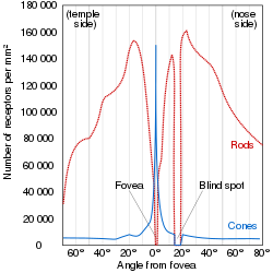
The blind spot in humans is located about 12–15° temporally and 1.5° below the horizontal and is roughly 7.5° high and 5.5° wide. [3]

Blind spot test

Demonstration of the blind spot
RL
Instructions: Close one eye and focus the other on the appropriate letter (R for right or L for left). Place your eye a distance from the screen approximately equal to three times the distance between the R and the L. Move your eye towards or away from the screen until you notice the other letter disappear. For example, close your right eye, look at the "L" with your left eye, and the "R" will disappear.

Night blind spot

Night blind spot Night blind spot.png
Night blind spot

It is estimated that once fully adapted to darkness, the rods are 10,000 times more sensitive to light than the cones, making them the primary receptors for night vision. Since the cones are concentrated near the fovea, the rods are also responsible for much of the peripheral vision. The concentration of cones in the fovea can make a night blind spot in the center of the field of vision. [4]

See also

Related Research Articles

<span class="mw-page-title-main">Retina</span> Part of the eye

The retina is the innermost, light-sensitive layer of tissue of the eye of most vertebrates and some molluscs. The optics of the eye create a focused two-dimensional image of the visual world on the retina, which then processes that image within the retina and sends nerve impulses along the optic nerve to the visual cortex to create visual perception. The retina serves a function which is in many ways analogous to that of the film or image sensor in a camera.

<span class="mw-page-title-main">Optic nerve</span> Second cranial nerve, which connects the eyes to the brain

In neuroanatomy, the optic nerve, also known as the second cranial nerve, cranial nerve II, or simply CN II, is a paired cranial nerve that transmits visual information from the retina to the brain. In humans, the optic nerve is derived from optic stalks during the seventh week of development and is composed of retinal ganglion cell axons and glial cells; it extends from the optic disc to the optic chiasma and continues as the optic tract to the lateral geniculate nucleus, pretectal nuclei, and superior colliculus.

<span class="mw-page-title-main">Eye</span> Organ that detects light and converts it into electro-chemical impulses in neurons

Eyes are organs of the visual system. They provide living organisms with vision, the ability to receive and process visual detail, as well as enabling several photo response functions that are independent of vision. Eyes detect light and convert it into electro-chemical impulses in neurons (neurones). In higher organisms, the eye is a complex optical system which collects light from the surrounding environment, regulates its intensity through a diaphragm, focuses it through an adjustable assembly of lenses to form an image, converts this image into a set of electrical signals, and transmits these signals to the brain through complex neural pathways that connect the eye via the optic nerve to the visual cortex and other areas of the brain. Eyes with resolving power have come in ten fundamentally different forms, and 96% of animal species possess a complex optical system. Image-resolving eyes are present in molluscs, chordates and arthropods.

<span class="mw-page-title-main">Macula</span> Oval-shaped pigmented area near the center of the retina

The macula (/ˈmakjʊlə/) or macula lutea is an oval-shaped pigmented area in the center of the retina of the human eye and in other animals. The macula in humans has a diameter of around 5.5 mm (0.22 in) and is subdivided into the umbo, foveola, foveal avascular zone, fovea, parafovea, and perifovea areas.

<span class="mw-page-title-main">Peripheral vision</span> Area of ones field of vision outside of the point of fixation

Peripheral vision, or indirect vision, is vision as it occurs outside the point of fixation, i.e. away from the center of gaze or, when viewed at large angles, in the "corner of one's eye". The vast majority of the area in the visual field is included in the notion of peripheral vision. "Far peripheral" vision refers to the area at the edges of the visual field, "mid-peripheral" vision refers to medium eccentricities, and "near-peripheral", sometimes referred to as "para-central" vision, exists adjacent to the center of gaze.

<span class="mw-page-title-main">Visual system</span> Body parts responsible for vision

The visual system comprises the sensory organ and parts of the central nervous system which gives organisms the sense of vision as well as enabling the formation of several non-image photo response functions. It detects and interprets information from the optical spectrum perceptible to that species to "build a representation" of the surrounding environment. The visual system carries out a number of complex tasks, including the reception of light and the formation of monocular neural representations, colour vision, the neural mechanisms underlying stereopsis and assessment of distances to and between objects, the identification of a particular object of interest, motion perception, the analysis and integration of visual information, pattern recognition, accurate motor coordination under visual guidance, and more. The neuropsychological side of visual information processing is known as visual perception, an abnormality of which is called visual impairment, and a complete absence of which is called blindness. Non-image forming visual functions, independent of visual perception, include the pupillary light reflex and circadian photoentrainment.

<span class="mw-page-title-main">Photoreceptor cell</span> Type of neuroepithelial cell

A photoreceptor cell is a specialized type of neuroepithelial cell found in the retina that is capable of visual phototransduction. The great biological importance of photoreceptors is that they convert light into signals that can stimulate biological processes. To be more specific, photoreceptor proteins in the cell absorb photons, triggering a change in the cell's membrane potential.

<span class="mw-page-title-main">Tetrachromacy</span> Type of color vision with four types of cone cells

Tetrachromacy is the condition of possessing four independent channels for conveying color information, or possessing four types of cone cell in the eye. Organisms with tetrachromacy are called tetrachromats.

<span class="mw-page-title-main">Rod cell</span> Photoreceptor cells that can function in lower light better than cone cells

Rod cells are photoreceptor cells in the retina of the eye that can function in lower light better than the other type of visual photoreceptor, cone cells. Rods are usually found concentrated at the outer edges of the retina and are used in peripheral vision. On average, there are approximately 92 million rod cells in the human retina. Rod cells are more sensitive than cone cells and are almost entirely responsible for night vision. However, rods have little role in color vision, which is the main reason why colors are much less apparent in dim light.

<span class="mw-page-title-main">Cone cell</span> Photoreceptor cells responsible for color vision made to function in bright light

Cone cells, or cones, are photoreceptor cells in the retinas of vertebrates' eyes. They respond differently to light of different wavelengths, and the combination of their responses is responsible for color vision. Cones function best in relatively bright light, called the photopic region, as opposed to rod cells, which work better in dim light, or the scotopic region. Cone cells are densely packed in the fovea centralis, a 0.3 mm diameter rod-free area with very thin, densely packed cones which quickly reduce in number towards the periphery of the retina. Conversely, they are absent from the optic disc, contributing to the blind spot. There are about six to seven million cones in a human eye, with the highest concentration being towards the macula.

In visual physiology, adaptation is the ability of the retina of the eye to adjust to various levels of light. Natural night vision, or scotopic vision, is the ability to see under low-light conditions. In humans, rod cells are exclusively responsible for night vision as cone cells are only able to function at higher illumination levels. Night vision is of lower quality than day vision because it is limited in resolution and colors cannot be discerned; only shades of gray are seen. In order for humans to transition from day to night vision they must undergo a dark adaptation period of up to two hours in which each eye adjusts from a high to a low luminescence "setting", increasing sensitivity hugely, by many orders of magnitude. This adaptation period is different between rod and cone cells and results from the regeneration of photopigments to increase retinal sensitivity. Light adaptation, in contrast, works very quickly, within seconds.

<span class="mw-page-title-main">Visual acuity</span> Clarity of vision

Visual acuity (VA) commonly refers to the clarity of vision, but technically rates an animal's ability to recognize small details with precision. Visual acuity depends on optical and neural factors. Optical factors of the eye influence the sharpness of an image on its retina. Neural factors include the health and functioning of the retina, of the neural pathways to the brain, and of the interpretative faculty of the brain.

<span class="mw-page-title-main">Fovea centralis</span> Small pit in the retina of the eye responsible for all central vision

The fovea centralis is a small, central pit composed of closely packed cones in the eye. It is located in the center of the macula lutea of the retina.

<span class="mw-page-title-main">Retinal ganglion cell</span> Type of cell within the eye

A retinal ganglion cell (RGC) is a type of neuron located near the inner surface of the retina of the eye. It receives visual information from photoreceptors via two intermediate neuron types: bipolar cells and retina amacrine cells. Retina amacrine cells, particularly narrow field cells, are important for creating functional subunits within the ganglion cell layer and making it so that ganglion cells can observe a small dot moving a small distance. Retinal ganglion cells collectively transmit image-forming and non-image forming visual information from the retina in the form of action potential to several regions in the thalamus, hypothalamus, and mesencephalon, or midbrain.

The visual field is "that portion of space in which objects are visible at the same moment during steady fixation of the gaze in one direction"; in ophthalmology and neurology the emphasis is on the structure inside the visual field and it is then considered “the field of functional capacity obtained and recorded by means of perimetry”.

<span class="mw-page-title-main">Optic disc</span> Optic nerve head, the point of exit for ganglion cell axons leaving the eye

The optic disc or optic nerve head is the point of exit for ganglion cell axons leaving the eye. Because there are no rods or cones overlying the optic disc, it corresponds to a small blind spot in each eye.

<span class="mw-page-title-main">Central retinal artery</span>

The central retinal artery branches off the ophthalmic artery, running inferior to the optic nerve within its dural sheath to the eyeball.

<span class="mw-page-title-main">Equine vision</span>

The equine eye is one of the largest of any land mammal. Its visual abilities are directly related to the animal's behavior; for example, it is active during both day and night, and it is a prey animal. Both the strengths and weaknesses of the horse's visual abilities should be taken into consideration when training the animal, as an understanding of the horse's eye can help to discover why the animal behaves the way it does in various situations.

<span class="mw-page-title-main">Bird vision</span> Senses for birds

Vision is the most important sense for birds, since good eyesight is essential for safe flight. Birds have a number of adaptations which give visual acuity superior to that of other vertebrate groups; a pigeon has been described as "two eyes with wings". Birds are theropod dinosaurs, and the avian eye resembles that of other reptiles, with ciliary muscles that can change the shape of the lens rapidly and to a greater extent than in the mammals. Birds have the largest eyes relative to their size in the animal kingdom, and movement is consequently limited within the eye's bony socket. In addition to the two eyelids usually found in vertebrates, bird's eyes are protected by a third transparent movable membrane. The eye's internal anatomy is similar to that of other vertebrates, but has a structure, the pecten oculi, unique to birds.

<span class="mw-page-title-main">Mammalian eye</span>

Mammals normally have a pair of eyes. Although mammalian vision is not so excellent as bird vision, it is at least dichromatic for most of mammalian species, with certain families possessing a trichromatic color perception.

References

  1. Brian A. Wandell (1 January 1995). Foundations of Vision. Sinauer Associates. ISBN   978-0-87893-853-7.
  2. Gregory, R., & Cavanagh, P. (2011). "The Blind Spot". Scholarpedia. Retrieved on 2011-05-21.
  3. MIL-STD-1472F, Military Standard, Human Engineering, Design Criteria For Military Systems, Equipment, And Facilities (23 Aug 1999) PDF
  4. "Chapter 17: Aeromedical Factors". Pilot's Handbook of Aeronautical Knowledge (FAA-H-8083-25C ed.). Federal Aviation Administration. 2023-07-17. p. 22.PD-icon.svg This article incorporates text from this source, which is in the public domain .