Heterotrimeric G protein

Last updated
Heterotrimeric G-protein GTPase
Identifiers
EC no. 3.6.5.1
CAS no. 9059-32-9
Databases
IntEnz IntEnz view
BRENDA BRENDA entry
ExPASy NiceZyme view
KEGG KEGG entry
MetaCyc metabolic pathway
PRIAM profile
PDB structures RCSB PDB PDBe PDBsum
Gene Ontology AmiGO / QuickGO
Search
PMC articles
PubMed articles
NCBI proteins
This heterotrimeric G protein is illustrated with its theoretical lipid anchors. GDP is black. Alpha chain is yellow. Beta and gamma chains are blue. G protein1.png
This heterotrimeric G protein is illustrated with its theoretical lipid anchors. GDP is black. Alpha chain is yellow. Beta and gamma chains are blue.
3D structure of a heterotrimeric G protein G-Protein.png
3D structure of a heterotrimeric G protein

Heterotrimeric G protein, also sometimes referred to as the "large" G proteins (as opposed to the subclass of smaller, monomeric small GTPases) are membrane-associated G proteins that form a heterotrimeric complex. The biggest non-structural difference between heterotrimeric and monomeric G protein is that heterotrimeric proteins bind to their cell-surface receptors, called G protein-coupled receptors (GPCR), directly. These G proteins are made up of alpha (α), beta (β) and gamma (γ) subunits. [1] The alpha subunit is attached to either a GTP or GDP, which serves as an on-off switch for the activation of G-protein.

Contents

When ligands bind a GPCR, the GPCR acquires GEF (guanine nucleotide exchange factor) ability, which activates the G-protein by exchanging the GDP on the alpha subunit to GTP. The binding of GTP to the alpha subunit results in a structural change and its dissociation from the rest of the G-protein. Generally, the alpha subunit binds membrane-bound effector proteins for the downstream signaling cascade, but the beta-gamma complex can carry out this function also. G-proteins are involved in pathways such as the cAMP/PKA pathway, ion channels, MAPK, PI3K.

There are four main families of G proteins: Gi/Go, Gq, Gs, and G12/13. [2]

Alpha subunits

G-protein's role in a G-protein coupled receptor activated pathway GPCR cycle.jpg
G-protein's role in a G-protein coupled receptor activated pathway

Reconstitution experiments carried out in the early 1980s showed that purified Gα subunits can directly activate effector enzymes. The GTP form of the α subunit of transducin (Gt) activates the cyclic GMP phosphodiesterase from retinal rod outer segments, [3] and the GTP form of the α subunit of the stimulatory G protein (Gs) activates hormone-sensitive adenylate cyclase. [4] [5] More than one type of G protein co-exist in the same tissue. For example, in adipose tissues, two different G-proteins with interchangeable beta-gamma complexes are used to activate or inhibit adenylyl cyclase. The alpha subunit of a stimulatory G protein activated by receptors for stimulatory hormones could stimulate adenylyl cyclase, which activates cAMP used for downstream signal cascades. While on the other hand, the alpha subunit of an inhibitory G protein activated by receptors of inhibitory hormones could inhibit adenylyl cyclase, which blocks downstream signal cascades.

Gα subunits consist of two domains, the GTPase domain, and the alpha-helical domain.

There exist at least 20 different Gα subunits, which are separated into four main groups. This nomenclature is based on their sequence homologies: [6]

G-protein familyα-subunitGeneSignal transductionUse/Receptors (examples)Effects (examples)
Gi-family (InterPro :  IPR001408 )
Gi/o αi, αo GNAO1, GNAI1, GNAI2, GNAI3 Inhibition of adenylate cyclase, opens K+-channels (via β/γ subunits), closes Ca2+-channelsMuscarinic M2 and M4, [7] chemokine receptors, α2-Adrenoreceptors, Serotonin 5-HT1 receptors, Histamine H3 and H4, Dopamine D2-like receptors, type 2 cannabinoid receptors (CB2) [8] Smooth muscle contraction, depress neuronal activity, interleukin secretion by human leukocytes [8]
Gtαt (Transducin) GNAT1, GNAT2 Activation of phosphodiesterase 6 Rhodopsin Vision
Ggustαgust (Gustducin) GNAT3 Activation of phosphodiesterase 6Taste receptorsTaste
Gzαz GNAZ Inhibition of adenylate cyclasePlateletsMaintaining the ionic balance of perilymphatic and endolymphatic cochlear fluids.
Gs-family (InterPro :  IPR000367 )
Gs αs GNAS Activation of adenylate cyclase Beta-adrenoreceptors; Serotonin 5-HT4, 5-HT6 and 5-HT7; Dopamine D1-like receptors, Histamine H2, Vasopressin V2, type 2 cannabinoid receptors [8] Increase heart rate, Smooth muscle relaxation, stimulate neuronal activity, interleukin secretion by human leukocytes [8]
Golfαolf GNAL Activation of adenylate cyclase olfactory receptors, Dopamine D1-like receptors [9] Smell
Gq-family (InterPro :  IPR000654 )
Gq αq, α11, α14, α15, α16 GNAQ, GNA11, GNA14 , GNA15 Activation of phospholipase C α1-Adrenoreceptors, Muscarinic M1, M3, and M5, [7] Histamine H1, Serotonin 5-HT2 , Vasopressin V1 receptorsSmooth muscle contraction, Ca2+ flux
G12/13-family (InterPro :  IPR000469 )
G12/13α12, α13 GNA12, GNA13 Activation of the Rho family of GTPases Cytoskeletal functions, Smooth muscle contraction

G beta-gamma complex

The β and γ subunits are closely bound to one another and are referred to as the G beta-gamma complex. Both beta and gamma subunits have different isoforms, and some combination of isoforms result in dimerization while other combinations do not. For example, beta1 binds both gamma subunits while beta3 binds neither. [10] Upon activation of the GPCR, the Gβγ complex is released from the Gα subunit after its GDP-GTP exchange.

Function

The free Gβγ complex can act as a signaling molecule itself, by activating other second messengers or by gating ion channels directly.

For example, the Gβγ complex, when bound to histamine receptors, can activate phospholipase A2. Gβγ complexes bound to muscarinic acetylcholine receptors, on the other hand, directly open G protein-coupled inward rectifying potassium channels (GIRKs). [11] When acetylcholine is the extracellular ligand in the pathway, the heart cell hyperpolarizes normally to decrease heart muscle contraction. When substances such as muscarine act as ligands, the dangerous amount of hyperpolarization leads to hallucination. Therefore, proper functioning of Gβγ plays a key role in our physiological well-being. The last function is activating L-type calcium channels, as in H3 receptor pharmacology.

Heterotrimeric G-proteins in plants

Heterotrimeric G-protein signaling in plants deviates from the metazoan model at various levels. For example, the presence of extra-Large G alpha, loss of G alpha and Regulator of G-protein signaling (RGS) in many plant lineages. [12] In addition, the G-proteins are not essential for the survival in dicotyledonous plants, while they are essential for the survival of monocotyledonous plants.

Related Research Articles

<span class="mw-page-title-main">Adenylyl cyclase</span> Enzyme with key regulatory roles in most cells

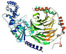
Adenylate cyclase is an enzyme with systematic name ATP diphosphate-lyase . It catalyzes the following reaction:

<span class="mw-page-title-main">G protein-coupled receptor</span> Class of cell surface receptors coupled to G-protein-associated intracellular signaling

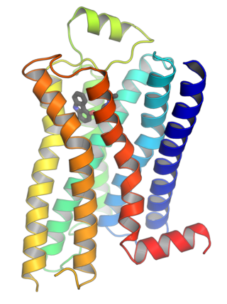
G protein-coupled receptors (GPCRs), also known as seven-(pass)-transmembrane domain receptors, 7TM receptors, heptahelical receptors, serpentine receptors, and G protein-linked receptors (GPLR), form a large group of evolutionarily related proteins that are cell surface receptors that detect molecules outside the cell and activate cellular responses. They are coupled with G proteins. They pass through the cell membrane seven times in the form of six loops of amino acid residues, which is why they are sometimes referred to as seven-transmembrane receptors. Ligands can bind either to the extracellular N-terminus and loops or to the binding site within transmembrane helices. They are all activated by agonists, although a spontaneous auto-activation of an empty receptor has also been observed.

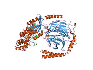
GTPases are a large family of hydrolase enzymes that bind to the nucleotide guanosine triphosphate (GTP) and hydrolyze it to guanosine diphosphate (GDP). The GTP binding and hydrolysis takes place in the highly conserved P-loop "G domain", a protein domain common to many GTPases.

<span class="mw-page-title-main">G protein</span> Type of proteins

G proteins, also known as guanine nucleotide-binding proteins, are a family of proteins that act as molecular switches inside cells, and are involved in transmitting signals from a variety of stimuli outside a cell to its interior. Their activity is regulated by factors that control their ability to bind to and hydrolyze guanosine triphosphate (GTP) to guanosine diphosphate (GDP). When they are bound to GTP, they are 'on', and, when they are bound to GDP, they are 'off'. G proteins belong to the larger group of enzymes called GTPases.

<span class="mw-page-title-main">Glucagon</span> Peptide hormone

Glucagon is a peptide hormone, produced by alpha cells of the pancreas. It raises the concentration of glucose and fatty acids in the bloodstream and is considered to be the main catabolic hormone of the body. It is also used as a medication to treat a number of health conditions. Its effect is opposite to that of insulin, which lowers extracellular glucose. It is produced from proglucagon, encoded by the GCG gene.

<span class="mw-page-title-main">Protein kinase A</span> Family of enzymes

In cell biology, protein kinase A (PKA) is a family of serine-threonine kinase whose activity is dependent on cellular levels of cyclic AMP (cAMP). PKA is also known as cAMP-dependent protein kinase. PKA has several functions in the cell, including regulation of glycogen, sugar, and lipid metabolism. It should not be confused with 5'-AMP-activated protein kinase.

A hormone receptor is a receptor molecule that binds to a specific hormone. Hormone receptors are a wide family of proteins made up of receptors for thyroid and steroid hormones, retinoids and Vitamin D, and a variety of other receptors for various ligands, such as fatty acids and prostaglandins. Hormone receptors are of mainly two classes. Receptors for peptide hormones tend to be cell surface receptors built into the plasma membrane of cells and are thus referred to as trans membrane receptors. An example of this is Actrapid. Receptors for steroid hormones are usually found within the protoplasm and are referred to as intracellular or nuclear receptors, such as testosterone. Upon hormone binding, the receptor can initiate multiple signaling pathways, which ultimately leads to changes in the behavior of the target cells.

<span class="mw-page-title-main">Opioid receptor</span> Group of biological receptors

Opioid receptors are a group of inhibitory G protein-coupled receptors with opioids as ligands. The endogenous opioids are dynorphins, enkephalins, endorphins, endomorphins and nociceptin. The opioid receptors are ~40% identical to somatostatin receptors (SSTRs). Opioid receptors are distributed widely in the brain, in the spinal cord, on peripheral neurons, and digestive tract.

<span class="mw-page-title-main">Alfred G. Gilman</span> American pharmacologist

Alfred Goodman Gilman was an American pharmacologist and biochemist. He and Martin Rodbell shared the 1994 Nobel Prize in Physiology or Medicine "for their discovery of G-proteins and the role of these proteins in signal transduction in cells."

Second messengers are intracellular signaling molecules released by the cell in response to exposure to extracellular signaling molecules—the first messengers. Second messengers trigger physiological changes at cellular level such as proliferation, differentiation, migration, survival, apoptosis and depolarization.

<span class="mw-page-title-main">G protein-gated ion channel</span>

G protein-gated ion channels are a family of transmembrane ion channels in neurons and atrial myocytes that are directly gated by G proteins.

<span class="mw-page-title-main">Beta-1 adrenergic receptor</span> Protein-coding gene in the species Homo sapiens

The beta-1 adrenergic receptor, also known as ADRB1, can refer to either the protein-encoding gene or one of the four adrenergic receptors. It is a G-protein coupled receptor associated with the Gs heterotrimeric G-protein that is expressed predominantly in cardiac tissue. In addition to cardiac tissue, beta-1 adrenergic receptors are also expressed in the cerebral cortex.

G<sub>s</sub> alpha subunit Mammalian protein found in Homo sapiens

The Gs alpha subunit is a subunit of the heterotrimeric G protein Gs that stimulates the cAMP-dependent pathway by activating adenylyl cyclase. Gsα is a GTPase that functions as a cellular signaling protein. Gsα is the founding member of one of the four families of heterotrimeric G proteins, defined by the alpha subunits they contain: the Gαs family, Gαi/Gαo family, Gαq family, and Gα12/Gα13 family. The Gs-family has only two members: the other member is Golf, named for its predominant expression in the olfactory system. In humans, Gsα is encoded by the GNAS complex locus, while Golfα is encoded by the GNAL gene.

Gq protein alpha subunit is a family of heterotrimeric G protein alpha subunits. This family is also commonly called the Gq/11 (Gq/G11) family or Gq/11/14/15 family to include closely related family members. G alpha subunits may be referred to as Gq alpha, Gαq, or Gqα. Gq proteins couple to G protein-coupled receptors to activate beta-type phospholipase C (PLC-β) enzymes. PLC-β in turn hydrolyzes phosphatidylinositol 4,5-bisphosphate (PIP2) to diacyl glycerol (DAG) and inositol trisphosphate (IP3). IP3 acts as a second messenger to release stored calcium into the cytoplasm, while DAG acts as a second messenger that activates protein kinase C (PKC).

Gi protein alpha subunit is a family of heterotrimeric G protein alpha subunits. This family is also commonly called the Gi/o family or Gi/o/z/t family to include closely related family members. G alpha subunits may be referred to as Gi alpha, Gαi, or Giα.

<span class="mw-page-title-main">G alpha subunit</span>

G alpha subunits are one of the three types of subunit of guanine nucleotide binding proteins, which are membrane-associated, heterotrimeric G proteins.

<span class="mw-page-title-main">Regulator of G protein signaling</span>

Regulators of G protein signaling (RGS) are protein structural domains or the proteins that contain these domains, that function to activate the GTPase activity of heterotrimeric G-protein α-subunits.

In the field of molecular biology, the cAMP-dependent pathway, also known as the adenylyl cyclase pathway, is a G protein-coupled receptor-triggered signaling cascade used in cell communication.

<span class="mw-page-title-main">G beta-gamma complex</span>

The G beta-gamma complex (Gβγ) is a tightly bound dimeric protein complex, composed of one Gβ and one Gγ subunit, and is a component of heterotrimeric G proteins. Heterotrimeric G proteins, also called guanosine nucleotide-binding proteins, consist of three subunits, called alpha, beta, and gamma subunits, or Gα, Gβ, and Gγ. When a G protein-coupled receptor (GPCR) is activated, Gα dissociates from Gβγ, allowing both subunits to perform their respective downstream signaling effects. One of the major functions of Gβγ is the inhibition of the Gα subunit.

<span class="mw-page-title-main">Protein–ligand complex</span>

A protein–ligand complex is a complex of a protein bound with a ligand that is formed following molecular recognition between proteins that interact with each other or with other molecules. Formation of a protein-ligand complex is based on molecular recognition between biological macromolecules and ligands, where ligand means any molecule that binds the protein with high affinity and specificity. Molecular recognition is not a process by itself since it is part of a functionally important mechanism involving the essential elements of life like in self-replication, metabolism, and information processing. For example DNA-replication depends on recognition and binding of DNA double helix by helicase, DNA single strand by DNA-polymerase and DNA segments by ligase. Molecular recognition depends on affinity and specificity. Specificity means that proteins distinguish the highly specific binding partner from less specific partners and affinity allows the specific partner with high affinity to remain bound even if there are high concentrations of less specific partners with lower affinity.

References

  1. Hurowitz EH, Melnyk JM, Chen YJ, Kouros-Mehr H, Simon MI, Shizuya H (April 2000). "Genomic characterization of the human heterotrimeric G protein alpha, beta, and gamma subunit genes". DNA Research. 7 (2): 111–20. doi: 10.1093/dnares/7.2.111 . PMID   10819326.
  2. Nature Reviews Drug Discovery GPCR Questionnaire Participants (July 2004). "The state of GPCR research in 2004". Nature Reviews. Drug Discovery (3 ed.). 3 (7): 575, 577–626. doi:10.1038/nrd1458. PMID   15272499. S2CID   33620092.
  3. Fung BK, Hurley JB, Stryer L (January 1981). "Flow of information in the light-triggered cyclic nucleotide cascade of vision". Proceedings of the National Academy of Sciences of the United States of America. 78 (1): 152–6. Bibcode:1981PNAS...78..152F. doi: 10.1073/pnas.78.1.152 . PMC   319009 . PMID   6264430.
  4. Cerione RA, Sibley DR, Codina J, Benovic JL, Winslow J, Neer EJ, Birnbaumer L, Caron MG, Lefkowitz RJ, et al. (August 1984). "Reconstitution of a hormone-sensitive adenylate cyclase system. The pure beta-adrenergic receptor and guanine nucleotide regulatory protein confer hormone responsiveness on the resolved catalytic unit". The Journal of Biological Chemistry. 259 (16): 9979–82. doi: 10.1016/S0021-9258(18)90913-0 . PMID   6088509.
  5. May DC, Ross EM, Gilman AG, Smigel MD (December 1985). "Reconstitution of catecholamine-stimulated adenylate cyclase activity using three purified proteins". The Journal of Biological Chemistry. 260 (29): 15829–33. doi: 10.1016/S0021-9258(17)36333-0 . PMID   2999139.
  6. Strathmann MP, Simon MI (July 1991). "G alpha 12 and G alpha 13 subunits define a fourth class of G protein alpha subunits". Proceedings of the National Academy of Sciences of the United States of America. 88 (13): 5582–6. Bibcode:1991PNAS...88.5582S. doi: 10.1073/pnas.88.13.5582 . PMC   51921 . PMID   1905812.
  7. 1 2 Qin K, Dong C, Wu G, Lambert NA (August 2011). "Inactive-state preassembly of G(q)-coupled receptors and G(q) heterotrimers". Nature Chemical Biology. 7 (10): 740–7. doi:10.1038/nchembio.642. PMC   3177959 . PMID   21873996.
  8. 1 2 3 4 Saroz, Yurii; Kho, Dan T.; Glass, Michelle; Graham, Euan Scott; Grimsey, Natasha Lillia (2019-10-19). "Cannabinoid Receptor 2 (CB 2 ) Signals via G-alpha-s and Induces IL-6 and IL-10 Cytokine Secretion in Human Primary Leukocytes". ACS Pharmacology & Translational Science. 2 (6): 414–428. doi: 10.1021/acsptsci.9b00049 . ISSN   2575-9108. PMC   7088898 . PMID   32259074.
  9. Zhuang, Xiaoxi; Belluscio, Leonardo; Hen, Rene (15 August 2000). "G OLFα Mediates Dopamine D 1 Receptor Signaling". The Journal of Neuroscience. 20 (16): RC91–RC91. doi:10.1523/JNEUROSCI.20-16-j0001.2000. ISSN   0270-6474. PMC   6772608 . PMID   10924528 . Retrieved 8 February 2024.
  10. Schmidt CJ, Thomas TC, Levine MA, Neer EJ (July 1992). "Specificity of G protein beta and gamma subunit interactions". The Journal of Biological Chemistry. 267 (20): 13807–10. doi: 10.1016/S0021-9258(19)49638-5 . PMID   1629181.
  11. Gulati S, Jin H, Masuho I, Orban T, Cai Y, Pardon E, Martemyanov KA, Kiser PD, Stewart PL, Ford CP, Steyaert J, Palczewski K (2018). "Targeting G protein-coupled receptor signaling at the G protein level with a selective nanobody inhibitor". Nature Communications. 9 (1): 1996. Bibcode:2018NatCo...9.1996G. doi:10.1038/s41467-018-04432-0. PMC   5959942 . PMID   29777099.
  12. Mohanasundaram, Boominathan; Dodds, Audrey; Kukshal, Vandna; Jez, Joseph M; Pandey, Sona (4 April 2022). "Distribution and the evolutionary history of G-protein components in plant and algal lineages". Plant Physiology: kiac153. doi:10.1093/plphys/kiac153. PMC   9237705 . PMID   35377452.