Presacral space

Last updated
Presacral space
Gray837.png
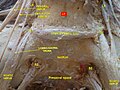
Sacral plexus of the right side. (Presacral space visible but not labeled.)
Anatomical terminology

In human anatomy, the presacral space is inside the pelvis, behind the rectum and in front of the coccyx and sacrum. Normally it is empty, or it contains a pocket of fat.

Pelvis lower part of the trunk of the human body between the abdomen and the thighs (sometimes also called pelvic region of the trunk

The pelvis is either the lower part of the trunk of the human body between the abdomen and the thighs or the skeleton embedded in it.

Rectum final section of intestine

The rectum is the final straight portion of the large intestine in humans and some other mammals, and the gut in others. The adult human rectum is about 12 centimetres (4.7 in) long, and begins at the rectosigmoid junction, the end of the sigmoid colon, at the level of the third sacral vertebra or the sacral promontory depending upon what definition is used. Its caliber is similar to that of the sigmoid colon at its commencement, but it is dilated near its termination, forming the rectal ampulla. It terminates at the level of the anorectal ring or the dentate line, again depending upon which definition is used. In humans, the rectum is followed by the anal canal which is about 4 centimetres (1.6 in) long, before the gastrointestinal tract terminates at the anal verge. The word rectum comes from the Latin rectum intestinum, meaning straight intestine.

Coccyx bone of the pelvis

The coccyx, commonly referred to as the tailbone, is the final segment of the vertebral column in all apes, and analogous structures in certain other mammals such as horses. In tailless primates since Nacholapithecus, the coccyx is the remnant of a vestigial tail. In animals with bony tails, it is known as tailhead or dock, in bird anatomy as tailfan. It comprises three to five separate or fused coccygeal vertebrae below the sacrum, attached to the sacrum by a fibrocartilaginous joint, the sacrococcygeal symphysis, which permits limited movement between the sacrum and the coccyx.

Contents

It is usually covered by sigmoid colon. [1]

Clinical significance

The presacral space may contain one of several kinds of tumor. The most common tumor here is sacrococcygeal teratoma. Others are retrorectal hamartoma (tailgut cyst), schwannoma, ganglioneuroma, and ependymoma. Also sometimes found here is an anterior meningocele, a relatively mild form of neural tube defect.

Sacrococcygeal teratoma (SCT) is a type of tumor known as a teratoma that develops at the base of the coccyx (tailbone) and is thought to be derived from the primitive streak. Sacrococcygeal teratomas are benign 75% of the time, malignant 12% of the time, and the remainder are considered "immature teratomas" that share benign and malignant features. Benign sacrococcygeal teratomas are more likely to develop in younger children who are less than 5 months old, and older children are more likely to develop malignant sacrococcygeal teratomas. The Currarino triad, due to an autosomal dominant mutation in the MNX1 gene, consists of a presacral mass, anorectal malformation and sacral dysgenesis.

Hamartoma A non cancerous growth made up of an abnormal mixture of cells and tissues normally found in the area of the body.

A hamartoma is a mostly benign, focal malformation that resembles a neoplasm in the tissue of its origin. While traditionally considered developmental malformation, many hamartomas have clonal chromosomal aberrations that are acquired through somatic mutations and on this basis are now considered to be neoplastic. It grows at the same rate as the surrounding tissue. It is composed of tissue elements normally found at that site, but they are growing in a disorganized manner. Hamartomas occur in many different parts of the body, and are most often asymptomatic incidentalomas.

Schwannoma Human disease

A schwannoma is a usually benign nerve sheath tumor composed of Schwann cells, which normally produce the insulating myelin sheath covering peripheral nerves.

Causes of increased pre-sacral space are: ulcerative colitis, granulomatous colitis, lymphogranuloma venereum, and in postirradiation changes, thrombosis of the inferior vena cava and tuberculous proctitis, tumor of the sacrum or posterior wall of the rectum. Carcinomatous deposits in the pelvis can also cause presacral space widening.

Ulcerative colitis inflammatory bowel disease that causes ulcers in the colon

Ulcerative colitis (UC) is a long-term condition that results in inflammation and ulcers of the colon and rectum. The primary symptoms of active disease are abdominal pain and diarrhea mixed with blood. Weight loss, fever, and anemia may also occur. Often, symptoms come on slowly and can range from mild to severe. Symptoms typically occur intermittently with periods of no symptoms between flares. Complications may include megacolon, inflammation of the eye, joints, or liver, and colon cancer.

Crohns disease type of inflammatory bowel disease

Crohn's disease is a type of inflammatory bowel disease (IBD) that may affect any part of the gastrointestinal tract from mouth to anus. Signs and symptoms often include abdominal pain, diarrhea, fever, and weight loss. Other complications may occur outside the gastrointestinal tract and include anemia, skin rashes, arthritis, inflammation of the eye, and tiredness. The skin rashes may be due to infections as well as pyoderma gangrenosum or erythema nodosum. Bowel obstruction may occur as a complication of chronic inflammation, and those with the disease are at greater risk of bowel cancer.

Lymphogranuloma venereum sexually transmitted disease caused by the invasive serovars L1, L2, L2a or L3 of Chlamydia trachomatis.

Lymphogranuloma venereum (LGV) is a sexually transmitted disease caused by the invasive serovars L1, L2, L2a, L2b or L3 of Chlamydia trachomatis.

Presacral edema is common in patients with heart failure who are confined to bed. [2]

Heart failure condition in which the heart is unable to provide sufficient pump action

Heart failure (HF), also known as chronic heart failure (CHF), is when the heart is unable to pump sufficiently to maintain blood flow to meet the body's needs. Signs and symptoms of heart failure commonly include shortness of breath, excessive tiredness, and leg swelling. The shortness of breath is usually worse with exercise, while lying down, and may wake the person at night. A limited ability to exercise is also a common feature. Chest pain, including angina, does not typically occur due to heart failure.

Related Research Articles

Levator ani

The levator ani is a broad, thin muscle, situated on either side of the pelvis. It is formed from three muscle components: the pubococcygeus, the iliococcygeus, and the puborectalis.

Sacrum bone of the pelvis

The sacrum, in human anatomy, is a large, triangular bone at the base of the spine that forms by the fusing of sacral vertebrae S1–S5 between 18 and 30 years of age.

Sigmoid colon part of the large intestine that is closest to the rectum and anus

The sigmoid colon is the part of the large intestine that is closest to the rectum and anus. It forms a loop that averages about 35–40 cm (13.78-15.75 in) in length. The loop is typically shaped like a Greek letter sigma (ς) or Latin letter S. This part of the colon normally lies within the pelvis, but on account of its freedom of movement it is liable to be displaced into the abdominal cavity.

Abdominal pain Stomach aches

Abdominal pain, also known as a stomach ache, is a symptom associated with both non-serious and serious medical issues.

Pelvic floor anatomical structure

The pelvic floor or pelvic diaphragm is composed of muscle fibers of the levator ani, the coccygeus muscle, and associated connective tissue which span the area underneath the pelvis. The pelvic diaphragm is a muscular partition formed by the levatores ani and coccygei, with which may be included the parietal pelvic fascia on their upper and lower aspects. The pelvic floor separates the pelvic cavity above from the perineal region below. Because, to accommodate the birth canal, a female's pelvic cavity is larger than a male's, the pelvic floor tends to be considered a part of female anatomy, but males have an equivalent pelvic floor.

Sacrotuberous ligament

The sacrotuberous ligament is situated at the lower and back part of the pelvis. It is flat, and triangular in form; narrower in the middle than at the ends.

Perineal hernia Human disease

Perineal hernia is a hernia involving the perineum. The hernia may contain fluid, fat, any part of the intestine, the rectum, or the bladder. It is known to occur in humans, dogs, and other mammals, and often appears as a sudden swelling to one side of the anus.

Median sacral artery

The median sacral artery is a small vessel that arises posterior to the abdominal aorta and superior to its bifurcation.

Superior hypogastric plexus

The superior hypogastric plexus is a plexus of nerves situated on the vertebral bodies anterior to the bifurcation of the abdominal aorta.

Pelvic cavity body cavity bounded by the bones of the pelvis

The pelvic cavity is a body cavity that is bounded by the bones of the pelvis. Its oblique roof is the pelvic inlet. Its lower boundary is the pelvic floor.

Superior rectal artery

The superior rectal artery is an artery that descends into the pelvis to supply blood to the rectum.

Internal iliac vein Large blood vessel of the pelvis

The internal iliac vein begins near the upper part of the greater sciatic foramen, passes upward behind and slightly medial to the internal iliac artery and, at the brim of the pelvis, joins with the external iliac vein to form the common iliac vein.

Urogenital triangle

The urogenital triangle is the anterior part of the perineum. In female mammals, it contains the vagina and associated parts of the external genitalia.

Anal triangle posterior part of the perineum

The anal triangle is the posterior part of the perineum. It contains the anal canal.

Uterosacral ligament

The uterosacral ligaments belong to the major ligaments of uterus.

Currarino syndrome

The Currarino syndrome is an inherited congenital disorder where either the sacrum is not formed properly, or there is a mass in the presacral space in front of the sacrum, and (3) there are malformations of the anus or rectum. It can also cause an anterior meningocele or a presacral teratoma.

The presacral fascia lines the anterior aspect of the sacrum, enclosing the sacral vessels and nerves. It continues anteriorly as the pelvic parietal fascia, covering the entire pelvic cavity.

References

  1. Linda Cardozo; David Staskin (2006). Textbook of female urology and urogynecology. CRC Press. pp. 1195–. ISBN   978-1-84184-358-2 . Retrieved 19 May 2011.
  2. Braunwald, Eugene; Loscalzo, Joseph (2015). "Edema". In Kasper, Dennis L.; Fauci, Anthony S.; Hauser, Stephen L.; Longo, Dan L.; Jameson, J. Larry. Harrison's Principles of Internal Medicine (19th ed.). New York: McGraw Hill Education. pp. 250–253. ISBN   978-0-07-180213-0.

Additional images