Tympanostomy tube

Last updated

Tympanostomy tube
Grommet.jpg
The grommet is less than 2 mm tall, smaller than a match head.
Other namesGrommet, T-tube, ear tube, pressure equalization tube, vent, PE tube, myringotomy tube

Tympanostomy tube, also known as a grommet,myringotomy tube, or pressure equalizing tube, is a small tube inserted into the eardrum via a surgical procedure called myringotomy to keep the middle ear aerated for a prolonged period of time, typically to prevent accumulation of fluid in the middle ear. [1] 
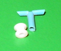
The tube itself is made in a variety of designs, most often shaped like a grommet for short-term use, or with long flanges and sometimes resembling a T-shape for long-term use. [2] Materials used to manufacture the tubes are often made from fluoroplastic or silicone, which have largely replaced the use of metal tubes made from stainless steel, titanium, or gold. [2]

Contents

Tympanostomy tubes of varying size and shape. Examples of short-term use grommet (left) and long-term use T-shape (right). Paukenroerchen.jpg
Tympanostomy tubes of varying size and shape. Examples of short-term use grommet (left) and long-term use T-shape (right).

Medical uses

Tympanostomy tube in the eardrum, draining fluid from the middle ear during an ear infection. Ear Tube.png
Tympanostomy tube in the eardrum, draining fluid from the middle ear during an ear infection.

Inserting tympanostomy tubes is one of the most common pediatric surgical procedures United States, with 9% of children having had tubes placed sometime in their lives. [1] [3] Tympanostomy tubes are typically placed in one or both eardrums to help children suffering from recurrent acute otitis media (ear infection) or persistent otitis media with effusion (sometimes called "glue ear"). [1] [4] [5]

Tympanostomy tubes work by improving drainage, allowing air to circulate in the middle ear, and offering a direct route for antibiotics to enter the middle ear. [2] [6] Tube placement has been shown to increase hearing in children with persistent otitis media with effusion and may lead to fewer ear infections for children with frequent ear infections. [1] [2] Once placed, short-term tubes are designed to stay in the eardrum for 6–15 months, whereas long-term tubes are designed to stay for 15–18 months. [2] [6] Tympanostomy tubes usually fall out on their own as the eardrum heals over time; however, they can sometimes get stuck in the eardrum and require surgical assistance for removal. [2]

Guidelines state that tubes are an option in:

While tympanostomy tubes are commonly used in children, they are seldom used in adults. Options for use in adults include:

Adverse effects

Otorrhea (ear discharge) is the most common complication of tympanostomy tube placement, affecting between 25–75% of children receiving this procedure. [2] [6] [9] [10] Saline washouts and antibiotic drops at the time of surgery are effective measures to reduce rates of otorrhea, which is why antibiotic ear drops are not routinely prescribed. [1] [6] [9] If children experience persistent ear drainage or have new discharge two weeks following surgery, antibiotic ear drops are an effective treatment and have been shown to work better than oral antibiotics. [1] [9] [10] Frequent use of ear drops in children may have an ototoxic effect, which is why antibiotic ear drop use following surgery should only be recommended by a trained healthcare professional. [9]

Potential risks of tympanostomy tube placement in children include going under general anesthesia to have the procedure as well as adverse effects following tube placement. [1] Estimates of these other adverse effects from tubes being in the eardrum include:

Long term effects include visible changes to the eardrum such as tympanosclerosis, cholesteatoma, focal atrophy, or retraction pockets. [1] [11] These changes usually resolve on their own and do not usually require medical treatment or result in hearing problems that are clinically significant. [1] [12]

Surgical intervention may be required in cases of persistent perforation or retained tympanostomy tubes. Persistent perforations are corrected via tympanoplasty with an 80-90% success rate. [1] Most tympanostomy tubes fall out on their own as the eardrum heals; however, when tubes remain after 2–3 years they are often removed to prevent complications. [13]

Procedure

Myringotomy with insertion of tympanostomy tubes is performed by an ENT doctor (otolaryngologist) and is one of the most common pediatric surgical procedures, accounting for more than 20% of all ambulatory pediatric surgeries in 2006. [1] Although myringotomy with tympanostomy tube insertion can be performed in-office under local anesthesia for adolescents and adults, children or any patient who may have difficulty lying still during the procedure require general anesthesia. [6] [14]

During the procedure, a small incision is made to the eardrum using either a myringotomy knife or a CO2 laser. [6] [15] The middle ear is then usually washed out thoroughly with saline before the tympanostomy tubes are placed. Antibiotic drops are commonly used during surgery once tubes are placed but are not routinely prescribed for use following surgery unless recommended by a doctor for individual reasons. [1] [6] [9]

Following surgery, it is suggested that children keep their ears dry for the first two weeks to help prevent complications. After two weeks children do not need to wear earplugs when swimming or to take other measures to prevent water from getting in their ears as there is minimal reduction in adverse effects. [1] [16] It is approximated that a child would need to wear ear plugs for 2.8 years to prevent one additional ear infection. [16]

Outcome

Tympanostomy tubes generally remain in the eardrum for six months to two years, and about 14% of children will require tympanostomy tubes more than once. [1] The eardrum usually closes without a residual hole at the tube site but in a small number of cases a perforation can persist. [1] For children with otitis media with effusion (glue ear), tympanostomy tubes decrease the prevalence of effusions by 33% and improve hearing by 5-12 decibels, within 1–3 months of the procedure. There is no long-term benefit for hearing by 12–24 months after the procedure. [1] [6] For children with frequent episodes of acute otitis media (ear infection), there is debate about the effectiveness of tympanostomy tubes in reducing rates of infections. There is consensus, however, on the beneficial role of tympanostomy tubes in allowing for drainage of infections and offering direct access to the middle ear with antibiotic drops. [1] This aids healthcare providers in identifying the cause of the infection so they can better treat it and decrease the need for systemic antibiotic use. [1] [6]

History

The first myringotomy dates back to 1649 when French anatomist Jean Riolan noticed an improvement in his hearing after intentionally perforating his eardrum with a spoon. [17] For nearly two hundred years, scientists would study and debate the potential benefits of myringotomy before German scientists Martell Frank and Gustav Lincke had the first documented use of tympanostomy tubes in 1845. These scientists used an approximately 6mm long gold tube in an attempt to prevent the eardrum from closing after myringotomy. [18] From 1845 to 1875, seven different types of tympanostomy tubes were manufactured and made of materials including rubber, silver, aluminum, and gold. These tubes were not widely used or accepted due to complications including falling into the middle ear, falling out of the ear, and the tubes getting plugged. [18] In 1952, tympanostomy tubes would make a return when American otolaryngologist Beverly Armstrong introduced them as a new treatment for chronic secretory otitis media. This would lead to numerous types of tympanostomy tubes being developed and studied throughout the 20th century. [18]

Today, silicone and fluoroplastic tympanostomy tubes are more commonly used than metal tubes made from stainless steel, titanium, or gold. [2] Research on ways to reduce biofilm formation on tympanostomy tubes, such as coating the tubes with antibiotics before placement to help prevent tube blockage or infection, has been started, but there is not enough data to determine its effectiveness. [2] Dissolvable tubes are also being explored as potential alternatives for current tube materials. [2]

Related Research Articles

<span class="mw-page-title-main">Otorhinolaryngology</span> Medical specialty

Otorhinolaryngology is a surgical subspeciality within medicine that deals with the surgical and medical management of conditions of the head and neck. Doctors who specialize in this area are called otorhinolaryngologists, otolaryngologists, head and neck surgeons, or ENT surgeons or physicians. Patients seek treatment from an otorhinolaryngologist for diseases of the ear, nose, throat, base of the skull, head, and neck. These commonly include functional diseases that affect the senses and activities of eating, drinking, speaking, breathing, swallowing, and hearing. In addition, ENT surgery encompasses the surgical management of cancers and benign tumors and reconstruction of the head and neck as well as plastic surgery of the face, scalp, and neck.

<span class="mw-page-title-main">Ménière's disease</span> Disorder of the inner ear

Ménière's disease (MD) is a disease of the inner ear that is characterized by potentially severe and incapacitating episodes of vertigo, tinnitus, hearing loss, and a feeling of fullness in the ear. Typically, only one ear is affected initially, but over time, both ears may become involved. Episodes generally last from 20 minutes to a few hours. The time between episodes varies. The hearing loss and ringing in the ears can become constant over time.

<span class="mw-page-title-main">Eardrum</span> Membrane separating the external ear from the middle ear

In the anatomy of humans and various other tetrapods, the eardrum, also called the tympanic membrane or myringa, is a thin, cone-shaped membrane that separates the external ear from the middle ear. Its function is to transmit sound from the air to the ossicles inside the middle ear, and thence to the oval window in the fluid-filled cochlea. The ear thereby converts and amplifies vibration in the air to vibration in cochlear fluid. The malleus bone bridges the gap between the eardrum and the other ossicles.

<span class="mw-page-title-main">Otitis media</span> Inflammation of the middle ear

Otitis media is a group of inflammatory diseases of the middle ear. One of the two main types is acute otitis media (AOM), an infection of rapid onset that usually presents with ear pain. In young children this may result in pulling at the ear, increased crying, and poor sleep. Decreased eating and a fever may also be present. The other main type is otitis media with effusion (OME), typically not associated with symptoms, although occasionally a feeling of fullness is described; it is defined as the presence of non-infectious fluid in the middle ear which may persist for weeks or months often after an episode of acute otitis media. Chronic suppurative otitis media (CSOM) is middle ear inflammation that results in a perforated tympanic membrane with discharge from the ear for more than six weeks. It may be a complication of acute otitis media. Pain is rarely present. All three types of otitis media may be associated with hearing loss. If children with hearing loss due to OME do not learn sign language, it may affect their ability to learn.

<span class="mw-page-title-main">Eustachian tube</span> Tube connecting middle ear to throat

The Eustachian tube, also called the auditory tube or pharyngotympanic tube, is a tube that links the nasopharynx to the middle ear, of which it is also a part. In adult humans, the Eustachian tube is approximately 35 mm (1.4 in) long and 3 

<span class="mw-page-title-main">Adenoidectomy</span> Surgical removal of the adenoid

Adenoidectomy is the surgical removal of the adenoid for reasons which include impaired breathing through the nose, chronic infections, or recurrent earaches. The effectiveness of removing the adenoids in children to improve recurrent nasal symptoms and/or nasal obstruction has not been well studied. The surgery is less commonly performed in adults in whom the adenoid is much smaller and less active than it is in children. It is most often done on an outpatient basis under general anesthesia. Post-operative pain is generally minimal and reduced by icy or cold foods. The procedure is often combined with tonsillectomy, for which the recovery time is an estimated 10–14 days, sometimes longer, mostly dependent on age.

<span class="mw-page-title-main">Conductive hearing loss</span> Medical condition

Conductive hearing loss (CHL) occurs when there is a problem transferring sound waves anywhere along the pathway through the outer ear, tympanic membrane (eardrum), or middle ear (ossicles). If a conductive hearing loss occurs in conjunction with a sensorineural hearing loss, it is referred to as a mixed hearing loss. Depending upon the severity and nature of the conductive loss, this type of hearing impairment can often be treated with surgical intervention or pharmaceuticals to partially or, in some cases, fully restore hearing acuity to within normal range. However, cases of permanent or chronic conductive hearing loss may require other treatment modalities such as hearing aid devices to improve detection of sound and speech perception.

A myringotomy is a surgical procedure in which an incision is created in the eardrum to relieve pressure caused by excessive buildup of fluid, or to drain pus from the middle ear. A tympanostomy tube may be inserted through the eardrum to keep the middle ear aerated for a prolonged time and to prevent reaccumulation of fluid. Without the insertion of a tube, the incision usually heals spontaneously within two to three weeks. Depending on the type, the tube is either naturally extruded in 6 to 12 months or removed during a minor procedure.

<span class="mw-page-title-main">Tympanoplasty</span> Surgical operation on the ear

Tympanoplasty is the surgical operation performed to reconstruct hearing mechanism of middle ear.

<span class="mw-page-title-main">Ear pain</span> Pain in the ear

Ear pain, also known as earache or otalgia, is pain in the ear. Primary ear pain is pain that originates from the ear. Secondary ear pain is a type of referred pain, meaning that the source of the pain differs from the location where the pain is felt.

<span class="mw-page-title-main">Mastoiditis</span> Middle ear disease

Mastoiditis is the result of an infection that extends to the air cells of the skull behind the ear. Specifically, it is an inflammation of the mucosal lining of the mastoid antrum and mastoid air cell system inside the mastoid process. The mastoid process is the portion of the temporal bone of the skull that is behind the ear. The mastoid process contains open, air-containing spaces. Mastoiditis is usually caused by untreated acute otitis media and used to be a leading cause of child mortality. With the development of antibiotics, however, mastoiditis has become quite rare in developed countries where surgical treatment is now much less frequent and more conservative, unlike former times.

<span class="mw-page-title-main">Otitis</span> Medical condition

Otitis is a general term for inflammation in ear or ear infection, inner ear infection, middle ear infection of the ear, in both humans and other animals. When infection is present, it may be viral or bacterial. When inflammation is present due to fluid build up in the middle ear and infection is not present it is considered Otitis media with effusion. It is subdivided into the following:

<span class="mw-page-title-main">Perforated eardrum</span> Injury leading to a hole in the eardrum

A perforated eardrum is a prick in the eardrum. It can be caused by infection, trauma, overpressure, inappropriate ear clearing, and changes in middle ear pressure. An otoscope can be used to view the eardrum to diagnose a perforation. Perforations may heal naturally or require surgery.

Jack Leon Paradise was a pediatrician, pediatric primary care researcher, and professor emeritus of pediatrics at University of Pittsburgh School of Medicine.

<span class="mw-page-title-main">Operating microscope</span> Optical microscope used in surgery

An operating microscope or surgical microscope is an optical microscope specifically designed to be used in a surgical setting, typically to perform microsurgery.

Myringoplasty is the closure of the perforation of pars tensa of the tympanic membrane. When myringoplasty is combined with removal of scar tissue, it is called tympanoplasty. The operation is performed with the patient supine and face turned to one side. The graft material most commonly used for the surgery is temporalis fascia. The tragal cartilage and tragal perichondrium are also used as the graft by some surgeons.

<span class="mw-page-title-main">Tympanosclerosis</span> Medical condition

Tympanosclerosis is a condition caused by hyalinization and subsequent calcification of subepithelial connective tissue of the tympanic membrane and middle ear, sometimes resulting in a detrimental effect to hearing.

<span class="mw-page-title-main">Tympanic membrane retraction</span> Medical condition

Tympanic membrane retraction describes a condition in which a part of the eardrum lies deeper within the ear than its normal position.

Eustachian tube dysfunction (ETD) is a disorder where pressure abnormalities in the middle ear result in symptoms.

Middle ear barotrauma (MEBT), also known to underwater divers as ear squeeze and reverse ear squeeze, is an injury caused by a difference in pressure between the external ear canal and the middle ear. It is common in underwater divers and usually occurs when the diver does not equalise sufficiently during descent or, less commonly, on ascent. Failure to equalise may be due to inexperience or eustachian tube dysfunction, which can have many possible causes. Unequalised ambient pressure increase during descent causes a pressure imbalance between the middle ear air space and the external auditory canal over the eardrum, referred to by divers as ear squeeze, causing inward stretching, serous effusion and haemorrhage, and eventual rupture. During ascent internal over-pressure is normally passively released through the eustachian tube, but if this does not happen the volume expansion of middle ear gas will cause outward bulging, stretching and eventual rupture of the eardrum known to divers as reverse ear squeeze. This damage causes local pain and hearing loss. Tympanic rupture during a dive can allow water into the middle ear, which can cause severe vertigo from caloric stimulation. This may cause nausea and vomiting underwater, which has a high risk of aspiration of vomit or water, with possibly fatal consequences.

References

  1. 1 2 3 4 5 6 7 8 9 10 11 12 13 14 15 16 17 18 19 20 21 22 23 24 25 26 27 Rosenfeld, Richard M.; Tunkel, David E.; Schwartz, Seth R.; Anne, Samantha; Bishop, Charles E.; Chelius, Daniel C.; Hackell, Jesse; Hunter, Lisa L.; Keppel, Kristina L.; Kim, Ana H.; Kim, Tae W.; Levine, Jack M.; Maksimoski, Matthew T.; Moore, Denee J.; Preciado, Diego A. (9 February 2022). "Clinical Practice Guideline: Tympanostomy Tubes in Children (Update)". Otolaryngology–Head and Neck Surgery: Official Journal of American Academy of Otolaryngology-Head and Neck Surgery. 166 (1_suppl): S1–S55. doi:10.1177/01945998211065662. ISSN   1097-6817. PMID   35138954. S2CID   246700402 via PubMed.
  2. 1 2 3 4 5 6 7 8 9 10 Nagar, Rashi R; Deshmukh, Prasad T (11 October 2022). "An Overview of the Tympanostomy Tube". Cureus. 14 (10): e30166. doi: 10.7759/cureus.30166 . ISSN   2168-8184. PMC   9647717 . PMID   36397911.
  3. Goodman, David C.; Morden, Nancy E.; Ralston, Shawn L.; Chang, Chiang-Hua; Parker, Devin M.; Weinstein, Shelsey J. (11 December 2013), "Common Surgical Procedures", The Dartmouth Atlas of Children’s Health Care in Northern New England [Internet], The Dartmouth Institute for Health Policy and Clinical Practice, retrieved 12 February 2024
  4. Venekamp, Roderick P; Mick, Paul; Schilder, Anne GM; Nunez, Desmond A (9 May 2018). Cochrane ENT Group (ed.). "Grommets (ventilation tubes) for recurrent acute otitis media in children". Cochrane Database of Systematic Reviews. 2019 (6): CD012017. doi:10.1002/14651858.CD012017.pub2. PMC   6494623 . PMID   29741289.
  5. "Glue ear". National Deaf Children's Society . Retrieved 16 February 2019.
  6. 1 2 3 4 5 6 7 8 9 10 11 12 Spaw, Mark; Camacho, Macario (2024), "Tympanostomy Tube", StatPearls, Treasure Island (FL): StatPearls Publishing, PMID   33351417 , retrieved 12 February 2024
  7. 1 2 3 Lieberthal, AS; Carroll, AE; Chonmaitree, T; Ganiats, TG; Hoberman, A; Jackson, MA; Joffe, MD; Miller, DT; Rosenfeld, RM; Sevilla, XD; Schwartz, RH; Thomas, PA; Tunkel, DE (March 2013). "The diagnosis and management of acute otitis media". Pediatrics. 131 (3): e964-99. doi: 10.1542/peds.2012-3488 . PMID   23439909.
  8. Canadian Society of Otolaryngology- Head and Neck Surgery (2023). "Otolaryngology: Nine tests and treatments to question in pediatric Orolaryngology". Choosing Wisely Canada. Retrieved 15 February 2024.
  9. 1 2 3 4 5 Syed, Mohammed Iqbal; Suller, Sharon; Browning, George G.; Akeroyd, Michael A. (2013). "Interventions for the prevention of postoperative ear discharge after insertion of ventilation tubes (grommets) in children". The Cochrane Database of Systematic Reviews (4): CD008512. doi:10.1002/14651858.CD008512.pub2. ISSN   1469-493X. PMID   23633358.
  10. 1 2 Venekamp, Roderick P.; Javed, Faisal; van Dongen, Thijs Ma; Waddell, Angus; Schilder, Anne Gm (2016). "Interventions for children with ear discharge occurring at least two weeks following grommet (ventilation tube) insertion". The Cochrane Database of Systematic Reviews. 2016 (11): CD011684. doi:10.1002/14651858.CD011684.pub2. ISSN   1469-493X. PMC   6465056 . PMID   27854381.
  11. 1 2 3 4 5 6 Kay, David J.; Nelson, Magalie; Rosenfeld, Richard M. (1 April 2001). "Meta-Analysis of Tympanostomy Tube Sequelae". Otolaryngology–Head and Neck Surgery. 124 (4): 374–380. doi:10.1067/mhn.2001.113941. ISSN   0194-5998. PMID   11283489. S2CID   25287112.
  12. Cayé-Thomasen, Per; Stangerup, Sven-Eric; Jørgensen, Gita; Drozdziewic, Dominika; Bonding, Per; Tos, Mirko (1 August 2008). "Myringotomy Versus Ventilation Tubes in Secretory Otitis Media". Otology & Neurotology. 29 (5): 649–657. doi:10.1097/mao.0b013e318173035b. ISSN   1531-7129. PMID   18520632. S2CID   42756454.
  13. Michel, Margaret; Nahas, Gabriel; Preciado, Diego (31 August 2020). "Retained Tympanostomy Tubes: Who, What, When, Why, and How to Treat?". Ear, Nose & Throat Journal: 014556132095049. doi: 10.1177/0145561320950490 . ISSN   0145-5613. PMID   32865460.
  14. "Ear tube insertion: MedlinePlus Medical Encyclopedia". medlineplus.gov. Retrieved 14 February 2024.
  15. Chang, Chan-Wei; Yang, Ya-Wen; Fu, Chong Yau; Shiao, An-Suey (1 January 2012). "Differences between children and adults with otitis media with effusion treated with CO2 laser myringotomy". Journal of the Chinese Medical Association. 75 (1): 29–35. doi: 10.1016/j.jcma.2011.10.001 . ISSN   1726-4901. PMID   22240534.
  16. 1 2 Moualed, Daniel; Masterson, Liam; Kumar, Sanjiv; Donnelly, Neil (27 January 2016). Cochrane ENT Group (ed.). "Water precautions for prevention of infection in children with ventilation tubes (grommets)". Cochrane Database of Systematic Reviews. 2016 (1): CD010375. doi:10.1002/14651858.CD010375.pub2. PMC   10612140 . PMID   26816299.
  17. Rimmer, Joanne; Giddings, Charles E.; Weir, Neil (19 March 2020). "The History of Myringotomy and Grommets". Ear, Nose & Throat Journal. 99 (1_suppl): 2S–7S. doi: 10.1177/0145561320914438 . ISSN   0145-5613. PMID   32189517.
  18. 1 2 3 Mudry, Albert (1 February 2013). "The tympanostomy tube: An ingenious invention of the mid 19th century". International Journal of Pediatric Otorhinolaryngology. 77 (2): 153–157. doi:10.1016/j.ijporl.2012.10.024. ISSN   0165-5876. PMID   23183195.