Paranasal sinuses are a group of four paired air-filled spaces that surround the nasal cavity. The maxillary sinuses are located under the eyes; the frontal sinuses are above the eyes; the ethmoidal sinuses are between the eyes and the sphenoidal sinuses are behind the eyes. The sinuses are named for the facial bones in which they are located.

In neuroanatomy, the mandibular nerve (V3) is the largest of the three divisions of the trigeminal nerve, the fifth cranial nerve (CN V). Unlike the other divisions of the trigeminal nerve (ophthalmic nerve, maxillary nerve) which contain only afferent fibers, the mandibular nerve contains both afferent and efferent fibers. These nerve fibers innervate structures of the lower jaw and face, such as the tongue, lower lip, and chin. The mandibular nerve also innervates the muscles of mastication.

The inferior alveolar nerve(IAN) is a branch of the mandibular nerve, which is itself the third branch of the trigeminal nerve. The inferior alveolar nerves supply sensation to the lower teeth.
Infiltration analgesia is deposition of an analgesic drug close to the apex of a tooth so that it can diffuse to reach the nerve entering the apical foramina. It is the most routinely used in dental local treatment.

The pterygopalatine ganglion is a parasympathetic ganglion found in the pterygopalatine fossa. It is largely innervated by the greater petrosal nerve ; and its postsinaptic axons project to the lacrimal glands and nasal mucosa. The flow of blood to the nasal mucosa, in particular the venous plexus of the conchae, is regulated by the pterygopalatine ganglion and heats or cools the air in the nose. It is one of four parasympathetic ganglia of the head and neck, the others being the submandibular ganglion, otic ganglion, and ciliary ganglion.

The pyramid-shaped maxillary sinus is the largest of the paranasal sinuses, and drains into the middle meatus of the nose through the osteomeatal complex.
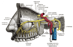
In neuroanatomy, the maxillary nerve (V2) is one of the three branches or divisions of the trigeminal nerve, the fifth (CN V) cranial nerve. It comprises the principal functions of sensation from the maxilla, nasal cavity, sinuses, the palate and subsequently that of the mid-face, and is intermediate, both in position and size, between the ophthalmic nerve and the mandibular nerve.

The maxillary first molar is the human tooth located laterally from both the maxillary second premolars of the mouth but mesial from both maxillary second molars.
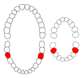
The mandibular first molar or six-year molar is the tooth located distally from both the mandibular second premolars of the mouth but mesial from both mandibular second molars. It is located on the mandibular (lower) arch of the mouth, and generally opposes the maxillary (upper) first molars and the maxillary 2nd premolar in normal class I occlusion. The function of this molar is similar to that of all molars in regard to grinding being the principal action during mastication, commonly known as chewing. There are usually five well-developed cusps on mandibular first molars: two on the buccal, two lingual, and one distal. The shape of the developmental and supplementary grooves, on the occlusal surface, are describes as being 'M' shaped. There are great differences between the deciduous (baby) mandibular molars and those of the permanent mandibular molars, even though their function are similar. The permanent mandibular molars are not considered to have any teeth that precede it. Despite being named molars, the deciduous molars are followed by permanent premolars.

In human anatomy, the pterygopalatine fossa is a fossa in the skull. A human skull contains two pterygopalatine fossae—one on the left side, and another on the right side. Each fossa is a cone-shaped paired depression deep to the infratemporal fossa and posterior to the maxilla on each side of the skull, located between the pterygoid process and the maxillary tuberosity close to the apex of the orbit. It is the indented area medial to the pterygomaxillary fissure leading into the sphenopalatine foramen. It communicates with the nasal and oral cavities, infratemporal fossa, orbit, pharynx, and middle cranial fossa through eight foramina.
The lingual nerve carries sensory innervation from the anterior two-thirds of the tongue. It contains fibres from both the mandibular division of the trigeminal nerve (CN V3) and from the facial nerve (CN VII). The fibres from the trigeminal nerve are for touch, pain and temperature (general sensation), and the ones from the facial nerve are for taste (special sensation).

The posterior superior alveolar artery is given off from the maxillary, frequently in conjunction with the infraorbital artery just as the trunk of the vessel is passing into the pterygopalatine fossa.
The anterior superior alveolar nerve (or anterior superior dental nerve), is a branch of the infraorbital nerve, itself a branch of the maxillary nerve (V2). It branches from the infraorbital nerve within the infraorbital canal before the infraorbital nerve exits through the infraorbital foramen. It descends in a canal in the anterior wall of the maxillary sinus, and divides into branches which supply the incisor and canine teeth.
The posterior superior alveolar branches arise from the trunk of the maxillary nerve just before it enters the infraorbital groove; they are generally two in number, but sometimes arise by a single trunk.

The infraorbital nerve is a branch of the maxillary nerve, itself a branch of the trigeminal nerve. It travels through the orbit and enters the infraorbital canal to exit onto the face through the infraorbital foramen. It provides sensory innervation to the skin and mucous membranes around the middle of the face.
Supernumerary roots is a condition found in teeth when there may be a larger number of roots than expected. The most common teeth affected are mandibular (lower) canines, premolars, and molars, especially third molars. Canines and most premolars, except for maxillary (upper) first premolars, usually have one root. Maxillary first premolars and mandibular molars usually have two roots. Maxillary molars usually have three roots. When an extra root is found on any of these teeth, the root is described as a supernumerary root. The clinical significance of this condition is associated with dentistry when accurate information regarding root canal anatomy is required when root canal treatment is required.
Dental anatomy is a field of anatomy dedicated to the study of human tooth structures. The development, appearance, and classification of teeth fall within its purview. Tooth formation begins before birth, and the teeth's eventual morphology is dictated during this time. Dental anatomy is also a taxonomical science: it is concerned with the naming of teeth and the structures of which they are made, this information serving a practical purpose in dental treatment.
Dental pertains to the teeth, including dentistry. Topics related to the dentistry, the human mouth and teeth include:
Inferior alveolar nerve block is a nerve block technique which induces anesthesia (numbness) in the areas of the mouth and face innervated by one of the inferior alveolar nerves which are paired on the left and right side. These areas are the skin and mucous membranes of the lower lip, the skin of the chin, the lower teeth and the labial gingiva of the anterior teeth, all unilaterally to the midline of the side on which the block is administered. However, depending on technique, the long buccal nerve may not be anesthetized by an IANB and therefore an area of buccal gingiva adjacent to the lower posterior teeth will retain normal sensation unless that nerve is anesthetized separately, via a (long) buccal nerve block. The inferior alveolar nerve is a branch of the mandibular nerve, the third division of the trigeminal nerve. This procedure attempts to anaesthetise the inferior alveolar nerve prior to it entering the mandibular foramen on the medial surface of the mandibular ramus.

In human anatomy, the mouth is the first portion of the alimentary canal that receives food and produces saliva. The oral mucosa is the mucous membrane epithelium lining the inside of the mouth.