Middle cerebral veins

Last updated
Middle cerebral veins
Gray's Anatomy plate 517 brain.png
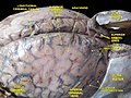
Outer surface of cerebral hemisphere, showing areas supplied by cerebral arteries. (Middle cerebral veins not labeled, but region drained is roughly equivalent to pink region.)
Lateral sulcus2.png
Lateral sulcus (Middle cerebral veins not visible, but veins run in lateral sulcus.)
Details
Drains to cavernous sinus, basal vein
Artery middle cerebral artery
Identifiers
Latin venae mediae cerebri (superficialis et profunda)
Anatomical terminology

The middle cerebral veins - the superficial middle cerebral vein and the deep middle cerebral vein - are two veins running along the lateral sulcus. The superficial middle cerebral vein is also known as the superficial Sylvian vein, and the deep middle cerebral vein is also known as the deep Sylvian vein. The lateral sulcus is also known as the Sylvian fissure.

Contents

Superficial middle cerebral vein

The superficial middle cerebral vein (superficial Sylvian vein) begins on the lateral surface of the hemisphere. It runs along the lateral sulcus [1] to empty into either the cavernous sinus, [1] [2] or sphenoparietal sinus. [1] It is adherent to the deep surface of the arachnoid mater bridging the lateral sulcus. It drains the adjacent cortex. [2]

Anastomoses

At its posterior extremity, [2] the superficial middle cerebral vein is connected with the superior sagittal sinus via the superior anastomotic vein, and with the transverse sinus via the inferior anastomotic vein. [1] [2]

Deep middle cerebral vein

The deep middle cerebral vein (deep Sylvian vein) receives tributaries from the insula and neighboring gyri, and runs in the lower part of the lateral sulcus. [3]

Additional images

Related Research Articles

<span class="mw-page-title-main">Oculomotor nerve</span> Cranial nerve III, for eye movements

The oculomotor nerve, also known as the third cranial nerve, cranial nerve III, or simply CN III, is a cranial nerve that enters the orbit through the superior orbital fissure and innervates extraocular muscles that enable most movements of the eye and that raise the eyelid. The nerve also contains fibers that innervate the intrinsic eye muscles that enable pupillary constriction and accommodation. The oculomotor nerve is derived from the basal plate of the embryonic midbrain. Cranial nerves IV and VI also participate in control of eye movement.

<span class="mw-page-title-main">Falx cerebri</span> Anatomical structure of the brain

The falx cerebri is a large, crescent-shaped fold of dura mater that descends vertically into the longitudinal fissure between the cerebral hemispheres of the human brain, separating the two hemispheres and supporting dural sinuses that provide venous and CSF drainage to the brain. It is attached to the crista galli anteriorly, and blends with the tentorium cerebelli posteriorly.

<span class="mw-page-title-main">Middle cerebral artery</span> Paired artery that supplies blood to the cerebrum

The middle cerebral artery (MCA) is one of the three major paired cerebral arteries that supply blood to the cerebrum. The MCA arises from the internal carotid artery and continues into the lateral sulcus where it then branches and projects to many parts of the lateral cerebral cortex. It also supplies blood to the anterior temporal lobes and the insular cortices.

<span class="mw-page-title-main">Straight sinus</span> Venous sinus draining the brain

The straight sinus, also known as tentorial sinus or the sinus rectus, is an area within the skull beneath the brain. It receives blood from the inferior sagittal sinus and the great cerebral vein, and drains into the confluence of sinuses.

<span class="mw-page-title-main">Superior sagittal sinus</span> Anatomical structure of the brain

The superior sagittal sinus, within the human head, is an unpaired area along the attached margin of the falx cerebri. It allows blood to drain from the lateral aspects of anterior cerebral hemispheres to the confluence of sinuses. Cerebrospinal fluid drains through arachnoid granulations into the superior sagittal sinus and is returned to venous circulation.

<span class="mw-page-title-main">Inferior sagittal sinus</span> Anatomical structure of the brain

The inferior sagittal sinus, within the human head, is an area beneath the brain which allows blood to drain outwards posteriorly from the center of the head. It drains to the straight sinus, which connects to the transverse sinuses. See diagram : labeled in the brain as "SIN. SAGITTALIS INF.".

<span class="mw-page-title-main">Inferior petrosal sinus</span>

The inferior petrosal sinuses are two small sinuses situated on the inferior border of the petrous part of the temporal bone, one on each side. Each inferior petrosal sinus drains the cavernous sinus into the internal jugular vein.

<span class="mw-page-title-main">Greater wing of sphenoid bone</span> Large part of the skull front behind the eye socket

The greater wing of the sphenoid bone, or alisphenoid, is a bony process of the sphenoid bone, positioned in the skull behind each eye. There is one on each side, extending from the side of the body of the sphenoid and curving upward, laterally, and backward.

<span class="mw-page-title-main">Transverse sinuses</span>

The transverse sinuses, within the human head, are two areas beneath the brain which allow blood to drain from the back of the head. They run laterally in a groove along the interior surface of the occipital bone. They drain from the confluence of sinuses to the sigmoid sinuses, which ultimately connect to the internal jugular vein. See diagram : labeled under the brain as "SIN. TRANS.".

<span class="mw-page-title-main">Inferior cerebral veins</span>

The inferior cerebral veins are veins that drain the undersurface of the cerebral hemispheres and empty into the cavernous and transverse sinuses.

<span class="mw-page-title-main">Squamous part of occipital bone</span>

The squamous part of occipital bone is situated above and behind the foramen magnum, and is curved from above downward and from side to side.

<span class="mw-page-title-main">Squamous part of the frontal bone</span> Upper two-thirds of the forward skull

The squamous part of the frontal bone is the superior portion when viewed in standard anatomical orientation. There are two surfaces of the squamous part of the frontal bone: the external surface, and the internal surface.

<span class="mw-page-title-main">Middle cranial fossa</span>

The middle cranial fossa is formed by the sphenoid bones, and the temporal bones. It lodges the temporal lobes, and the pituitary gland. It is deeper than the anterior cranial fossa, is narrow medially and widens laterally to the sides of the skull. It is separated from the posterior cranial fossa by the clivus and the petrous crest.

<span class="mw-page-title-main">Superior cerebral veins</span>

The superior cerebral veins are several cerebral veins that drain the superolateral and superomedial surfaces of the cerebral hemispheres into the superior sagittal sinus. There are 8-12 cerebral veins. They are predominantly found in the sulci between the gyri, but can also be found running across the gyri.

<span class="mw-page-title-main">Basal vein</span>

The basal vein is a vein in the brain. It is formed at the anterior perforated substance by the union of

<span class="mw-page-title-main">Cerebral veins</span> Veins which drain blood from the cerebrum

In human anatomy, the cerebral veins are blood vessels in the cerebral circulation which drain blood from the cerebrum of the human brain. They are divisible into external and internal groups according to the outer or inner parts of the hemispheres they drain into.

The cistern of lateral cerebral fossa is an elongated subarachnoid cistern formed by arachnoid mater bridging the lateral sulcus between the frontal, temporal, and parietal opercula. The cistern contains the middle cerebral artery (MCA) and its branches, and the two middle cerebral veins (MCVs).

<span class="mw-page-title-main">Outline of human anatomy</span> Overview of and topical guide to human anatomy

The following outline is provided as an overview of and topical guide to human anatomy:

The superior anastomotic vein, also known as the vein of Trolard, is a superficial cerebral vein grouped with the superior cerebral veins. The vein was eponymously named after the 18th century anatomist Jean Baptiste Paulin Trolard. The vein anastomoses with the middle cerebral vein and the superior sagittal sinus.

The inferior anastomotic vein is one of several superficial cerebral veins. It forms an anastomosis between the superficial middle cerebral vein and transverse sinus, opening into either at either end.

References

  1. 1 2 3 4 PD-icon.svgOne or more of the preceding sentences incorporates text in the public domain from page 652 of  the 20th edition of Gray's Anatomy (1918)
  2. 1 2 3 4 Sinnatamby, Chummy (2011). Last's Anatomy (12th ed.). p. 473. ISBN   978-0-7295-3752-0.
  3. PD-icon.svgOne or more of the preceding sentences incorporates text in the public domain from page 653 of  the 20th edition of Gray's Anatomy (1918)