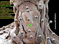
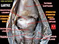
Cricothyroid ligament | |
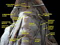
---|---|
Details | |
Identifiers | |
Latin | ligamentum cricothyroideum |
FMA | 55233 |
Anatomical terminology |
The cricothyroid ligament (also known as the cricothyroid membrane or cricovocal membrane) is a ligament in the neck. It connects the cricoid cartilage to the thyroid cartilage. It prevents these cartilages from moving too far apart. It is cut during an emergency cricothyrotomy to treat upper airway obstruction.
The cricothyroid ligament is composed of two parts:
The conus elasticus (which means elastic cone in Latin) is the lateral portion of the cricothyroid ligament. [1] The lateral portions are thinner and lie close under the mucous membrane of the larynx; they extend from the upper border of the cricoid cartilage to the lower margin of the vocal ligaments, with which they are continuous. [1] The vocal ligaments may therefore be regarded as the free borders of each conus elasticus. [1] They extend from the vocal processes of the arytenoid cartilages to the angle of the thyroid cartilage about midway between its upper and lower borders.
The prelaryngeal lymph node (also known as the Delphian lymph node) sits anterior to the median cricothyroid ligament. [2]
The cricothyroid ligament prevents the cricoid cartilage and the thyroid cartilage from moving too far apart.
The cricothyroid ligament is cut during an emergency cricothyrotomy. This kind of surgical intervention is necessary during airway obstruction above the level of vocal folds.
The cricothyroid ligament is named after the two structures it connects: the cricoid cartilage and the thyroid cartilage. It is also known as the cricothyroid membrane, and the cricovocal membrane. [3] The various parts of the cricothyroid ligament have been named in many different ways, which can cause confusion.
The cricothyroid ligament can be found in many other animals, such as cats, [4] dogs, [4] and horses. [5] The trachea can be accessed through the cricothyroid ligament, such as for aspiration. [4] It can be an important landmark. [5]
The larynx, commonly called the voice box, is an organ in the top of the neck involved in breathing, producing sound and protecting the trachea against food aspiration. The opening of larynx into pharynx known as the laryngeal inlet is about 4–5 centimeters in diameter. The larynx houses the vocal cords, and manipulates pitch and volume, which is essential for phonation. It is situated just below where the tract of the pharynx splits into the trachea and the esophagus. The word 'larynx' comes from the Ancient Greek word lárunx ʻlarynx, gullet, throat.ʼ
The trachea, also known as the windpipe, is a cartilaginous tube that connects the larynx to the bronchi of the lungs, allowing the passage of air, and so is present in almost all animals with lungs. The trachea extends from the larynx and branches into the two primary bronchi. At the top of the trachea the cricoid cartilage attaches it to the larynx. The trachea is formed by a number of horseshoe-shaped rings, joined together vertically by overlying ligaments, and by the trachealis muscle at their ends. The epiglottis closes the opening to the larynx during swallowing.
Tracheal intubation, usually simply referred to as intubation, is the placement of a flexible plastic tube into the trachea (windpipe) to maintain an open airway or to serve as a conduit through which to administer certain drugs. It is frequently performed in critically injured, ill, or anesthetized patients to facilitate ventilation of the lungs, including mechanical ventilation, and to prevent the possibility of asphyxiation or airway obstruction.
The recurrent laryngeal nerve (RLN) is a branch of the vagus nerve that supplies all the intrinsic muscles of the larynx, with the exception of the cricothyroid muscles. There are two recurrent laryngeal nerves, right and left. The right and left nerves are not symmetrical, with the left nerve looping under the aortic arch, and the right nerve looping under the right subclavian artery then traveling upwards. They both travel alongside the trachea. Additionally, the nerves are among the few nerves that follow a recurrent course, moving in the opposite direction to the nerve they branch from, a fact from which they gain their name.
The posterior cricoarytenoid muscle is a intrinsic muscle of the larynx. It arises from the cricoid cartilage; it inserts onto the arytenoid cartilage of the same side. It is innervated by the recurrent laryngeal nerve. Each acts to open the vocal folds by pulling the vocal fold of the same side laterally. It participates in the production of sounds.
The cricoid cartilage, or simply cricoid or cricoid ring, is the only complete ring of cartilage around the trachea. It forms the back part of the voice box and functions as an attachment site for muscles, cartilages, and ligaments involved in opening and closing the airway and in producing speech.
The thyroid cartilage is the largest of the nine cartilages that make up the laryngeal skeleton, the cartilage structure in and around the trachea that contains the larynx. It does not completely encircle the larynx.
The cricothyroid muscle is the only tensor muscle of the larynx aiding with phonation. It is innervated by the superior laryngeal nerve. Its action tilts the thyroid forward to help tense the vocal cords, thus increasing the pitch of the voice.
The arytenoid cartilages are a pair of small three-sided pyramids which form part of the larynx. They are the site of attachment of the vocal cords. Each is pyramidal or ladle-shaped and has three surfaces, a base, and an apex. The arytenoid cartilages allow for movement of the vocal cords by articulating with the cricoid cartilage. They may be affected by arthritis, dislocations, or sclerosis.
The sternohyoid muscle is a bilaterally paired, long, thin, narrow strap muscle of the anterior neck. It is one of the infrahyoid muscles. It is innervated by the ansa cervicalis. It acts to depress the hyoid bone.
The thyrohyoid muscle is a small skeletal muscle of the neck. Above, it attaches onto the greater cornu of the hyoid bone; below, it attaches onto the oblique line of the thyroid cartilage. It is innervated by fibres derived from the cervical spinal nerve 1 that run with the hypoglossal nerve to reach this muscle. The thyrohyoid muscle depresses the hyoid bone and elevates the larynx during swallowing. By controlling the position and shape of the larynx, it aids in making sound.
The thyrohyoid membrane is a broad, fibro-elastic sheet of the larynx. It connects the upper border of the thyroid cartilage to the hyoid bone.
The thyrocervical trunk is a short artery of the neck. It arises from the subclavian artery, then promptly divides into its branches: the inferior thyroid artery, suprascapular artery, and (sometimes) the transverse cervical artery.
The anterior jugular vein is a vein in the neck.
The superior laryngeal nerve is a branch of the vagus nerve. It arises from the middle of the inferior ganglion of vagus nerve and additionally also receives a sympathetic branch from the superior cervical ganglion.
The portion of the cavity of the larynx above the vestibular fold is called the laryngeal vestibule; it is wide and triangular in shape, its base or anterior wall presenting, however, about its center the backward projection of the tubercle of the epiglottis. It contains the vestibular folds, and between these and the vocal folds are the laryngeal ventricles.
The carina of trachea is a ridge of cartilage at the base of the trachea separating the openings of the left and right main bronchi.
The infraglottic cavity is the portion of the larynx situated inferior to the glottis. It is situated between the vocal cords, and the inferior border of the cricoid cartilage where it is continuous with the trachea.
The laryngeal saccules are soft tissue masses that are extensions of the laryngeal ventricles in the larynx. Their function is not well understood, but they may lubricate the vocal cords, and increase the resonance of vocalisation. They may be involved in airway disease and airway obstruction. They may be surgically removed using a laryngeal sacculectomy.
The malleus, or hammer, is a hammer-shaped small bone or ossicle of the middle ear. It connects with the incus, and is attached to the inner surface of the eardrum. The word is Latin for 'hammer' or 'mallet'. It transmits the sound vibrations from the eardrum to the incus (anvil).
This article incorporates text in the public domain from page 1078 of the 20th edition of Gray's Anatomy (1918)