Corniculate cartilages

Last updated
Corniculate cartilages
Gray952.png
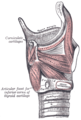
Ligaments of the larynx. Posterior view. (Corniculate cartilage labeled at center right.)
Gray955.png
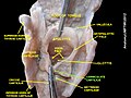
The entrance to the larynx, viewed from behind. (Corniculate cartilage labeled at bottom right.)
Details
Identifiers
Latin cartilagines corniculatae
TA98 A06.2.05.001
TA2 997
FMA 55110
Anatomical terminology

The corniculate cartilages (cartilages of Santorini) are two small conical nodules consisting of elastic cartilage, which articulate with the summits of the arytenoid cartilages and serve to prolong them posteriorly and medially.

Contents

They are situated in the posterior parts of the aryepiglottic folds of mucous membrane, and are sometimes fused with the arytenoid cartilages.

Eponym

It is named by Giovanni Domenico Santorini. [1] [2] The word "Corniculate" has a Latin root "cornu". Cornu means horn like projections. The projections of Corniculate cartilage look like "horns" hence the name. [3]

Additional images

Related Research Articles

<span class="mw-page-title-main">Larynx</span> Voice box, an organ in the neck of amphibians, reptiles, and mammals

The larynx, commonly called the voice box, is an organ in the top of the neck involved in breathing, producing sound and protecting the trachea against food aspiration. The opening of larynx into pharynx known as the laryngeal inlet is about 4–5 centimeters in diameter. The larynx houses the vocal cords, and manipulates pitch and volume, which is essential for phonation. It is situated just below where the tract of the pharynx splits into the trachea and the esophagus. The word 'larynx' comes from the Ancient Greek word lárunx ʻlarynx, gullet, throat.ʼ

<span class="mw-page-title-main">Hyoid bone</span> Bone situated in the neck between the chin and the thyroid cartilage

The hyoid bone is a horseshoe-shaped bone situated in the anterior midline of the neck between the chin and the thyroid cartilage. At rest, it lies between the base of the mandible and the third cervical vertebra.

<span class="mw-page-title-main">Rima glottidis</span> Opening between the true vocal cords and the arytenoid cartilages of the larynx

The rima glottidis is the opening between the two true vocal cords anteriorly, and the two arytenoid cartilages posteriorly. It is part of the larynx.

<span class="mw-page-title-main">Lateral cricoarytenoid muscle</span>

The lateral cricoarytenoid is an intrinsic muscle of the larynx. It attaches at the cricoid cartilage anteriorly, and at the arytenoid cartilage of the same side posteriorly. It is innervated by the recurrent laryngeal nerve. It acts to close the rima glottidis, thus closing the airway.

<span class="mw-page-title-main">Posterior cricoarytenoid muscle</span> Muscle of the larynx

The posterior cricoarytenoid muscle is a intrinsic muscle of the larynx. It arises from the cricoid cartilage; it inserts onto the arytenoid cartilage of the same side. It is innervated by the recurrent laryngeal nerve. Each acts to open the vocal folds by pulling the vocal fold of the same side laterally. It participates in the production of sounds.

<span class="mw-page-title-main">Cricoid cartilage</span> Complete ring of cartilage around the trachea

The cricoid cartilage, or simply cricoid or cricoid ring, is the only complete ring of cartilage around the trachea. It forms the back part of the voice box and functions as an attachment site for muscles, cartilages, and ligaments involved in opening and closing the airway and in producing speech.

<span class="mw-page-title-main">Thyroid cartilage</span> Largest of nine cartilages that make up the laryngeal skeleton

The thyroid cartilage is the largest of the nine cartilages that make up the laryngeal skeleton, the cartilage structure in and around the trachea that contains the larynx. It does not completely encircle the larynx.

<span class="mw-page-title-main">Arytenoid cartilage</span> Part of the larynx, to which the vocal folds (vocal cords) are attached

The arytenoid cartilages are a pair of small three-sided pyramids which form part of the larynx. They are the site of attachment of the vocal cords. Each is pyramidal or ladle-shaped and has three surfaces, a base, and an apex. The arytenoid cartilages allow for movement of the vocal cords by articulating with the cricoid cartilage. They may be affected by arthritis, dislocations, or sclerosis.

<span class="mw-page-title-main">Arytenoid muscle</span> Muscle of the larynx

The arytenoid muscle or interarytenoid muscle is a composite intrinsic muscle of the larynx, consisting of a transverse part and an oblique part - the two parts may be considered as separate muscles: an unpaired transverse arytenoid muscle, and a bilaterally paired oblique arytenoid muscle.

<span class="mw-page-title-main">Thyroarytenoid muscle</span>

The thyroarytenoid muscle is a broad, thin muscle that forms the body of the vocal fold and that supports the wall of the ventricle and its appendix. It functions to shorten the vocal folds.

<span class="mw-page-title-main">Thyrohyoid membrane</span> Elastic membrane in the larynx

The thyrohyoid membrane is a broad, fibro-elastic sheet of the larynx. It connects the upper border of the thyroid cartilage to the hyoid bone.

<span class="mw-page-title-main">Cuneiform cartilages</span> Type of cartilage in the human larynx

In the human larynx, the cuneiform cartilages are two small, elongated pieces of yellow elastic cartilage, placed one on either side, in the aryepiglottic fold.

<span class="mw-page-title-main">Aryepiglottic muscle</span>

The aryepiglottic muscle or aryepiglotticus muscle is an intrinsic muscle of the larynx.

<span class="mw-page-title-main">Oblique arytenoid</span>

The oblique arytenoid is bilaterally paired intrinsic muscle of the larynx. It is superficial to the transverse arytenoid; the oblique and transverse arytenoids are often considered two parts of a single muscle - the interarytenoid muscle.

<span class="mw-page-title-main">Laryngeal vestibule</span>

The portion of the cavity of the larynx above the vestibular fold is called the laryngeal vestibule; it is wide and triangular in shape, its base or anterior wall presenting, however, about its center the backward projection of the tubercle of the epiglottis. It contains the vestibular folds, and between these and the vocal folds are the laryngeal ventricles.

<span class="mw-page-title-main">Aryepiglottic fold</span> Folds near the larynx

The aryepiglottic folds are triangular folds of mucous membrane of the larynx. They enclose ligamentous and muscular fibres. They extend from the lateral borders of the epiglottis to the arytenoid cartilages, hence the name 'aryepiglottic'. They contain the aryepiglottic muscles and form the upper borders of the quadrangular membrane. They have a role in growling as a form of phonation. They may be narrowed and cause stridor, or be shortened and cause laryngomalacia.

<span class="mw-page-title-main">Giovanni Domenico Santorini</span> Italian anatomist (1681–1737)

Giovanni Domenico Santorini was an Italian anatomist. He was a native of Venice, earning his medical doctorate at Pisa in 1701. He is remembered for conducting anatomical dissections of the human body.

<span class="mw-page-title-main">Transverse arytenoid</span> Muscle in the larynx

The transverse arytenoid is an unpaired intrinsic muscle of the larynx. It is situated deep to the two oblique arytenoids; the oblique and transverse arytenoids are often considered two parts of a single muscle - the interarytenoid (arytenoid) muscle.

<span class="mw-page-title-main">Muscular process of arytenoid cartilage</span> Human anatomical structure

The muscular process of arytenoid cartilage is the posterolateral projection of the lateral angle of the base of the arytenoid cartilage. The muscular process gives insertion to the posterior cricoarytenoid muscles behind, and to the lateral cricoarytenoid muscles in front.

<span class="mw-page-title-main">Vocal process</span>

In the human larynx, the vocal process is the anterior angle of the base of the arytenoid cartilage, as it projects horizontally forward and gives attachment to the vocal ligament.

References

PD-icon.svgThis article incorporates text in the public domain from page 1075 of the 20th edition of Gray's Anatomy (1918)

  1. synd/3088 at Who Named It?
  2. G. D. Santorini. Observationes anatomicae. Venetiis, apus J. B. Recurti, 1724; Leiden, 1939.
  3. "Farlex free dictionary:Corniculate".