Aryepiglottic fold

Last updated
Aryepiglottic fold
Gray955.png
The entrance to the larynx, viewed from behind (aryepiglottic fold labeled at center right)
Gray956.png
Laryngoscopic view of interior of larynx (aryepiglottic fold labeled at center right)
Details
System Larynx
Identifiers
Latin plica aryepiglottica
TA98 A06.2.09.003
TA2 3193
FMA 55448
Anatomical terminology

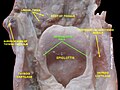
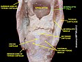
The aryepiglottic folds are triangular folds of mucous membrane of the larynx. They enclose ligamentous and muscular fibres. They extend from the lateral borders of the epiglottis to the arytenoid cartilages, hence the name 'aryepiglottic'. They contain the aryepiglottic muscles and form the upper borders of the quadrangular membrane. They have a role in growling as a form of phonation. They may be narrowed and cause stridor, or be shortened and cause laryngomalacia.

Contents

Structure

The aryepiglottic folds are triangular. They are narrow in front, wide behind, and slope obliquely downward and backward. They originate from the lateral borders of the epiglottis. [1] They insert into the arytenoid cartilages. [1]

In front, they are bounded by the epiglottis. Behind, they are bounded by the apices of the arytenoid cartilages, the corniculate cartilages, and the interarytenoid notch. [2] Within the posterior part of each aryepiglottic fold exists a cuneiform cartilage which forms a whitish prominence, the cuneiform tubercle.

The aryepiglottic folds contain the aryepiglottic muscles. They form the upper borders of the quadrangular membrane, and the lateral borders of the laryngeal inlet. [1]

Function

Phonation

Under certain circumstances, the aryepiglottic folds take part in phonation, for instance in the singing technique of vocal growl, such as practiced by Louis Armstrong and other jazz singers. The approximation of the aryepiglottic folds during vocalization may establish sustained co-oscillations, at relatively low frequencies, producing the growl or growling effect. [3]

Clinical significance

Stridor

If the aryepiglottic folds narrow the laryngeal inlet, they may cause stridor. [1] [4]

Laryngomalacia

The aryepiglottic folds are shortened in laryngomalacia. [5] They may be surgically removed to prevent problems eating and shortness of breath. [5]

Related Research Articles

The term phonation has slightly different meanings depending on the subfield of phonetics. Among some phoneticians, phonation is the process by which the vocal folds produce certain sounds through quasi-periodic vibration. This is the definition used among those who study laryngeal anatomy and physiology and speech production in general. Phoneticians in other subfields, such as linguistic phonetics, call this process voicing, and use the term phonation to refer to any oscillatory state of any part of the larynx that modifies the airstream, of which voicing is just one example. Voiceless and supra-glottal phonations are included under this definition.

<span class="mw-page-title-main">Larynx</span> Voice box, an organ in the neck of amphibians, reptiles, and mammals

The larynx, commonly called the voice box, is an organ in the top of the neck involved in breathing, producing sound and protecting the trachea against food aspiration. The opening of larynx into pharynx known as the laryngeal inlet is about 4–5 centimeters in diameter. The larynx houses the vocal cords, and manipulates pitch and volume, which is essential for phonation. It is situated just below where the tract of the pharynx splits into the trachea and the esophagus. The word 'larynx' comes from the Ancient Greek word lárunx ʻlarynx, gullet, throat.ʼ

<span class="mw-page-title-main">Epiglottis</span> Leaf-shaped flap in the throat that prevents food from entering the windpipe and the lungs

The epiglottis is a leaf-shaped flap in the throat that prevents food and water from entering the trachea and the lungs. It stays open during breathing, allowing air into the larynx. During swallowing, it closes to prevent aspiration of food into the lungs, forcing the swallowed liquids or food to go along the esophagus toward the stomach instead. It is thus the valve that diverts passage to either the trachea or the esophagus.

<span class="mw-page-title-main">Rima glottidis</span> Opening between the true vocal cords and the arytenoid cartilages of the larynx

The rima glottidis is the opening between the two true vocal cords anteriorly, and the two arytenoid cartilages posteriorly. It is part of the larynx.

<span class="mw-page-title-main">Lateral cricoarytenoid muscle</span>

The lateral cricoarytenoid is an intrinsic muscle of the larynx. It attaches at the cricoid cartilage anteriorly, and at the arytenoid cartilage of the same side posteriorly. It is innervated by the recurrent laryngeal nerve. It acts to close the rima glottidis, thus closing the airway.

<span class="mw-page-title-main">Posterior cricoarytenoid muscle</span> Muscle of the larynx

The posterior cricoarytenoid muscle is a intrinsic muscle of the larynx. It arises from the cricoid cartilage; it inserts onto the arytenoid cartilage of the same side. It is innervated by the recurrent laryngeal nerve. Each acts to open the vocal folds by pulling the vocal fold of the same side laterally. It participates in the production of sounds.

<span class="mw-page-title-main">Cricothyroid ligament</span>

The cricothyroid ligament is a ligament in the neck. It connects the cricoid cartilage to the thyroid cartilage. It prevents these cartilages from moving too far apart. It is cut during an emergency cricothyrotomy to treat upper airway obstruction.

<span class="mw-page-title-main">Arytenoid cartilage</span> Part of the larynx, to which the vocal folds (vocal cords) are attached

The arytenoid cartilages are a pair of small three-sided pyramids which form part of the larynx. They are the site of attachment of the vocal cords. Each is pyramidal or ladle-shaped and has three surfaces, a base, and an apex. The arytenoid cartilages allow for movement of the vocal cords by articulating with the cricoid cartilage. They may be affected by arthritis, dislocations, or sclerosis.

<span class="mw-page-title-main">Laryngeal inlet</span>

The laryngeal inlet is the opening that connects the pharynx and the larynx.

<span class="mw-page-title-main">Laryngomalacia</span> Medical condition

Laryngomalacia is the most common cause of chronic stridor in infancy, in which the soft, immature cartilage of the upper larynx collapses inward during inhalation, causing airway obstruction. It can also be seen in older patients, especially those with neuromuscular conditions resulting in weakness of the muscles of the throat. However, the infantile form is much more common. Laryngomalacia is one of the most common laryngeal congenital disease in infancy and public education about the signs and symptoms of the disease is lacking.

<span class="mw-page-title-main">Thyroarytenoid muscle</span>

The thyroarytenoid muscle is a broad, thin muscle that forms the body of the vocal fold and that supports the wall of the ventricle and its appendix. It functions to shorten the vocal folds.

<span class="mw-page-title-main">Cuneiform cartilages</span> Type of cartilage in the human larynx

In the human larynx, the cuneiform cartilages are two small, elongated pieces of yellow elastic cartilage, placed one on either side, in the aryepiglottic fold.

<span class="mw-page-title-main">Aryepiglottic muscle</span>

The aryepiglottic muscle or aryepiglotticus muscle is an intrinsic muscle of the larynx.

<span class="mw-page-title-main">Oblique arytenoid</span>

The oblique arytenoid is bilaterally paired intrinsic muscle of the larynx. It is superficial to the transverse arytenoid; the oblique and transverse arytenoids are often considered two parts of a single muscle - the interarytenoid muscle.

<span class="mw-page-title-main">Superior laryngeal nerve</span> Branch of the vagus nerve

The superior laryngeal nerve is a branch of the vagus nerve. It arises from the middle of the inferior ganglion of vagus nerve and additionally also receives a sympathetic branch from the superior cervical ganglion.

<span class="mw-page-title-main">Laryngeal vestibule</span>

The portion of the cavity of the larynx above the vestibular fold is called the laryngeal vestibule; it is wide and triangular in shape, its base or anterior wall presenting, however, about its center the backward projection of the tubercle of the epiglottis. It contains the vestibular folds, and between these and the vocal folds are the laryngeal ventricles.

<span class="mw-page-title-main">Corniculate cartilages</span>

The corniculate cartilages are two small conical nodules consisting of elastic cartilage, which articulate with the summits of the arytenoid cartilages and serve to prolong them posteriorly and medially.

<span class="mw-page-title-main">Vestibular fold</span> Part of the larynx

The vestibular fold is one of two thick folds of mucous membrane, each enclosing a narrow band of fibrous tissue, the vestibular ligament, which is attached in front to the angle of the thyroid cartilage immediately below the attachment of the epiglottis, and behind to the antero-lateral surface of the arytenoid cartilage, a short distance above the vocal process.

<span class="mw-page-title-main">Quadrangular membrane</span> Anatomical term for specific tissue in the throat

The quadrangular membrane is a layer of submucosa. It extends between the lateral margin of the epiglottis, and the apex and fovea triangularis of the ipsilateral arytenoid cartilage. It has free superior and inferior borders.

<span class="mw-page-title-main">Pharynx</span> Part of the throat that is behind the mouth and nasal cavity

The pharynx is the part of the throat behind the mouth and nasal cavity, and above the esophagus and trachea. It is found in vertebrates and invertebrates, though its structure varies across species. The pharynx carries food to the esophagus and air to the larynx. The flap of cartilage called the epiglottis stops food from entering the larynx.

References

PD-icon.svgThis article incorporates text in the public domain from page 1079 of the 20th edition of Gray's Anatomy (1918)

  1. 1 2 3 4 Reidenbach, Martina Maria (1998). "Aryepiglottic fold: Normal topography and clinical implications". Clinical Anatomy . 11 (4): 223–235. doi:10.1002/(SICI)1098-2353(1998)11:4<223::AID-CA1>3.0.CO;2-S. ISSN   1098-2353. PMID   9652536.
  2. Olesiak, Doctor (2023-07-23). "An Overview Of Throat Anatomy" . Retrieved 2023-07-29.
  3. Sakakibara, Ken-Ichi; Fuks, Leonardo; Imagawa, Hiroshi; Tayama, Niro (2004). "Growl Voice in Ethnic and Pop Styles" (PDF). Proceedings of the International Symposium on Musical Acoustics. Retrieved 19 June 2013.
  4. Peron, Didier L.; Graffino, Donatella B.; Zenker, David O. (1988). "The redundant aryepiglottic fold: Report of a new cause of stridor". The Laryngoscope . 98 (6): 659–663. doi:10.1288/00005537-198806000-00016. ISSN   1531-4995. PMID   3374242.
  5. 1 2 Polonovski, Jean-Michel; Contencin, Philippe; Francois, Martine; Viala, Paul; Narcy, Philippe (1990-08-01). "Aryepiglottic Fold Excision for the Treatment of Severe Laryngomalacia". Annals of Otology, Rhinology & Laryngology. 99 (8): 625–627. doi:10.1177/000348949009900807. ISSN   0003-4894. PMID   2382935.

Bibliography

  • Hals-Nasen-Ohren-Heilkunde, 12. Auflage, 2005, S.239–45. ISBN   978-3-540-21969-9 (in German)
  • Schuhmacher GH, Aumüller G, Topographische Anatomie des Menschen, Elsevier, 2004, ISBN   3-437-41367-8 (in German)