Thyrohyoid membrane

Last updated
Thyrohyoid membrane
Larynx external en.svg
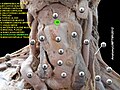
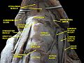
The ligaments of the larynx. Antero-lateral view.
Details
System skeletal
Identifiers
Latin membrana thyrohyoidea, membrana hyothyreoidea
TA98 A06.2.02.013
TA2 1651
FMA 55132
Anatomical terminology

The thyrohyoid membrane (or hyothyroid membrane) is a broad, fibro-elastic sheet of the larynx. It connects the upper border of the thyroid cartilage to the hyoid bone.

Contents

Structure

The thyrohyoid membrane is attached below to the upper border of the thyroid cartilage and to the front of its superior cornu, and above to the upper margin of the posterior surface of the body and greater cornu of the hyoid bone. [1] It passes behind the posterior surface of the body of the hyoid. It is separated from the hyoid bone by a mucous bursa, which allows for the upward movement of the larynx during swallowing. [1]

Its middle thicker part is termed the median thyrohyoid ligament. [1] Its lateral thinner portions are pierced by the superior laryngeal vessels and the internal branch of the superior laryngeal nerve. [1] Its anterior surface is in relation with the thyrohyoid muscle, sternohyoid muscle, and omohyoid muscles, and with the body of the hyoid bone. It is pierced by the superior laryngeal nerve. [2] It is also pierced the superior thyroid artery, where there is a thickening of the membrane. [2]

Clinical significance

Superior laryngeal artery

The thyrohyoid membrane needs to be manipulated to access the superior thyroid artery. [2]

History

The thyrohyoid membrane refers to the two structures it connects: the thyroid cartilage and the hyoid bone. It may also be known as the hyothyroid membrane, where the two structures are reversed. [3]

Additional images

Related Research Articles

Larynx Voice box, an organ in the neck of amphibians, reptiles, and mammals

The larynx, commonly called the voice box, is an organ in the top of the neck involved in breathing, producing sound and protecting the trachea against food aspiration. The opening of larynx into pharynx known as the laryngeal inlet is about 4–5 centimeters in diameter. The larynx houses the vocal cords, and manipulates pitch and volume, which is essential for phonation. It is situated just below where the tract of the pharynx splits into the trachea and the esophagus. The word larynx comes from a similar Ancient Greek word.

Neck Part of the body on many vertebrates that connects the head with the torso

The neck is the part of the body on many vertebrates that connects the head with the torso and provides the mobility and movements of the head. The structures of the human neck are anatomically grouped into four compartments; vertebral, visceral and two vascular compartments. Within these compartments, the neck houses the cervical vertebrae and cervical part of the spinal cord, upper parts of the respiratory and digestive tracts, endocrine glands, nerves, arteries and veins. Muscles of the neck are described separately from the compartments. They bound the neck triangles.

Articles related to anatomy include:

Hyoid bone

The hyoid bone is a horseshoe-shaped bone situated in the anterior midline of the neck between the chin and the thyroid cartilage. At rest, it lies at the level of the base of the mandible in the front and the third cervical vertebra (C3) behind.

External carotid artery Major artery of the head and neck

The external carotid artery is a major artery of the head and neck. It arises from the common carotid artery when it splits into the external and internal carotid artery. External carotid artery supplies blood to the face and neck.

Common carotid artery One of the two arteries that supply the head and neck with blood

In anatomy, the left and right common carotid arteries (carotids) are arteries that supply the head and neck with oxygenated blood; they divide in the neck to form the external and internal carotid arteries.

Sternothyroid muscle

The sternothyroid muscle, or sternothyroideus, is an infrahyoid muscle in the neck. It acts to depress the hyoid bone. It is below the sternohyoid muscle. It is shorter and wider than the sternohyoid.

Thyrohyoid muscle

The thyrohyoid muscle is a small skeletal muscle on the neck which depresses the hyoid and elevates the larynx.

Hyoglossus

The hyoglossus, thin and quadrilateral, arises from the side of the body and from the whole length of the greater cornu of the hyoid bone, and passes almost vertically upward to enter the side of the tongue, between the styloglossus and the inferior longitudinal muscle of the tongue. It forms a part of the floor of submandibular triangle.

Pharyngeal arch Embryonic precursor structures in vertebrates

The pharyngeal arches, also known as visceral arches, are structures seen in the embryonic development of vertebrates that are recognisable precursors for many structures. In fish, the arches are known as the branchial arches, or gill arches.

Lateral thyrohyoid ligament

The lateral thyrohyoid ligament is a round elastic cord, which forms the posterior border of the thyrohyoid membrane and passes between the tip of the superior cornu of the thyroid cartilage and the extremity of the greater cornu of the hyoid bone. The internal branch of the superior laryngeal nerve typical lies lateral to this ligament.

Lingual artery

The lingual artery arises from the external carotid artery between the superior thyroid artery and facial artery. It can be located easily in the tongue.

Superior thyroid artery

The superior thyroid artery arises from the external carotid artery just below the level of the greater cornu of the hyoid bone and ends in the thyroid gland.

Inferior thyroid artery Artery of the neck

The inferior thyroid artery is an artery in the neck. It arises from the thyrocervical trunk and passes upward, in front of the vertebral artery and longus colli muscle. It then turns medially behind the carotid sheath and its contents, and also behind the sympathetic trunk, the middle cervical ganglion resting upon the vessel.

Superior laryngeal nerve

The superior laryngeal nerve is a branch of the vagus nerve. It arises from the middle of the inferior ganglion of vagus nerve and in its course receives a branch from the superior cervical ganglion of the sympathetic nervous system.

Deep cervical fascia

The deep cervical fascia lies under cover of the platysma, and invests the muscles of the neck; it also forms sheaths for the carotid vessels, and for the structures situated in front of the vertebral column. Its attachment to the hyoid bone prevents the formation of a dewlap.

Carotid triangle

The carotid triangle is a portion of the anterior triangle of the neck.

Muscular triangle

The inferior carotid triangle, is bounded, in front, by the median line of the neck from the hyoid bone to the sternum; behind, by the anterior margin of the sternocleidomastoid; above, by the superior belly of the omohyoid.

Median thyrohyoid ligament

The median thyrohyoid ligament is the thicker, middle part of the thyrohyoid membrane. Its lateral thinner portions are pierced by the superior laryngeal vessels and the internal branch of the superior laryngeal nerve. Its anterior surface is in relation with the thyrohyoideus, sternohyoideus, and omohyoideus muscles, and with the body of the hyoid bone.

Outline of human anatomy Overview of and topical guide to human anatomy

The following outline is provided as an overview of and topical guide to human anatomy:

References

PD-icon.svgThis article incorporates text in the public domain from page 1076 of the 20th edition of Gray's Anatomy (1918)

  1. 1 2 3 4 Coleman, Lee; Zakowski, Mark; Gold, Julian A.; Ramanathan, Sivam (2013-01-01), Hagberg, Carin A. (ed.), "Chapter 1 - Functional Anatomy of the Airway", Benumof and Hagberg's Airway Management (Third Edition), Philadelphia: W.B. Saunders, pp. 3–20.e2, ISBN   978-1-4377-2764-7 , retrieved 2021-01-06
  2. 1 2 3 Barral, Jean-Pierre; Croibier, Alain (2011). "22 - Vessels of the thyroid". Visceral vascular manipulations. Edinburgh: Churchill Livingstone / Elsevier. pp. 166–181. doi:10.1016/B978-0-7020-4351-2.00022-3. ISBN   978-0-7020-4351-2. OCLC   805158376.
  3. Bruyn, G. W. (1983-12-01). "Superior Laryngeal Neuralgia". Cephalalgia. 3 (4): 235–240. doi:10.1046/j.1468-2982.1983.0304235.x. ISSN   0333-1024.