Laryngeal ventricle

Last updated
Laryngeal ventricle
Gray954.png
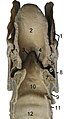
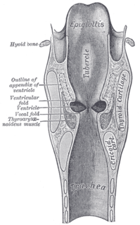
Coronal section of larynx and upper part of trachea, with Ventricle labeled at center left.
Details
Identifiers
Latin ventriculus laryngis
TA A06.2.09.010
FMA 64171
Anatomical terminology

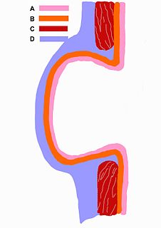
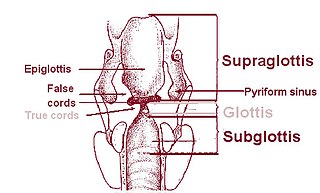
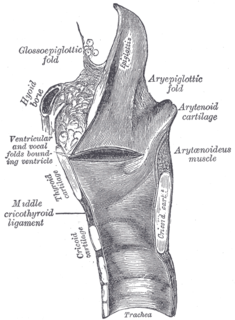
The laryngeal ventricle, (also called the ventricle of the larynx, laryngeal sinus, or Morgagni's sinus) [1] is a fusiform fossa, situated between the vestibular and vocal folds on either side, and extending nearly their entire length. There is also a sinus of Morgagni in the pharynx.

In anatomy, a fossa is a depression or hollow, usually in a bone, such as the hypophyseal fossa. Some examples include:

Vestibular fold

The vestibular fold is one of two thick folds of mucous membrane, each enclosing a narrow band of fibrous tissue, the vestibular ligament, which is attached in front to the angle of the thyroid cartilage immediately below the attachment of the epiglottis, and behind to the antero-lateral surface of the arytenoid cartilage, a short distance above the vocal process.

In the pharynx, the sinus of Morgagni is the enclosed space between the upper border of the superior pharyngeal constrictor muscle, the base of the skull and the pharyngeal aponeurosis.

Contents

The fossa is bounded, above, by the free crescentic edge of the vestibular ligament; below, by the straight margin of the vocal fold and laterally, by the mucous membrane covering the corresponding thyroarytenoid muscle.

Mucous membrane the protective layer, which lines the interior of hollow organs

A mucous membrane or mucosa is a membrane that lines various cavities in the body and covers the surface of internal organs. It consists of one or more layers of epithelial cells overlying a layer of loose connective tissue. It is mostly of endodermal origin and is continuous with the skin at various body openings such as the eyes, ears, inside the nose, inside the mouth, lip, vagina, the urethral opening and the anus. Some mucous membranes secrete mucus, a thick protective fluid. The function of the membrane is to stop pathogens and dirt from entering the body and to prevent bodily tissues from becoming dehydrated.

Thyroarytenoid muscle

The thyroarytenoid muscle is a broad, thin muscle that forms the body of the vocal fold and that supports the wall of the ventricle and its appendix. It functions to relax the vocal folds.

The anterior part of the ventricle leads up by a narrow opening into a pouch-like diverticulum, a mucous membranous sac of variable size called the appendix of the laryngeal ventricle. The appendix (also called the laryngeal saccule, pouch or Hilton's pouch) extends vertically from the laryngeal ventricle. It runs between the vestibular fold, thyroarytenoid muscle, and thyroid cartilage, and is conical, bending slightly backward. It is covered in roughly seventy mucous glands. The muscles surrounding the appendix compress it until mucus is secreted to lubricate the vocal folds.

Diverticulum medical or biological term for an outpouching of a hollow (or a fluid-filled) structure in the body

A diverticulum is the medical or biological term for an outpouching of a hollow structure in the body. Depending upon which layers of the structure are involved, they are described as being either true or false.

Thyroid cartilage

The thyroid cartilage is the largest of the nine cartilages that make up the laryngeal skeleton, the cartilage structure in and around the trachea that contains the larynx. It does not completely encircle the larynx; only the cricoid cartilage does.

Additional images

Related Research Articles

Larynx voice box, an organ in the neck of amphibians, reptiles, and mammals

The larynx, commonly called the voice box, is an organ in the top of the neck of tetrapods involved in breathing, producing sound, and protecting the trachea against food aspiration. The larynx houses the vocal folds, and manipulates pitch and volume, which is essential for phonation. It is situated just below where the tract of the pharynx splits into the trachea and the esophagus. The word larynx comes from a similar Ancient Greek word.

Articles related to anatomy include:

Epiglottis thin flap in the oesophagus

The epiglottis is a flap in the throat that keeps food from entering the windpipe and the lungs. The flap is made of elastic cartilage covered with a mucous membrane, attached to the entrance of the larynx. It projects obliquely upwards behind the tongue and the hyoid bone, pointing dorsally. It stands open during breathing, allowing air into the larynx. During swallowing, it closes to prevent aspiration and forcing the swallowed liquids or food to go along the esophagus instead. It is thus the valve that diverts passage to either the trachea or the esophagus.

Recurrent laryngeal nerve

The recurrent laryngeal nerve (RLN) is a branch of the vagus nerve that supplies all the intrinsic muscles of the larynx, with the exception of the cricothyroid muscles. There are two recurrent laryngeal nerves, right and left, in the human body. The right and left nerves are not symmetrical, with the left nerve looping under the aortic arch, and the right nerve looping under the right subclavian artery then traveling upwards. They both travel alongside of the trachea. Additionally, the nerves are one of few nerves that follow a recurrent course, moving in the opposite direction to the nerve they branch from, a fact from which they gain their name.

Anal canal part of large intestine

The anal canal is the terminal part of the large intestine. It is situated between the rectum and anus, below the level of the pelvic diaphragm. In humans it is approximately 2.5 to 4 cm (0.98-1.58 in) long. It lies in the anal triangle of perineum in between the right and left ischioanal fossa.

Rima glottidis Organ

The rima glottidis is the opening between the true vocal cords and the arytenoid cartilages of the larynx.

Posterior cricoarytenoid muscle

The posterior cricoarytenoid muscles are small, paired muscles that extend from the posterior cricoid cartilage to the arytenoid cartilages in the larynx.

Thyrohyoid membrane

The thyrohyoid membrane is a broad, fibro-elastic sheet of the larynx. It is attached below to the upper border of the thyroid cartilage and to the front of its superior cornu, and above to the upper margin of the posterior surface of the body and greater cornua of the hyoid bone, thus passing behind the posterior surface of the body of the hyoid. It is separated from the hyoid bone by a mucous bursa, which facilitates the upward movement of the larynx during swallowing.

Superior thyroid artery

The superior thyroid artery arises from the external carotid artery just below the level of the greater cornu of the hyoid bone and ends in the thyroid gland.

Oblique arytenoid

The oblique arytenoid, the more superficial Arytenoid muscle, forms two fasciculi, which pass from the base of one cartilage to the apex of the opposite one, and therefore cross each other like the limbs of the letter X; a few fibers are continued around the lateral margin of the cartilage, and are prolonged into the aryepiglottic fold; they are sometimes described as a separate muscle, the Aryepiglotticus.

Inferior thyroid artery Artery of the neck

The inferior thyroid artery is an artery in the neck. It arises from the thyrocervical trunk and passes upward, in front of the vertebral artery and longus colli muscle. It then turns medially behind the carotid sheath and its contents, and also behind the sympathetic trunk, the middle cervical ganglion resting upon the vessel.

Superior laryngeal nerve

The superior laryngeal nerve is a branch of the vagus nerve. It arises from the middle of the inferior ganglion of vagus nerve and in its course receives a branch from the superior cervical ganglion of the sympathetic nervous system.

Laryngeal vestibule

The portion of the cavity of the larynx above the vocal folds is called the laryngeal vestibule; it is wide and triangular in shape, its base or anterior wall presenting, however, about its center the backward projection of the tubercle of the epiglottis. It contains the vestibular folds, and between these and the vocal folds are the laryngeal ventricles.

Piriform sinus

On either side of the laryngeal orifice in humans is a recess, termed the piriform sinus, which is bounded medially by the aryepiglottic fold, laterally by the thyroid cartilage and thyrohyoid membrane. The fossae are involved in speech.

Laryngocele congenital disorder of respiratory system

Laryngocele refers to a congenital anomalous air sac communicating with the cavity of the larynx, which may bulge outward on the neck.

Outline of human anatomy Overview of and topical guide to human anatomy

The following outline is provided as an overview of and topical guide to human anatomy:

Laryngeal cavity

The laryngeal cavity extends from the laryngeal inlet downwards to the lower border of the cricoid cartilage where it is continuous with that of the trachea.

Pharynx part of the throat that is behind the mouth and nasal cavity

The pharynx is the part of the throat behind the mouth and nasal cavity and above the oesophagus and larynx, or the tubes going down to the stomach and the lungs. It is found in vertebrates and invertebrates, though its structure varies across species.

References

This article incorporates text in the public domain from page 1080 of the 20th edition of Gray's Anatomy (1918)

The public domain consists of all the creative works to which no exclusive intellectual property rights apply. Those rights may have expired, been forfeited, expressly waived, or may be inapplicable.

<i>Grays Anatomy</i> English-language textbook of human anatomy

Gray's Anatomy is an English language textbook of human anatomy originally written by Henry Gray and illustrated by Henry Vandyke Carter. Earlier editions were called Anatomy: Descriptive and Surgical, Anatomy of the Human Body and Gray's Anatomy: Descriptive and Applied, but the book's name is commonly shortened to, and later editions are titled, Gray's Anatomy. The book is widely regarded as an extremely influential work on the subject, and has continued to be revised and republished from its initial publication in 1858 to the present day. The latest edition of the book, the 41st, was published in September 2015.

  1. Medical Definition of Laryngeal sinus in lexic.us. Updated 05 Mar 2000
General Electric American industrial company

General Electric Company (GE) is an American multinational conglomerate incorporated in New York and headquartered in Boston. As of 2018, the company operates through the following segments: aviation, healthcare, power, renewable energy, digital industry, additive manufacturing, venture capital and finance, lighting, and oil and gas.