Muscular triangle

Last updated
Muscular triangle
Musculi coli base, my edits for tringles, MuscularTRI.svg
Muscular triangle
Gray1210.png
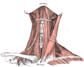
Side of neck, showing chief surface markings. (Nerves are yellow, arteries are red.)
Details
Identifiers
Latin trigonum musculare
TA A01.2.02.005
FMA 61601
Anatomical terminology

The inferior carotid triangle (or muscular triangle), is bounded, in front, by the median line of the neck from the hyoid bone to the sternum; behind, by the anterior margin of the sternocleidomastoid; above, by the superior belly of the omohyoid.

Hyoid bone horseshoe-shaped bone situated in the anterior midline of the neck between the chin and the thyroid cartilage

The hyoid bone is a horseshoe-shaped bone situated in the anterior midline of the neck between the chin and the thyroid cartilage. At rest, it lies at the level of the base of the mandible in the front and the third cervical vertebra (C3) behind.

Contents

It is covered by the integument, superficial fascia, platysma, and deep fascia, ramifying in which are some of the branches of the supraclavicular nerves.

Beneath these superficial structures are the sternohyoid and sternothyroid, which, together with the anterior margin of the sternocleidomastoid, conceal the lower part of the common carotid artery.

Common carotid artery

In anatomy, the left and right common carotid arteries (carotids) are arteries that supply the head and neck with oxygenated blood; they divide in the neck to form the external and internal carotid arteries.

This vessel is enclosed within its sheath, together with the internal jugular vein and vagus nerve; the vein lies lateral to the artery on the right side of the neck, but overlaps it below on the left side; the nerve lies between the artery and vein, on a plane posterior to both.

Internal jugular vein paired vein collecting the blood from the brain, the superficial parts of the face, and the neck

The internal jugular vein is a paired jugular vein that collects blood from the brain and the superficial parts of the face and neck. The vein runs in the carotid sheath with the common carotid artery and vagus nerve.

Vagus nerve tenth cranial nerve

The vagus nerve, historically cited as the pneumogastric nerve, is the tenth cranial nerve or CN X, and interfaces with the parasympathetic control of the heart, lungs, and digestive tract. The vagus nerves are paired but are normally referred to in the singular. It is the longest nerve of the autonomic nervous system in the human body.

In front of the sheath are a few descending filaments from the ansa cervicalis; behind the sheath are the inferior thyroid artery, the recurrent nerve, and the sympathetic trunk; and on its medial side, the esophagus, the trachea, the thyroid gland, and the lower part of the larynx.

Ansa cervicalis

The ansa cervicalis is a loop of nerves that are part of the cervical plexus. It lies superficial to the internal jugular vein in the carotid triangle. Its name means "handle of the neck" in Latin.

Inferior thyroid artery Artery of the neck

The inferior thyroid artery is an artery in the neck. It arises from the thyrocervical trunk and passes upward, in front of the vertebral artery and longus colli muscle. It then turns medially behind the carotid sheath and its contents, and also behind the sympathetic trunk, the middle cervical ganglion resting upon the vessel.

Sympathetic trunk paired bundle of nerve fibers that run from the base of the skull to the coccyx

The sympathetic trunks are a paired bundle of nerve fibers that run from the base of the skull to the coccyx.

By cutting into the upper part of this space, and slightly displacing the sternocleidomastoid, the common carotid artery may be tied below the omohyoid.

Sternocleidomastoid muscle A paired muscle of the neck that traverses the neck between the manubrium sterni and the mastoid process.

The sternocleidomastoid muscle is one of the largest and most superficial cervical muscles. The primary actions of the muscle are rotation of the head to the opposite side and flexion of the neck. The sternocleidomastoid is innervated by the accessory nerve.

See also

Related Research Articles

Subclavian artery major arteries of the upper thorax, below the clavicle

In human anatomy, the subclavian arteries are paired major arteries of the upper thorax, below the clavicle. They receive blood from the aortic arch. The left subclavian artery supplies blood to the left arm and the right subclavian artery supplies blood to the right arm, with some branches supplying the head and thorax. On the left side of the body, the subclavian comes directly off the aortic arch, while on the right side it arises from the relatively short brachiocephalic artery when it bifurcates into the subclavian and the right common carotid artery.

External carotid artery major artery of the head and neck

The external carotid artery is a major artery of the head and neck. It arises from the common carotid artery when it splits into the external and internal carotid artery. It supplies blood to the face and neck.

Omohyoid muscle

The omohyoid muscle is a muscle that depresses the hyoid. It is located in the front of the neck and consists of two bellies separated by an intermediate tendon. Its superior belly serves as the most lateral member of the infrahyoid muscles, located lateral to both the sternothyroid and thyrohyoid muscles. Its name derives from the Greek "omos" meaning shoulder, giving one of its attachments, and "hyoid", giving the other attachment - the hyoid bone.

The vertebral arteries are major arteries of the neck. Typically, the vertebral arteries originate from the subclavian arteries. Each vessel courses superiorly along each side of the neck, merging within the skull to form the single, midline basilar artery. As the supplying component of the vertebrobasilar vascular system, the vertebral arteries provide supply blood to the upper spinal cord, brainstem, cerebellum, and posterior part of brain.

Facial artery

The facial artery is a branch of the external carotid artery that supplies structures of the superficial face.

Sternothyroid muscle

The Sternothyroideus is a muscle in the neck. It is shorter and wider than the sternohyoideus, beneath which it is situated.

Carotid sheath

The carotid sheath is an anatomical term for the fibrous connective tissue that surrounds the vascular compartment of the neck. It is part of the deep cervical fascia of the neck, below the superficial cervical fascia meaning the subcutaneous adipose tissue immediately beneath the skin.

Occipital artery

The occipital artery arises from the external carotid artery opposite the facial artery. Its path is below the posterior belly of digastric to the occipital region. This artery supplies blood to the back of the scalp and sterno-mastoid muscles, and deep muscles in the back and neck.

Posterior triangle of the neck

The posterior triangle is a region of the neck.

Superior thyroid artery

The superior thyroid artery arises from the external carotid artery just below the level of the greater cornu of the hyoid bone and ends in the thyroid gland.

Transverse cervical artery Artery of the neck

The transverse cervical artery is an artery in the neck and a branch of the thyrocervical trunk, running at a higher level than the suprascapular artery.

Anterior triangle of the neck

The anterior triangle is a region of the neck.

Deep cervical fascia

The deep cervical fascia lies under cover of the platysma, and invests the muscles of the neck; it also forms sheaths for the carotid vessels, and for the structures situated in front of the vertebral column. Its attachment to the hyoid bone prevents the formation of a dewlap.

Submental triangle

The submental triangle is a division of the anterior triangle of the neck.

Submandibular triangle

The submandibular triangle corresponds to the region of the neck immediately beneath the body of the mandible.

Carotid triangle

The carotid triangle is a portion of the anterior triangle of the neck.

Subclavian triangle

The subclavian triangle, the smaller division of the posterior triangle, is bounded, above, by the inferior belly of the omohyoideus; below, by the clavicle; its base is formed by the posterior border of the sternocleidomastoideus.

References

This article incorporates text in the public domain from page 563 of the 20th edition of Gray's Anatomy (1918)