The scapula, also known as the shoulder blade, is the bone that connects the humerus with the clavicle. Like their connected bones, the scapulae are paired, with each scapula on either side of the body being roughly a mirror image of the other. The name derives from the Classical Latin word for trowel or small shovel, which it was thought to resemble.
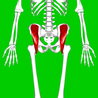
The iliacus is a flat, triangular muscle which fills the iliac fossa. It forms the lateral portion of iliopsoas, providing flexion of the thigh and lower limb at the acetabulofemoral joint.

The popliteal fossa is a shallow depression located at the back of the knee joint. The bones of the popliteal fossa are the femur and the tibia. Like other flexion surfaces of large joints, it is an area where blood vessels and nerves pass relatively superficially, and with an increased number of lymph nodes.

In human anatomy, the pterygopalatine fossa is a fossa in the skull. A human skull contains two pterygopalatine fossae—one on the left side, and another on the right side. Each fossa is a cone-shaped paired depression deep to the infratemporal fossa and posterior to the maxilla on each side of the skull, located between the pterygoid process and the maxillary tuberosity close to the apex of the orbit. It is the indented area medial to the pterygomaxillary fissure leading into the sphenopalatine foramen. It communicates with the nasal and oral cavities, infratemporal fossa, orbit, pharynx, and middle cranial fossa through eight foramina.
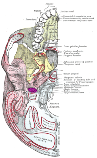
The jugular fossa is a deep depression (fossa) in the inferior part of the temporal bone at the base of the skull. It lodges the bulb of the internal jugular vein.

The canine fossa is a depression lateral to the incisive fossa of the maxilla in the musculoskeletal anatomy of the human head. It is larger and deeper than the comparable incisive fossa, and it is separated from it by a vertical ridge, the canine eminence, corresponding to the socket of the canine tooth.

The pterygoid processes of the sphenoid, one on either side, descend perpendicularly from the regions where the body and the greater wings of the sphenoid bone unite.

The maxillary artery supplies deep structures of the face. It branches from the external carotid artery just deep to the neck of the mandible.

The middle cranial fossa is formed by the sphenoid bones, and the temporal bones. It lodges the temporal lobes, and the pituitary gland. It is deeper than the anterior cranial fossa, is narrow medially and widens laterally to the sides of the skull. It is separated from the posterior cranial fossa by the clivus and the petrous crest.

The infratemporal fossa is an irregularly shaped cavity that is a part of the skull. It is situated below and medial to the zygomatic arch. It is not fully enclosed by bone in all directions. It contains superficial muscles, including the lower part of the temporalis muscle, the lateral pterygoid muscle, and the medial pterygoid muscle. It also contains important blood vessels such as the middle meningeal artery, the pterygoid plexus, and the retromandibular vein, and nerves such as the mandibular nerve (CN V3) and its branches.

The lesser petrosal nerve is the general visceral efferent (GVE) nerve conveying pre-ganglionic parasympathetic secretomotor fibers for the parotid gland from the tympanic plexus to the otic ganglion. It passes out of the tympanic cavity through the petrous part of the temporal bone into the middle cranial fossa of the cranial cavity, then exits the cranial cavity through its own canaliculus to reach the infratemporal fossa.

The subarcuate fossa is a shallow depression upon the internal surface of the petrous part of the temporal bone forming the wall of the posterior cranial fossa. The fossa accommodates the flocculus of the cerebellum. It is situated lateral/posterior to the internal auditory meatus.

The pterygoid canal is a passage in the sphenoid bone of the skull leading from just anterior to the foramen lacerum in the middle cranial fossa to the pterygopalatine fossa.

The mandibular fossa, also known as the glenoid fossa in some dental literature, is the depression in the temporal bone that articulates with the mandible.
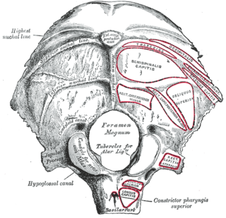
Behind either condyle of the lateral parts of occipital bone is a depression, the condyloid fossa, which receives the posterior margin of the superior facet of the atlas when the head is bent backward; the floor of this fossa is sometimes perforated by the condyloid canal, through which an emissary vein passes from the transverse sinus.
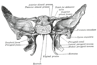
The pterygoid fossa is an anatomical term for the fossa formed by the divergence of the lateral pterygoid plate and the medial pterygoid plate of the sphenoid bone.

The infraspinousfossa of the scapula is much larger than the supraspinatous fossa; toward its vertebral margin a shallow concavity is seen at its upper part; its center presents a prominent convexity, while near the axillary border is a deep groove which runs from the upper toward the lower part.

The supraspinous fossa of the posterior aspect of the scapula is smaller than the infraspinous fossa, concave, smooth, and broader at its vertebral than at its humeral end. Its medial two-thirds give origin to the Supraspinatus.

The navicular fossa is a short dilated portion of the male urethra within the glans penis just proximal to the external urethral meatus. The roof of the fossa is especially dilated, forming a lacuna; medical instruments being inserted into the male urethra should initially be directed towards the floor of the fossa so as not to get snagged at the fossa. It is one of three dilations of the male urethra.

A cranial fossa is formed by the floor of the cranial cavity.