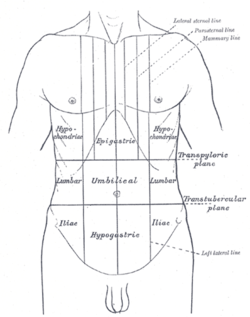
The umbilical region is one of the nine regions of the abdomen. It is the region that surrounds the area around the umbilicus and is placed approximately halfway between the xiphoid process and the pubic symphysis. This region of the abdomen contains part of the stomach, the head of the pancreas, the duodenum, a section of the transverse colon and the lower aspects of the left and right kidney. The upper three regions, from left to right, are the left hypochondriac, epigastric, and right hypochondriac regions. The middle three regions, from left to right, are the left lumbar, umbilical, and right lumbar regions. The bottom three regions, from left to right, are the left inguinal, hypogastric, and right inguinal regions.

A hernia is the abnormal exit of tissue or an organ, such as the bowel, through the wall of the cavity in which it normally resides. The term is also used for the normal development of the intestinal tract, referring to the retraction of the intestine from the extra-embryonal navel coelom into the abdomen in the healthy embryo at about 7½ weeks.

In anatomy, the epigastrium is the upper central region of the abdomen. It is located between the costal margins and the subcostal plane. Pain may be referred to the epigastrium from damage to structures derived from the foregut.

The external iliac arteries are two major arteries which bifurcate off the common iliac arteries anterior to the sacroiliac joint of the pelvis.

The abdominal internal oblique muscle, also internal oblique muscle or interior oblique, is an abdominal muscle in the abdominal wall that lies below the external oblique muscle and just above the transverse abdominal muscle.

The ventral body cavity is a human body cavity that is in the anterior (front) aspect of the human body. It is made up of the thoracic cavity, and the abdominopelvic cavity. The abdominopelvic cavity is further divided into the abdominal cavity and pelvic cavity, but there is no physical barrier between the two. The abdominal cavity contains digestive organs, spleen and the kidneys, the pelvic cavity contains the urinary bladder, internal reproductive organs, and rectum.

The abdominal external oblique muscle is the largest and outermost of the three flat abdominal muscles of the lateral anterior abdomen.

The abdomen is the part of the body between the thorax (chest) and pelvis, in humans and in other vertebrates. The abdomen is the front part of the abdominal segment of the torso. The area occupied by the abdomen is called the abdominal cavity. In arthropods, it is the posterior tagma of the body; it follows the thorax or cephalothorax.

The femoral nerve is a nerve in the thigh that supplies skin on the upper thigh and inner leg, and the muscles that extend the knee. It is the largest branch of the lumbar plexus.

The ilioinguinal nerve is a branch of the first lumbar nerve (L1). It separates from the first lumbar nerve along with the larger iliohypogastric nerve. It emerges from the lateral border of the psoas major just inferior to the iliohypogastric, and passes obliquely across the quadratus lumborum and iliacus. The ilioinguinal nerve then perforates the transversus abdominis near the anterior part of the iliac crest, and communicates with the iliohypogastric nerve between the transversus and the internal oblique muscle.

The obturator nerve in human anatomy arises from the ventral divisions of the second, third, and fourth lumbar nerves in the lumbar plexus; the branch from the third is the largest, while that from the second is often very small.

In anatomy, the division of the abdomen into regions can employ a nine-region scheme. The hypochondrium refers to the two hypochondriac regions in the upper third of the abdomen; the left hypochondrium and right hypochondrium. They are located on the lateral sides of the abdominal wall respectively, inferior to (below) the thoracic cage, being separated by the epigastrium.
The transpyloric plane, also known as Addison's plane, is an imaginary horizontal plane, located halfway between the suprasternal notch of the manubrium and the upper border of the symphysis pubis at the level of the first lumbar vertebrae, L1. It lies roughly a hand's breadth beneath the xiphisternum or midway between the xiphisternum and the umbilicus. The plane in most cases cuts through the pylorus of the stomach, the tips of the ninth costal cartilages and the lower border of the first lumbar vertebra.

The iliac fossa is a large, smooth, concave surface on the internal surface of the ilium.

The following outline is provided as an overview of and topical guide to human anatomy:
The interspinous plane is an anatomical transverse plane that passes through the anterior superior iliac spines. It separates the lateral lumbar region from the inguinal region and the umbilical region from the pubic region.
Supracristal plane is an anatomical transverse plane lying at the upper most part of the pelvis, the iliac crest. This is usually at the level of the L4 vertebrae. It passes through the umbilical region and the left and right lumbar regions.

Anatomical terminology is a form of scientific terminology used by anatomists, zoologists, and health professionals such as doctors, physicians, and pharmacists.

The human abdomen is divided into quadrants and regions by anatomists and physicians for the purposes of study, diagnosis, and treatment. The division into four quadrants allows the localisation of pain and tenderness, scars, lumps, and other items of interest, narrowing in on which organs and tissues may be involved. The quadrants are referred to as the left lower quadrant, left upper quadrant, right upper quadrant and right lower quadrant. These terms are not used in comparative anatomy, since most other animals do not stand erect.
This page is based on this
Wikipedia article Text is available under the
CC BY-SA 4.0 license; additional terms may apply.
Images, videos and audio are available under their respective licenses.