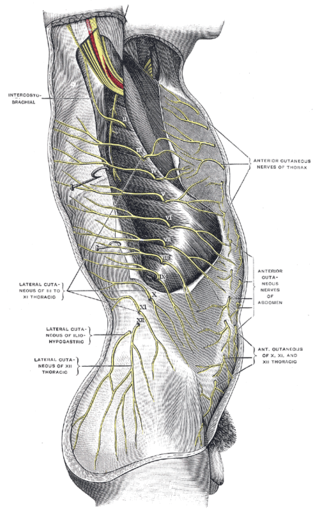
The anterior divisions of the seventh, eighth, ninth, tenth, and eleventh thoracic intercostal nerves are continued anteriorly from the intercostal spaces into the abdominal wall; hence they are named thoraco-abdominal nerves (or thoracicoabdominal intercostal nerves).
They have the same arrangement as the upper ones as far as the anterior ends of the intercostal spaces, where they pass behind the costal cartilages, and between the obliquus internus and transversus abdominis, to the sheath of the rectus abdominis, which they perforate.
They supply the rectus abdominis and end as the anterior cutaneous branches of the abdomen; they supply the skin of the front of the abdomen.
The lower intercostal nerves supply the intercostales and abdominal muscles; the last three send branches to the serratus posterior inferior. About the middle of their course they give off lateral cutaneous branches.
These pierce the intercostales externi and the obliquus externus abdominis, in the same line as the lateral cutaneous branches of the upper thoracic nerves, and divide into anterior and posterior branches, which are distributed to the skin of the abdomen and back; the anterior branches supply the digitations of the obliquus externus abdominis, and extend downward and forward nearly as far as the margin of the rectus abdominis; the posterior branches pass backward to supply the skin over the latissimus dorsi.

The rectus abdominis muscle, also known as the "abdominal muscle" or simply the "abs", is a pair of segmented skeletal muscle on the ventral aspect of a person's abdomen. The paired muscle is separated at the midline by a band of dense connective tissue called the linea alba, and the connective tissue defining each lateral margin of the rectus abdominus is the linea semilunaris. The muscle extends from the pubic symphysis, pubic crest and pubic tubercle inferiorly, to the xiphoid process and costal cartilages of the 5th–7th ribs superiorly.

The transverse abdominal muscle (TVA), also known as the transverse abdominis, transversalis muscle and transversus abdominis muscle, is a muscle layer of the anterior and lateral abdominal wall, deep to the internal oblique muscle. It is thought by most fitness instructors to be a significant component of the core.

The abdominal internal oblique muscle, also internal oblique muscle or interior oblique, is an abdominal muscle in the abdominal wall that lies below the external oblique muscle and just above the transverse abdominal muscle.
The abdominal external oblique muscle is the largest and outermost of the three flat abdominal muscles of the lateral anterior abdomen.

The intercostal nerves are part of the somatic nervous system, and arise from the anterior rami of the thoracic spinal nerves from T1 to T11. The intercostal nerves are distributed chiefly to the thoracic pleura and abdominal peritoneum, and differ from the anterior rami of the other spinal nerves in that each pursues an independent course without plexus formation.

In human anatomy, the inferior epigastric artery is an artery that arises from the external iliac artery. It is accompanied by the inferior epigastric vein; inferiorly, these two inferior epigastric vessels together travel within the lateral umbilical fold The inferior epigastric artery then traverses the arcuate line of rectus sheath to enter the rectus sheath, then anastomoses with the superior epigastric artery within the rectus sheath.

The femoral nerve is a nerve in the thigh that supplies skin on the upper thigh and inner leg, and the muscles that extend the knee. It is the largest branch of the lumbar plexus.

The iliohypogastric nerve is a nerve that originates from the lumbar plexus that supplies sensation to skin over the lateral gluteal and hypogastric regions and motor to the internal oblique muscles and transverse abdominal muscles.
The ilioinguinal nerve is a branch of the first lumbar nerve (L1). It separates from the first lumbar nerve along with the larger iliohypogastric nerve. It emerges from the lateral border of the psoas major just inferior to the iliohypogastric, and passes obliquely across the quadratus lumborum and iliacus. The ilioinguinal nerve then perforates the transversus abdominis near the anterior part of the iliac crest, and communicates with the iliohypogastric nerve between the transversus and the internal oblique muscle.

The obturator nerve in human anatomy arises from the ventral divisions of the second, third, and fourth lumbar nerves in the lumbar plexus; the branch from the third is the largest, while that from the second is often very small.

The conjoint tendon is a sheath of connective tissue formed from the lower part of the common aponeurosis of the abdominal internal oblique muscle and the transversus abdominis muscle, joining the muscle to the pelvis. It forms the medial part of the posterior wall of the inguinal canal.

The lumbar arteries are arteries located in the lower back or lumbar region. The lumbar arteries are in parallel with the intercostals.

The intercostobrachial nerve is the name applied to the lateral cutaneous branch of the second intercostal nerve. It arises anterior to the long thoracic nerve. It provides sensory innervation to the skin of the axilla, and a variable region of the medial side of the upper arm.
The subcostal nerve is a mixed motor and sensory nerve contributing to the lumbar plexus. It runs along the lower border of the twelfth rib, often gives a communicating branch to the first lumbar nerve, and passes under the lateral lumbocostal arch.
The subcostal arteries, so named because they lie below the last ribs, constitute the lowest pair of branches derived from the thoracic aorta, and are in series with the intercostal arteries.
The arcuate line of rectus sheath is a line of demarcation corresponding to the free inferior margin of the posterior layer of the rectus sheath inferior to which only the anterior layer of the rectus sheath is present and the rectus abdominis muscle is therefore in direct contact with the transversalis fascia. The arcuate line is concave inferior-wards.

The intercostal arteries are a group of arteries passing within an intercostal space. There are 9 anterior and 11 posterior intercostal arteries on each side of the body. The anterior intercostal arteries are branches of the internal thoracic artery and its terminal branch - the musculophrenic artery. The posterior intercostal arteries are branches of the supreme intercostal artery and thoracic aorta.
Lateral cutaneous branch can refer to:
The rectus sheath is a tough fibrous compartment formed by the aponeuroses of the transverse abdominal muscle, and the internal and external oblique muscles. It contains the rectus abdominis and pyramidalis muscles, as well as vessels and nerves.
The rectus abdominis muscle is crossed by three fibrous bands called the tendinous intersections or tendinous inscriptions. One is usually situated at the level of the umbilicus, one at the extremity of the xiphoid process, and the third about midway between the two.
This page is based on this
Wikipedia article Text is available under the
CC BY-SA 4.0 license; additional terms may apply.
Images, videos and audio are available under their respective licenses.

