Related Research Articles

The aorta is the main and largest artery in the human body, originating from the left ventricle of the heart, branching upwards immediately after, and extending down to the abdomen, where it splits at the aortic bifurcation into two smaller arteries. The aorta distributes oxygenated blood to all parts of the body through the systemic circulation.

Aortic dissection (AD) occurs when an injury to the innermost layer of the aorta allows blood to flow between the layers of the aortic wall, forcing the layers apart. In most cases, this is associated with a sudden onset of agonizing chest or back pain, often described as "tearing" in character. Vomiting, sweating, and lightheadedness may also occur. Damage to other organs may result from the decreased blood supply, such as stroke, lower extremity ischemia, or mesenteric ischemia. Aortic dissection can quickly lead to death from insufficient blood flow to the heart or complete rupture of the aorta.

Bicuspid aortic valve (BAV) is a form of heart disease in which two of the leaflets of the aortic valve fuse during development in the womb resulting in a two-leaflet (bicuspid) valve instead of the normal three-leaflet (tricuspid) valve. BAV is the most common cause of heart disease present at birth and affects approximately 1.3% of adults. Normally, the mitral valve is the only bicuspid valve and this is situated between the heart's left atrium and left ventricle. Heart valves play a crucial role in ensuring the unidirectional flow of blood from the atria to the ventricles, or from the ventricle to the aorta or pulmonary trunk. BAV is normally inherited.

An aortic aneurysm is an enlargement (dilatation) of the aorta to greater than 1.5 times normal size. Typically, there are no symptoms except when the aneurysm dissects or ruptures, which causes sudden, severe pain in the abdomen and lower back.

A thoracic aortic aneurysm is an aortic aneurysm that presents primarily in the thorax.

Coarctation of the aorta (CoA) is a congenital condition whereby the aorta is narrow, usually in the area where the ductus arteriosus inserts. The word coarctation means "pressing or drawing together; narrowing". Coarctations are most common in the aortic arch. The arch may be small in babies with coarctations. Other heart defects may also occur when coarctation is present, typically occurring on the left side of the heart. When a patient has a coarctation, the left ventricle has to work harder. Since the aorta is narrowed, the left ventricle must generate a much higher pressure than normal in order to force enough blood through the aorta to deliver blood to the lower part of the body. If the narrowing is severe enough, the left ventricle may not be strong enough to push blood through the coarctation, thus resulting in a lack of blood to the lower half of the body. Physiologically its complete form is manifested as interrupted aortic arch.

Aortic valve repair or aortic valve reconstruction is the reconstruction of both form and function of a dysfunctional aortic valve. Most frequently it is used for the treatment of aortic regurgitation. It can also become necessary for the treatment of aortic aneurysm, less frequently for congenital aortic stenosis.

The aortic arch, arch of the aorta, or transverse aortic arch is the part of the aorta between the ascending and descending aorta. The arch travels backward, so that it ultimately runs to the left of the trachea.

The ascending aorta (AAo) is a portion of the aorta commencing at the upper part of the base of the left ventricle, on a level with the lower border of the third costal cartilage behind the left half of the sternum.
Interrupted aortic arch is a very rare heart defect in which the aorta is not completely developed. There is a gap between the ascending and descending thoracic aorta. In a sense it is the complete form of a coarctation of the aorta. Almost all patients also have other cardiac anomalies, including a ventricular septal defect (VSD), aorto-pulmonary window, and truncus arteriosus. There are three types of interrupted aortic arch, with type B being the most common. Interrupted aortic arch is often associated with DiGeorge syndrome.
Valve-sparing aortic root replacement is a cardiac surgery procedure which is used to treat Aortic aneurysms and to prevent Aortic dissection. It involves replacement of the aortic root without replacement of the aortic valve. Two similar procedures were developed, one by Sir Magdi Yacoub, and another by Tirone David.
The Bentall procedure is a type of cardiac surgery involving composite graft replacement of the aortic valve, aortic root, and ascending aorta, with re-implantation of the coronary arteries into the graft. This operation is used to treat combined disease of the aortic valve and ascending aorta, including lesions associated with Marfan syndrome. The Bentall procedure was first described in 1968 by Hugh Bentall and Antony De Bono. It is considered a standard for individuals who require aortic root replacement, and the vast majority of individuals who undergo the surgery receive mechanical valves.

Annuloaortic ectasia is characterized by pure aortic valve regurgitation and aneurysmal dilatation of the ascending aorta. Men are more likely than women to develop idiopathic annuloaortic ectasia, which usually manifests in the fourth or sixth decades of life. Additional factors that contribute to this condition include osteogenesis imperfecta, inflammatory aortic diseases, intrinsic valve disease, Loeys-Dietz syndrome, Marfan syndrome, and operated congenital heart disease.

Endovascular aneurysm repair (EVAR) is a type of minimally-invasive endovascular surgery used to treat pathology of the aorta, most commonly an abdominal aortic aneurysm (AAA). When used to treat thoracic aortic disease, the procedure is then specifically termed TEVAR for "thoracic endovascular aortic/aneurysm repair." EVAR involves the placement of an expandable stent graft within the aorta to treat aortic disease without operating directly on the aorta. In 2003, EVAR surpassed open aortic surgery as the most common technique for repair of AAA, and in 2010, EVAR accounted for 78% of all intact AAA repair in the United States.

Acute aortic syndrome (AAS) describes a range of severe, painful, potentially life-threatening abnormalities of the aorta. These include aortic dissection, intramural thrombus, and penetrating atherosclerotic aortic ulcer. AAS can be caused by a lesion on the wall of the aorta that involves the tunica media, often in the descending aorta. It is possible for AAS to lead to acute coronary syndrome. The term was introduced in 2001.

Familial aortic dissection or FAD refers to the splitting of the wall of the aorta in either the arch, ascending or descending portions. FAD is thought to be passed down as an autosomal dominant disease and once inherited will result in dissection of the aorta, and dissecting aneurysm of the aorta, or rarely aortic or arterial dilation at a young age. Dissection refers to the actual tearing open of the aorta. However, the exact gene(s) involved has not yet been identified. It can occur in the absence of clinical features of Marfan syndrome and of systemic hypertension. Over time this weakness, along with systolic pressure, results in a tear in the aortic intima layer thus allowing blood to enter between the layers of tissue and cause further tearing. Eventually complete rupture of the aorta occurs and the pleural cavity fills with blood. Warning signs include chest pain, ischemia, and hemorrhaging in the chest cavity. This condition, unless found and treated early, usually results in death. Immediate surgery is the best treatment in most cases. FAD is not to be confused with PAU and IMH, both of which present in ways similar to that of familial aortic dissection.
Double aortic arch is a relatively rare congenital cardiovascular malformation. DAA is an anomaly of the aortic arch in which two aortic arches form a complete vascular ring that can compress the trachea and/or esophagus. Most commonly there is a larger (dominant) right arch behind and a smaller (hypoplastic) left aortic arch in front of the trachea/esophagus. The two arches join to form the descending aorta which is usually on the left side. In some cases the end of the smaller left aortic arch closes and the vascular tissue becomes a fibrous cord. Although in these cases a complete ring of two patent aortic arches is not present, the term ‘vascular ring’ is the accepted generic term even in these anomalies.

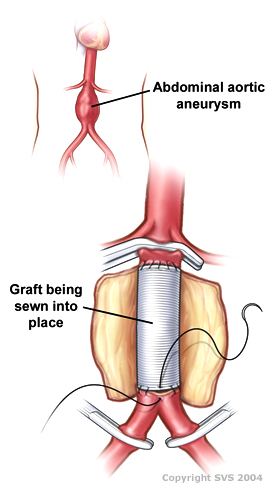
Open aortic surgery (OAS), also known as open aortic repair (OAR), describes a technique whereby an abdominal, thoracic or retroperitoneal surgical incision is used to visualize and control the aorta for purposes of treatment, usually by the replacement of the affected segment with a prosthetic graft. OAS is used to treat aneurysms of the abdominal and thoracic aorta, aortic dissection, acute aortic syndrome, and aortic ruptures. Aortobifemoral bypass is also used to treat atherosclerotic disease of the abdominal aorta below the level of the renal arteries. In 2003, OAS was surpassed by endovascular aneurysm repair (EVAR) as the most common technique for repairing abdominal aortic aneurysms in the United States.

Kurudamannil Abraham Abraham was an Indian interventional cardiologist and a medical writer. He was a Chief Cardiologist at the Southern Railway Headquarters Hospital, Chennai, and Chief Medical Director of the Southern Railways, where he worked for 25 years.
Giovanni J. Ughi, engineer and scientist, contributed to the invention of multimodality optical coherence tomography (OCT) and Laser-induced fluorescence molecular imaging, pioneering a first-in-man study of coronary arteries during his work at Massachusetts General Hospital and Harvard Medical School. The results of his work, combining two imaging technologies, may better identify dangerous coronary plaques, responsible for coronary artery disease and myocardial infarction.
References
- ↑ O'Rourke, Michael; Farnsworth, Alan; O'Rourke, John (2008). "Aortic Dimensions and Stiffness in Normal Adults". J Am Coll Cardiol Img. 1 (6): 749–751. doi: 10.1016/j.jcmg.2008.08.002 . PMID 19356511.
- ↑ Sugawara, Jun (2008). "Age-Associated Elongation of the Ascending Aorta". Adults J Am Coll Cardiol Img. 1 (6): 739–748. doi: 10.1016/j.jcmg.2008.06.010 . PMID 19356510.